. 2019 Jun;80(3):295-305.
doi: 10.1055/s-0038-1669937. Epub 2018 Sep 6.

Pure Endoscopic Lateral Orbitotomy Approach to the Cavernous Sinus, Posterior, and Infratemporal Fossae: Anatomic Study

Affiliations

Pure Endoscopic Lateral Orbitotomy Approach to the Cavernous Sinus, Posterior, and Infratemporal Fossae: Anatomic Study

Lili Laleva et al. J Neurol Surg B Skull Base. 2019 Jun.

Abstract

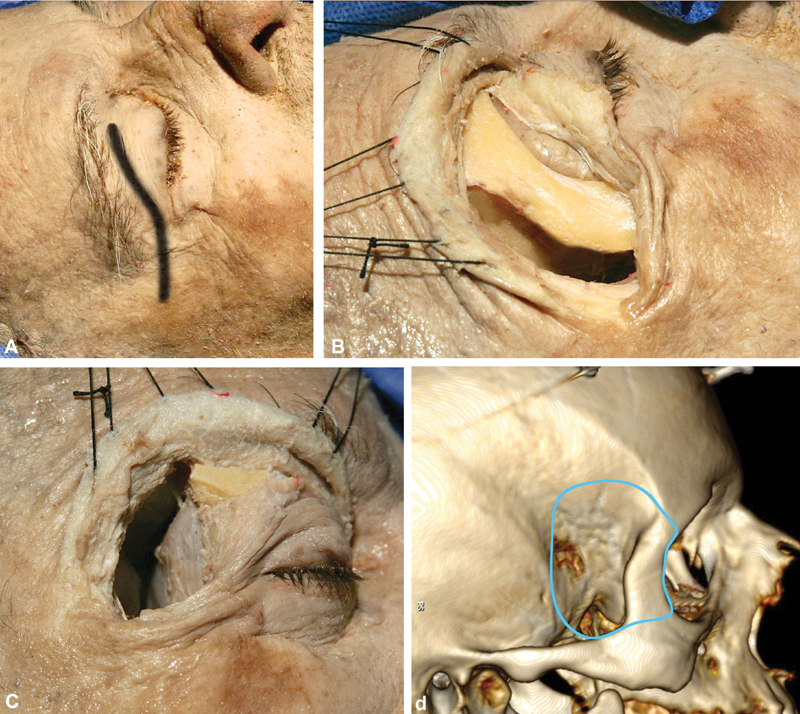
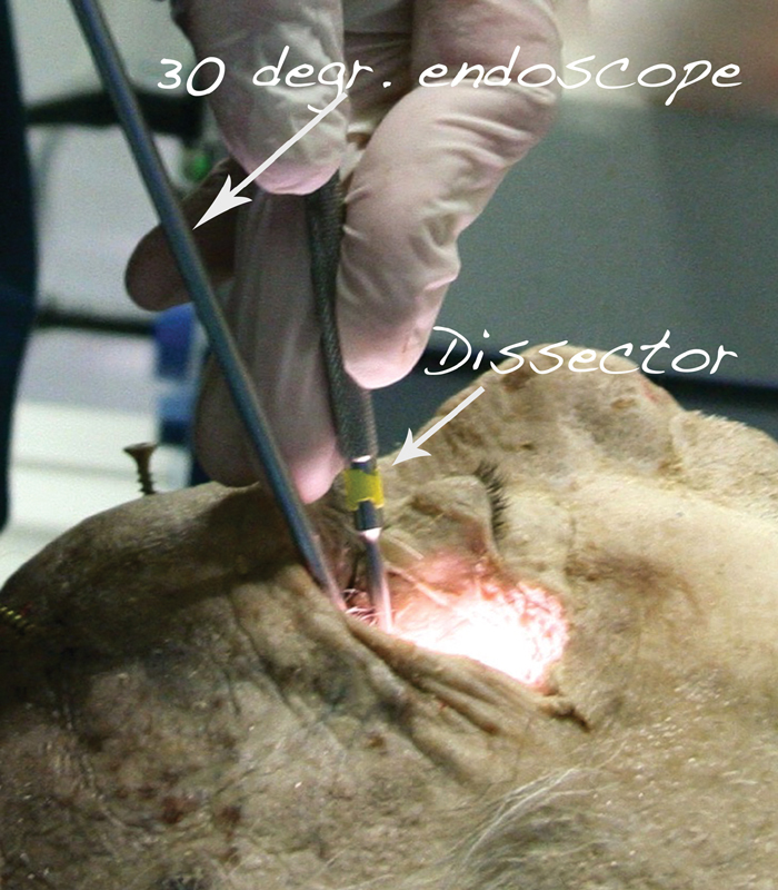
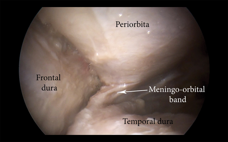
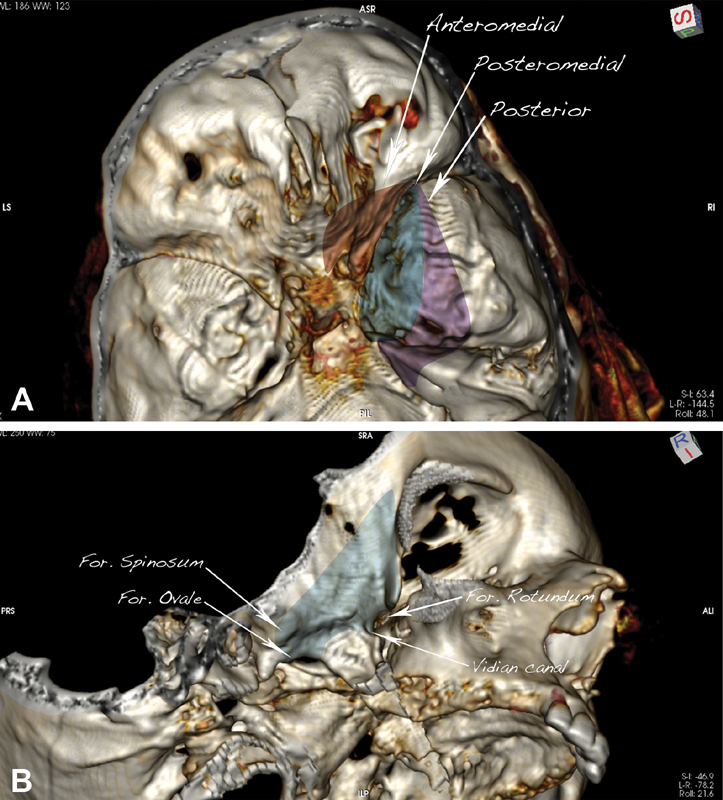
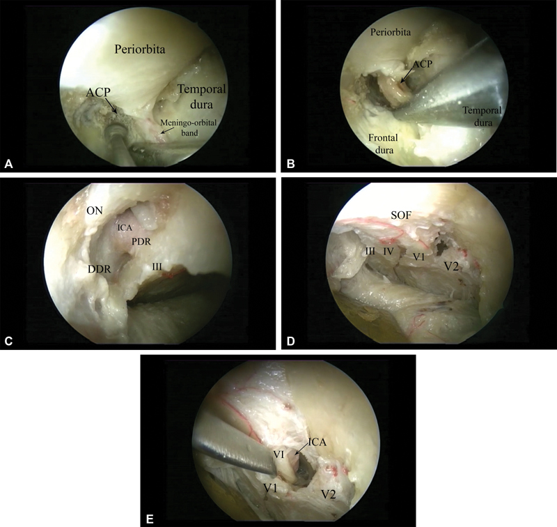
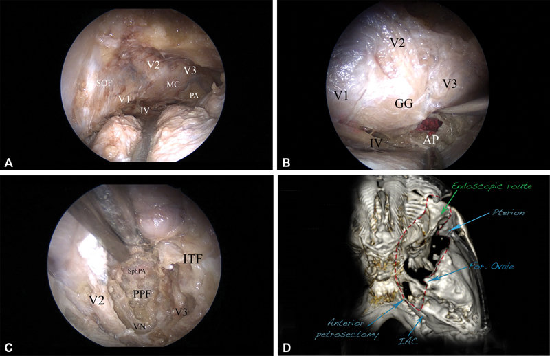
Objective The aim of this anatomic study is to describe a fully endoscopic lateral orbitotomy extradural approach to the cavernous sinus, posterior, and infratemporal fossae. Material and Methods Three prefixed latex-injected head specimens (six orbital exposures) were used in the study. Before and after dissection, a computed tomography scan was performed on each cadaver head and a neuronavigation system was used to guide the approach. The extent of bone removal and the area of exposure of the targeted corridor were evaluated with the aid of OsiriX software (Pixmeo, Bernex, Switzerland). Results The lateral orbital approach offers four main endoscopic extradural routes: the anteromedial, posteromedial, posterior, and inferior. The anteromedial route allows a direct route to the optic canal by removal of the anterior clinoid process, whereas the posteromedial route allows for exposure of the lateral wall of the cavernous sinus. The posterior route is targeted to Meckel's cave and provides access to the posterior cranial fossa by exposure and drilling of the petrous apex, whereas the inferior route gives access to the pterygopalatine and infratemporal fossae by drilling the floor of the middle cranial fossa and the bone between the second and third branches of the trigeminal nerve. Conclusion The lateral orbitotomy endoscopic approach provides direct access to the cavernous sinus, posterior, and infratemporal fossae. Advantages of the approach include a favorable angle of attack, minimal brain retraction, and the possibility of dissection within the two dural layers of the cavernous sinus without entering its neurovascular compartment.

Keywords: anatomy; endoscopic surgery; lateral orbital approach; skull base surgery.


Conflict of interest statement

Conflict of Interest All authors certify that they have no affiliations with or involvement in any organization or entity with any financial interest (such as honoraria; educational grants; participation in speakers' bureaus; membership, employment, consultancies, stock ownership, or other equity interest; and expert testimony or patent-licensing arrangements) or nonfinancial interest (such as personal or professional relationships, affiliations, knowledge, or beliefs) in the subject matter or materials discussed in this manuscript.

This article does not contain any studies with human participants performed by any of the authors.

Figures

Fig. 1
Fig. 1
Macroscopic part of the approach. ( A ) The skin incision is through the eyelid, starting from the midpupillary line and extending 1 cm behind the lateral orbital ridge, along the zygomatic arch. ( B ) Exposure of the lateral orbital rim in strict subperiosteal fashion. The lateral orbital rim is removed by drilling of the sphenoid ridge and exposure of the frontal, temporal dura and periorbita, followed by two osteotomies, using an oscillating saw, just above the frontozygomatic suture superiorly and at the level of the zygomatic arch. ( C ) Macroscopic view of the surgical field after the osteotomies and after the bone removal extending to the pterion is completed. The frontal, temporal lobes, as well as the periorbital, are visualized. ( D ) Three-dimensional reconstruction using the OsiriX software (version 5.8.1, Pixmeo, Bernex, Switzerland), presenting the limits of the craniotomy (the visualized screws are the markers for the neuronavigation).
Fig. 2
Fig. 2
Position of head, endoscope, and instruments in the laboratory settings.
Fig. 3
Fig. 3
Right lateral orbital approach. Endoscopic view of the right meningo-orbital band.
Fig. 4
Fig. 4
The surgical routes described in the study. ( A ) Anteromedial corridor (dark red), targeted to the optic canal; posteromedial (dark blue), targeted to the lateral cavernous sinus; posterior (purple), targeted to Meckel's cave and the petrous apex. ( B ) Inferior corridor (light blue), targeted to the pterygopalatine and infratemporal fossae.
Fig. 5
Fig. 5
Anatomical structures exposed through the different corridors (right side) ( A ) Drilling of the anterior clinoid process (ACP) using the anteromedial route; ( B ) removal of the anterior clinoid process with pituitary rongeur. ( C ) Endoscopic view via anteromedial route after removal of the anterior clinoid process and decompression of the optic nerve. The third nerve and the clinoid segment of the ICA, together with the proximal and distal dural rings as well as the optic nerve, are visualized. ( D ) Posteromedial route, exposing the lateral wall of the cavernous sinus. The III, IV, V1, and V2 nerves are seen. ( E ) The V1 nerve is elevated, and the abducens nerve, which is located medial to it, overlying the cavernous carotid artery, is exposed. Abbreviations: ACP, anterior clinoid process; DDR, distal dural ring; ICA, internal carotid artery; III, cranial nerve III; PDR, proximal dural ring; SOF, superior orbital fissure.
Fig. 6
Fig. 6
The posterolateral route and exposure to the posterior fossa, right side. ( A ) The whole cavernous sinus is seen, including the Meckel's cave (MC), V1, V2, V3, and IV cranial nerves, as well as the petrous apex (PA). ( B ) The mandibular division of the trigeminal nerve is elevated, and the anterior petrosectomy (AP) is done, exposing the corridor of the posterior fossa. The dura is incised, and the pons, as well as the trigeminal nerve, is seen. ( C ) The inferior route (light blue, Fig. 4B ), exposing the pterygopalatine fossa (PPF) and the infratemporal fossa (ITF). The bone between V2 and V3, as well as the floor of the middle cranial fossa, is removed, completely unroofing V2 and V3 and thus opening access to the PPF and the ITF. The vidian nerve (VN), as well as the vidian canal, are revealed. The sphenopalatine artery (SphPA) is seen in the PPF. ( D ) Postoperative computed tomography (CT) scan-based three-dimensional (3D) reconstruction presenting the area of bone removal (red dashed line), corresponding to the exposure to the posterior fossa (anterior petrosectomy) and to the infratemporal fossa (temporal fossa floor removal). 3D reconstruction using the OsiriX software (version 5.8.1, Pixmeo, Bernex, Switzerland). Abbreviations: AP, anterior petrosectomy; ITF, infratemporal fossa; MC Meckel's cave; PA, petrous apex; PPF, pterygopalatine fossa; SphPA, sphenopalatine artery; VN, vidian nerve.

References

    1. Al-Mefty O. Supraorbital-pterional approach to skull base lesions. Neurosurgery. 1987;21(04):474–477. - PubMed
    1. Hakuba A, Liu S, Nishimura S. The orbitozygomatic infratemporal approach: a new surgical technique. Surg Neurol. 1986;26(03):271–276. - PubMed
    1. Ikeda K, Yamashita J, Hashimoto M, Futami K. Orbitozygomatic temporopolar approach for a high basilar tip aneurysm associated with a short intracranial internal carotid artery: a new surgical approach. Neurosurgery. 1991;28(01):105–110. - PubMed
    1. Jane J A, Park T S, Pobereskin L H, Winn H R, Butler A B. The supraorbital approach: technical note. Neurosurgery. 1982;11(04):537–542. - PubMed
    1. Aziz K M, Froelich S C, Cohen P L, Sanan A, Keller J T, van Loveren H R. The one-piece orbitozygomatic approach: the MacCarty burr hole and the inferior orbital fissure as keys to technique and application. Acta Neurochir (Wien) 2002;144(01):15–24. - PubMed