. 2019 Jul;24(4):817-824.
doi: 10.1007/s12192-019-01009-8. Epub 2019 May 30.

The endoplasmic reticulum unfolded protein response varies depending on the affected region of the tissue but independently from the source of stress

Affiliations

The endoplasmic reticulum unfolded protein response varies depending on the affected region of the tissue but independently from the source of stress

Jessica Perochon et al. Cell Stress Chaperones. 2019 Jul.

Abstract

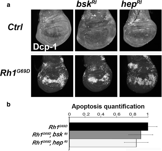
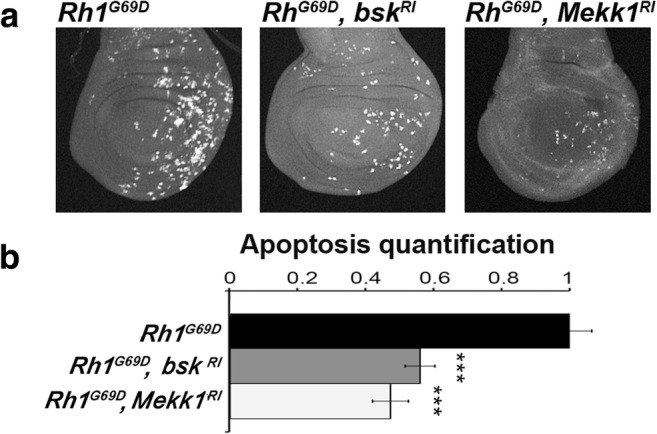
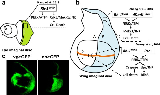
Accumulation of unfolded proteins and calcium dyshomeostasis induces endoplasmic reticulum (ER) stress, which can be resolved by the unfolded protein response (UPR). We have previously reported that activation of the PERK/ATF4 branch of the UPR, by overexpressing Presenilin in part of the vestigial domain of Drosophila wing imaginal discs, induces both a caspase-dependent apoptosis and a Slpr/JNK/Dilp8-dependent developmental delay that allows compensation of cell death in the tissue. Recently, dDad1 depletion in Drosophila in engrailed-expressing cells of wing imaginal discs was also reported to activate the PERK/ATF4 branch but induced Mekk1/JNK-dependent apoptosis. Here, we assessed whether the stressed cell location in the wing imaginal disc could explain these differences in response to chronic ER stress or whether the stress source could be responsible for the signaling discrepancy. To address this question, we overexpressed a Rhodopsin-1 mutant prone to aggregate either in vestigial- or engrailed-expressing cells. We observed similar responses to the Presenilin overexpression in the vestigial domain and to the dDad1 depletion in the engrailed domain. Therefore, the consequences of a PERK/ATF4 branch activation depend on the position of the cell in the Drosophila wing imaginal disc, suggesting interactions of PERK signaling with developmental pathways involved in the determination or maintenance of wing domains.

Keywords: Apoptosis; Homeostasis; PERK; UPR; Wing imaginal disc.


Figures

Fig. 1
Fig. 1
Rh-1G69D expression at the wing disc dorso-ventral boundary induces both cell death and JNK pathway activation in a PERK/ATF4-dependent manner. a Projections from confocal stacks of male third-instar larval wing imaginal discs immunostained with an anti-activated Dcp-1 antibody (top) and expressing the TRE reporter of JNK signaling (bottom). From left to right, genotypes are vg-Gal4, TRE-red-2L/+ (Ctrl), vg-Gal4, TRE-red-2L/+; UAS-Rh1G69D/+ (Rh1G69D), vg-Gal4, TRE-red-2L/+; UAS-Rh1G69D/UAS-Atf4-RNAi (Rh1G69D, Atf4RI) and vg-Gal4, TRE-red-2L/+; UAS-Rh1G69D/UAS-Perk-RNAi (Rh1G69D, PerkRI). b Quantification of apoptosis and JNK reporter stainings on wing discs of the previous genotypes. Error bars indicate S.E.M. Statistical difference with the control is indicated by * if p < 5% or *** if p < 10−3 (Mann-Whitney U test)
Fig. 2
Fig. 2
Rh-1G69D expression-induced apoptosis in the wing disc dorso-ventral boundary is JNK independent. a Projections from confocal stacks of male third-instar larval wing imaginal discs immunostained with an anti-activated Dcp-1 antibody. From left to right, genotypes of control discs on the top row are vg-GAL4/+, vg-GAL4/UAS-bsk-RNAi, and vg-GAL4/UAS-hep-RNAi. From left to right, wing imaginal discs expressing ectopically Rh1G69D (bottom row) display the following genotypes: vg-GAL4/+; UAS-Rh1G69D/+, vg-GAL4/UAS-bsk-RNAi; UAS-Rh1G69D/+ and vg-GAL4/UAS-hep-RNAi; UAS-Rh1G69D/+. b Quantification of apoptosis stainings on wing discs for the previously described genotypes allowing Rh-1G69D ectopic expression. Error bars indicate S.E.M. The Mann-Whitney U test revealed no statistic difference
Fig. 3
Fig. 3
Dilp8 expression in wing imaginal disc cells expressing Rh-1G69D depends on the Rac1/Slpr/JNK pathway. a Fluorescence intensity of GFP using a fire lookup table (ImageJ) reflecting the expression of a dilp8::EGFP reporter transgene in projections from confocal stacks of male third-instar larval wing imaginal discs. (Top row) From left to right, control genotypes are vg-GAL4/+; dilp8MI00727/+, vg-GAL4/UAS-Mekk1-RNAi; dilp8MI00727/+, vg-GAL4/UAS-slpr-RNAi; dilp8MI00727/+ and vg-GAL4/UAS-bsk-RNAi; dilp8MI00727/+. (Bottom row) From left to right, Rh-1G69D expressing wing disc genotypes are vg-GAL4/+; dilp8MI00727/UAS-Rh1G69D, vg-GAL4/UAS-Mekk1-RNAi; dilp8MI00727/UAS-Rh1G69D, vg-GAL4/UAS-slpr-RNAi; dilp8MI00727/UAS-Rh1G69D and vg-GAL4/UAS-bsk-RNAi; dilp8MI00727/UAS-Rh1G69D. b Quantification of Dilp8::EGFP was performed for the previously described genotypes allowing Rh-1G69D ectopic expression. Error bars represent the S.E.M. The asterisks indicate significant differences between the indicated genotype and its control (p < 10−3, Mann-Whitney U test). cdilp8 RNA levels measured by quantitative reverse PCR. Data represent mean ± S.E.M. of three independent experiments. RNA was extracted from control (vg-GAL4/+, open bar), from ER stressed (vg-GAL4/+; UAS-Rh1G69D/+, black bar), or from Rac1-depleted ER-stressed (vg-GAL4/UAS-rac1-RNAi; UAS-Rh1G69D/+, gray bar) wing imaginal discs. ** indicates a significant difference with the control (p < 1%, Student’s t test)
Fig. 4
Fig. 4
Rh-1G69D expression in the wing posterior compartment activates both JNK signaling and apoptosis in a PERK/ATF4-dependent manner. a Projections from confocal stacks of male third-instar larval wing imaginal discs expressing the TRE reporter of JNK signaling (top) and immunostained with an anti-activated Dcp-1 antibody (bottom). From left to right, genotypes are en-GAL4, TRE-red-2L/+; UAS-Rh1G69D/+ (Rh1G69D), en-GAL4, TRE-red-2L/+; UAS-Rh1G69D/UAS-Atf4-RNAi (Rh1G69D, Atf4RI) and en-GAL4, TRE-red-2L/+; UAS-Rh1G69D/UAS-Perk-RNAi (Rh1G69D, PerkRI). b Quantification of apoptosis (top) and JNK reporter (bottom) stainings on wing discs of the previous genotypes. Error bars indicate S.E.M. Statistical difference with the control is indicated by * if p < 5% or *** if p < 10−3 (Mann-Whitney U test)
Fig. 5
Fig. 5
Rh-1G69D expression in the wing posterior compartment induces a Mekk1/JNK-dependent apoptosis. a Projections from confocal stacks of male third-instar larval wing imaginal discs immunostained with an anti-activated Dcp-1 antibody. From left to right, genotypes of wing imaginal discs expressing ectopically Rh1G69D are en-GAL4/+; UAS-Rh1G69D/+, en-GAL4/UAS-bsk-RNAi; UAS-Rh1G69D/+ and en-GAL4/UAS-Mekk1-RNAi; UAS-Rh1G69D/+. b Quantification of apoptosis stainings on wing discs of the previous genotypes. Error bars indicate S.E.M. Statistical difference with the control is indicated by *** (p < 10−3, Mann-Whitney U test)
Fig. 6
Fig. 6
Proposed model of ER stress pathways in Drosophila. Mutated aggregation-prone Rh1 induces different pathways depending on the tissue location. As in the compartment posterior to the morphogenetic furrow of the eye imaginal disc (a, green domain), ectopic Rh1G69D triggers a JNK-dependent cell death in the posterior compartment (blue domain) but not on the dorso-ventral frontier (orange line) of the wing imaginal disc (b). Both mutant Rh1G69D and depletion of dDad1, which is essential to N-glycosylation, induce the same unfolded protein response to ER stress in the posterior compartment of wing discs. On the dorso-ventral frontier, mutant Rh1G69D activates a different unfolded protein response to ER stress, which is identical to the signaling induced by overexpressing Psn. The exact pattern of expression of vg-GAL4 (left) and en-GAL4 (right), which drive the different transgenes used in this study, is revealed with a UAS-GFP reporter transgene (c). Our data together with the literature strongly suggest that the ER stress source is not crucial to specify the cell response, while cell location is

References

    1. Aliee M, Röper J-C, Landsberg KP, Pentzold C, Widmann TJ, Jülicher F, Dahmann C. Physical mechanisms shaping the Drosophila dorsoventral compartment boundary. Curr Biol. 2012;22:967–976. doi: 10.1016/j.cub.2012.03.070. - DOI - PubMed
    1. Allen D, Seo J. ER stress activates the TOR pathway through Atf6. J Mol Signal. 2018;13:1. doi: 10.5334/1750-2187-13-1. - DOI - PMC - PubMed
    1. Andersen DS, Colombani J, Palmerini V, Chakrabandhu K, Boone E, Röthlisberger M, Toggweiler J, Basler K, Mapelli M, Hueber AO, Léopold P. The Drosophila TNF receptor Grindelwald couples loss of cell polarity and neoplastic growth. Nature. 2015;522:482–486. doi: 10.1038/nature14298. - DOI - PubMed
    1. Bergmann TJ, Fregno I, Fumagalli F, Rinaldi A, Bertoni F, Boersema PJ, Picotti P, Molinari M. Chemical stresses fail to mimic the unfolded protein response resulting from luminal load with unfolded polypeptides. J Biol Chem. 2018;293:5600–5612. doi: 10.1074/jbc.RA117.001484. - DOI - PMC - PubMed
    1. Bravo R, Parra V, Gatica D, et al. Endoplasmic reticulum and the unfolded protein response: dynamics and metabolic integration. Int Rev Cell Mol Biol. 2013;301:215–290. doi: 10.1016/B978-0-12-407704-1.00005-1. - DOI - PMC - PubMed

MeSH terms

LinkOut - more resources