. 2019 Jul 2;58(26):2906-2920.
doi: 10.1021/acs.biochem.8b01316. Epub 2019 Jun 18.

A Central Region of NF-κB Essential Modulator Is Required for IKKβ-Induced Conformational Change and for Signal Propagation

Affiliations

A Central Region of NF-κB Essential Modulator Is Required for IKKβ-Induced Conformational Change and for Signal Propagation

Robert Shaffer et al. Biochemistry. .

Abstract

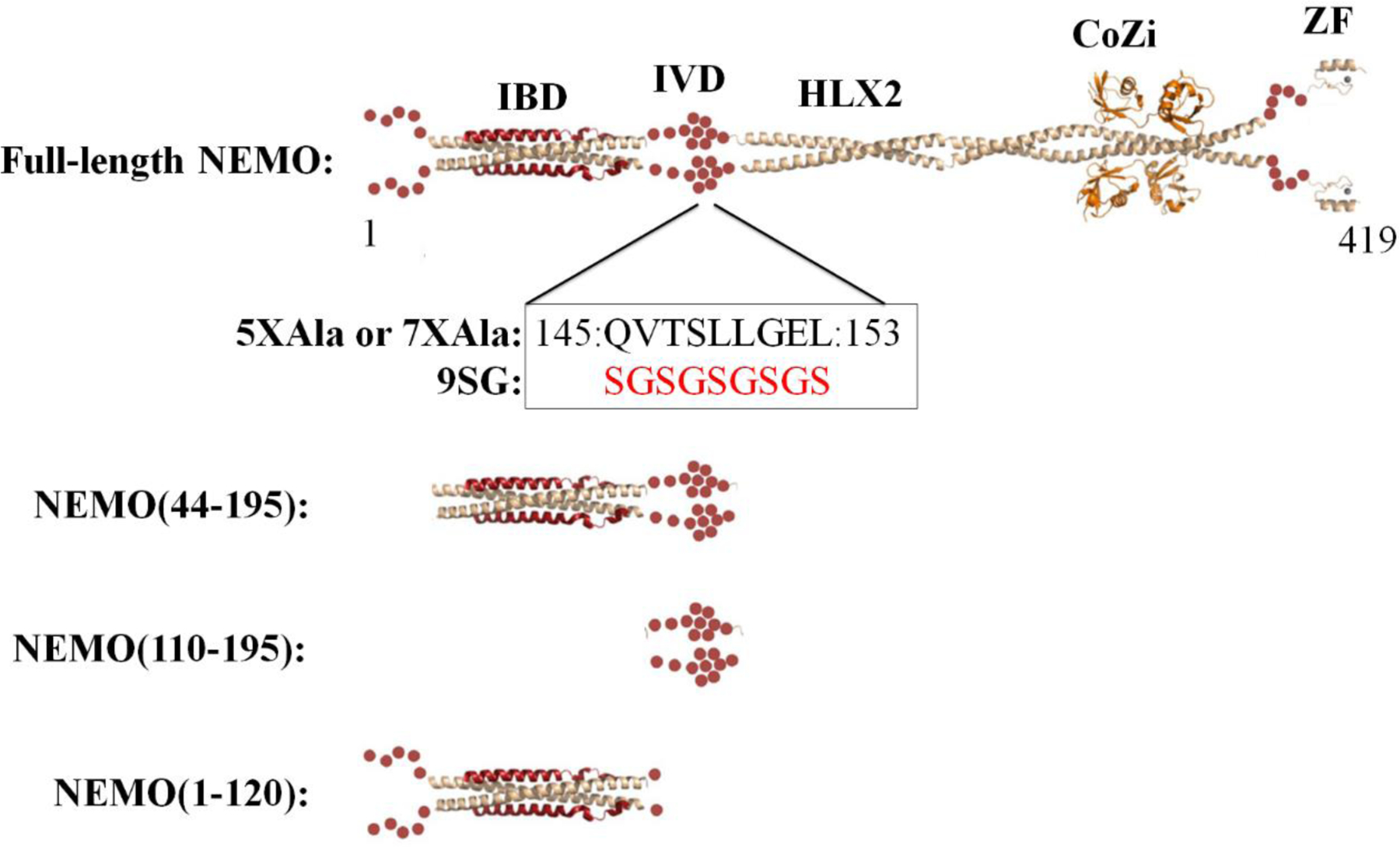
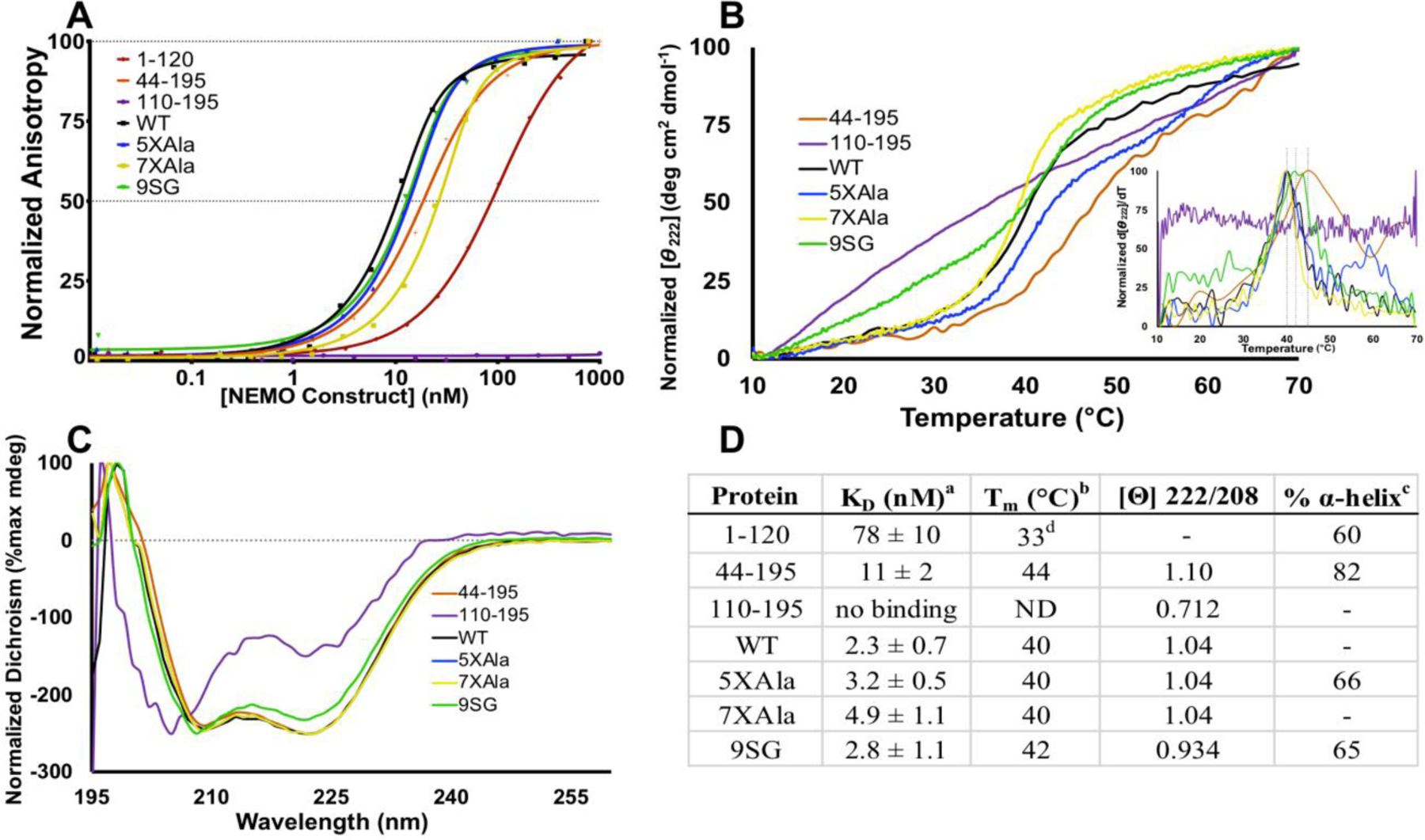
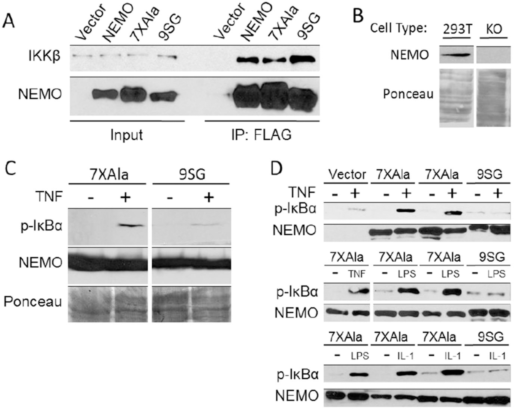
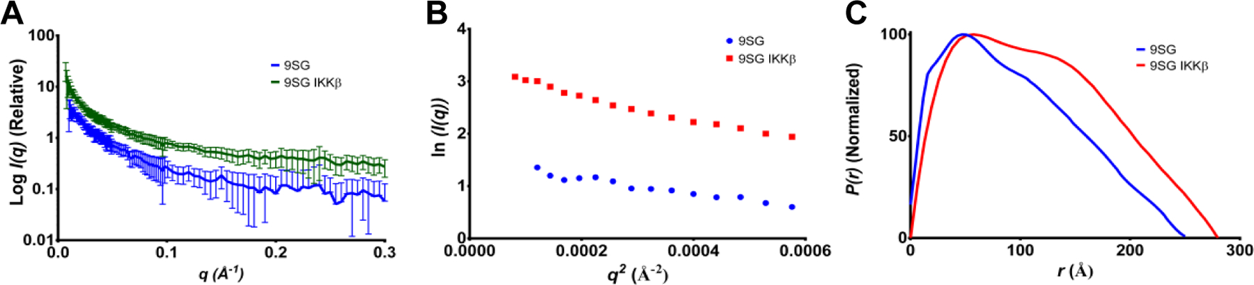
NF-κB essential modulator (NEMO) regulates NF-κB signaling by acting as a scaffold for the kinase IKKβ to direct its activity toward the NF-κB inhibitor, IκBα. Here, we show that a highly conserved central region of NEMO termed the intervening domain (IVD, amino acids 112-195) plays a key role in NEMO function. We determined a structural model of full-length NEMO by small-angle X-ray scattering and show that full-length, wild-type NEMO becomes more compact upon binding of a peptide comprising the NEMO binding domain of IKKβ (amino acids 701-745). Mutation of conserved IVD residues (9SG-NEMO) disrupts this conformational change in NEMO and abolishes the ability of NEMO to propagate NF-κB signaling in cells, although the affinity of 9SG-NEMO for IKKβ compared to that of the wild type is unchanged. On the basis of these results, we propose a model in which the IVD is required for a conformational change in NEMO that is necessary for its ability to direct phosphorylation of IκBα by IKKβ. Our findings suggest a molecular explanation for certain disease-associated mutations within the IVD and provide insight into the role of conformational change in signaling scaffold proteins.


Figures

Figure 1.
Figure 1.
Domain map of NEMO with existing experimentally determined structures of individual NEMO domains and constructs used in this study. NEMO in beige ribbon; IKKβ(701–745) in red ribbon; linear di-ubiquitin in orange ribbon; red spheres represent structurally uncharacterized regions of NEMO. IBD, IKKβ binding domain; IVD, intervening domain; HLX2, coiled-coil 2; CoZi, leucine zipper; ZF, zinc finger.
Figure 2.
Figure 2.
SAXS model of 5XAla-NEMO. (A) Scattering curve for 5XAla-NEMO (blue), with theoretical scattering (red) calculated from the model shown in panel D. Inset: the error-weighted residual difference plot, Δ/σ = [I exp(q) − cI mod(q)]/σ(q) versus q for model fitting to experimental scattering. (B) Guinier plot. Linearity at low q is not necessarily expected for a partially flexible, extended protein,. (C) Distance-distribution function generated by GNOM. (D) Shape reconstruction generated by DAMMIN (gray envelope) with superposed NEMO model (blue) generated from three NEMO domains by BUNCH. “N-term” and “C-term” labels denote NEMO N terminus and C terminus, respectively; the Intervening Domain is denoted “IVD.” (E) Guinier plots showing lack of aggregation with increasing protein concentration for WT-NEMO.
Figure 3.
Figure 3.
Alignment of the core sequence of the NEMO Intervening Domain. Shown is the region that spans residues 141 to 161 of human NEMO. Residues in blue are identical to the corresponding position in human NEMO. The full alignment is shown in Figure S2.
Figure 4.
Figure 4.
The IVD contributes to NEMO-IKKβ binding affinity and NEMO thermal stability. (A) Fluorescence anisotropy binding assays. The indicated NEMO proteins were titrated from 0.01 nM – 1000 nM, with FITC-labeled IKKβ(701–745) kept constant at 15 nM. Results are representative of three independent experiments performed with triplicate samples. (B) The indicated NEMO proteins were subjected to thermal denaturation, as monitored by CD. An increase in signal at Θ = 222 nm corresponds to loss of secondary structure. Inset: first derivative of melting curve used to identify Tm value. (C) CD spectra of NEMO constructs determined at 10 °C. The negative peak with minima at 208 and 222 nm is indicative of α-helical content. (D) Table of key results. aKD for binding FITC-IKKβ(701–745) measured using the FA binding assay. bDetermined by CD, monitoring change in Θ222 as temperature was increased at 1 °C/min. c The % α-helical content was determined from the CD spectrum in aqueous buffer compared with that in 90% TFE.d The Tm for NEMO(1–120) was reported previously, and is shown here for reference.
Figure 5.
Figure 5.
5XAla-NEMO and WT-NEMO become more compact upon IKKβ-binding. (A) SAXS scattering curves for 5XAla-NEMO, WT-NEMO, and NEMO(44–195), with and without IKKβ(701–745). Data for unliganded NEMO(44–195) and WT and NEMO(44–195), complexed with IKKβ in a 1:1.2 ratio and subjected to SEC-SAXS. Theoretical scattering curves calculated from the resulting structural models for the unbound proteins are overlaid in red. (B) Guinier plots for the data sets shown in (A). (C) Distance-distribution functions generated by GNOM for the data sets shown in (A). (D) Shape reconstruction for unbound NEMO(44–195) generated by DAMMIF. (E) Shape reconstruction for NEMO(44–195) in orange superposed on the shape reconstruction for 5XAla-NEMO (gray), from Figure 2.
Figure 6.
Figure 6.
The 9SG mutation abolishes the ability of NEMO to function in signal-induced activation of IκB kinase. (A) The indicated FLAG-tagged NEMO constructs were transfected into 293T cells, and extracts were immunoprecipitated (IP) with anti-FLAG beads. Immunoprecipitates were then analyzed by anti-FLAG (bottom) and anti-IKKβ (top) Western blotting. In the Input lanes, 4% of the extract used in the immunoprecipitations was analyzed by Western blotting. (B) Whole cell extracts from wild-type and NEMO-knockout 293T cells were analyzed by anti-NEMO Western blotting (top) or Ponceau staining for total protein (bottom). (C) 293T NEMO knockout cells were transfected with plasmids for the expression of 7XAla- or 9SG-NEMO. Cells that were either untreated (−) or treated with TNFα (+) were analyzed by Western blotting for phospho-IκBα or NEMO or by Ponceau staining. (D) Mouse NEMO knockout fibroblasts were stably transduced with retroviral vectors for the indicated NEMO proteins. Stable cell lines were then untreated (−) or treated (+) with the indicated compounds. Extracts were analyzed by Western blotting for the indicated proteins.
Figure 7.
Figure 7.
The IVD-region mutations present in 9SG-NEMO abolish the ability of IKKβ(701–745) binding to induce a more compact conformation. (A) SAXS scattering curve for 9SG-NEMO with (green) and without (blue) IKKβ(701–745). (B) Guinier plot for 9SG-NEMO. Linearity at low q is not necessarily expected for a partially flexible, extended protein,. (C) Distance-distribution function generated by GNOM.
Scheme 1.
Scheme 1.
Simplified scheme illustrating how NEMO mediates the phosphorylation of IκB by IKKα/β in response to activation of an upstream receptor (in this example, TNFR1), leading to ubiquitination and degradation of IκB, releasing NF-κB to translocate to the nucleus where it stimulates expression of target genes. Abbreviations: TNF, tumor necrosis factor; TNFR1, p55 TNF receptor; TRAF6, TNF receptor associated factor 6; RIP, receptor-interacting serine/threonine-protein kinase 1; TAK1, TGF β activated kinase 1, a.k.a. mitogen-activated protein kinase kinase kinase 7; p50/p65, a dimer comprising one form of the transcription factor NF-κB; IκB, inhibitor of κB; NEMO, NF-κB essential modulator; IKKα/β, inhibitor of κB kinase α and β; Ub, ubiquitin. The red asterisks represent sites of phosphorylation.

References

    1. Shaw AS, and Filbert EL (2009) Scaffold proteins and immune-cell signalling. Nat Rev Immunol 9, 47–56. - PubMed
    1. Cortese MS, Uversky VN, and Keith Dunker A (2008) Intrinsic disorder in scaffold proteins: Getting more from less. Prog Biophys Mol Biol 98, 85–106. - PMC - PubMed
    1. Pan CQ, Sudol M, Sheetz M, and Low BC (2012) Modularity and functional plasticity of scaffold proteins as p(l)acemakers in cell signaling. Cell Signal 24, 2143–2165. - PubMed
    1. Dhanasekaran DN, Kashef K, Lee CM, Xu H, and Reddy EP (2007) Scaffold proteins of MAP-kinase modules. Oncogene 26, 3185–3202. - PubMed
    1. Vaquero J, Nguyen Ho-Bouldoires TH, Clapéron A, and Fouassier L (2017) Role of the PDZ-scaffold protein NHERF1/EBP50 in cancer biology: from signaling regulation to clinical relevance. Oncogene 3067–3079. - PubMed

Publication types