

Forceful closure: cytoskeletal networks in embryonic wound repair
- PMID: 31145669
- PMCID: PMC6724689
- DOI: 10.1091/mbc.E18-04-0248
Forceful closure: cytoskeletal networks in embryonic wound repair
Abstract
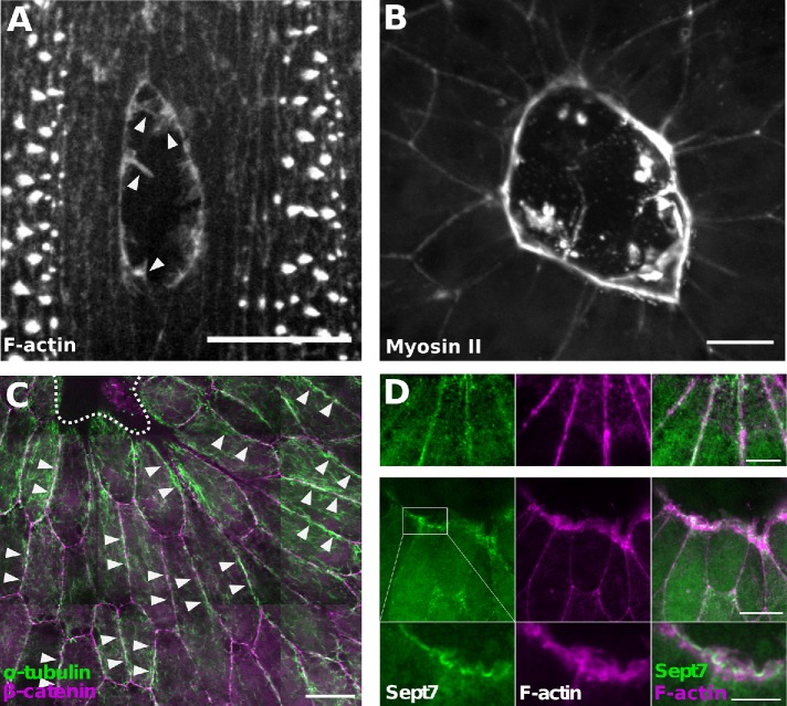
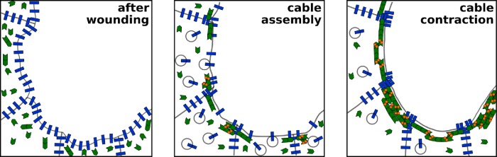
Embryonic tissues heal wounds rapidly and without scarring, in a process conserved across species and driven by collective cell movements. The mechanisms of coordinated cell movement during embryonic wound closure also drive tissue development and cancer metastasis; therefore, embryonic wound repair has received considerable attention as a model of collective cell migration. During wound closure, a supracellular actomyosin cable at the wound edge coordinates cells, while actin-based protrusions contribute to cell crawling and seamless wound healing. Other cytoskeletal networks are reorganized during wound repair: microtubules extend into protrusions and along cell-cell boundaries as cells stretch into damaged regions, septins accumulate at the wound margin, and intermediate filaments become polarized in the cells adjacent to the wound. Thus, diverse cytoskeletal networks work in concert to maintain tissue structure, while also driving and organizing cell movements to promote rapid repair. Understanding the signals that coordinate the dynamics of different cytoskeletal networks, and how adhesions between cells or with the extracellular matrix integrate forces across cells, will be important to elucidate the mechanisms of efficient embryonic wound healing and may have far-reaching implications for developmental and cancer cell biology.
Figures

Similar articles
-
Actin and myosin dynamics are independent during Drosophila embryonic wound repair.Mol Biol Cell. 2019 Nov 1;30(23):2901-2912. doi: 10.1091/mbc.E18-11-0703. Epub 2019 Sep 25. Mol Biol Cell. 2019. PMID: 31553671 Free PMC article.
-
Integrin-based adhesions promote cell-cell junction and cytoskeletal remodelling to drive embryonic wound healing.J Cell Sci. 2024 Mar 1;137(5):jcs261138. doi: 10.1242/jcs.261138. Epub 2023 Dec 15. J Cell Sci. 2024. PMID: 37970744
-
Rap1 coordinates cell-cell adhesion and cytoskeletal reorganization to drive collective cell migration in vivo.Curr Biol. 2023 Jul 10;33(13):2587-2601.e5. doi: 10.1016/j.cub.2023.05.009. Epub 2023 May 26. Curr Biol. 2023. PMID: 37244252
-
Tension (re)builds: Biophysical mechanisms of embryonic wound repair.Mech Dev. 2017 Apr;144(Pt A):43-52. doi: 10.1016/j.mod.2016.11.004. Epub 2016 Dec 15. Mech Dev. 2017. PMID: 27989746 Review.
-
Coordinating cell movements in vivo: junctional and cytoskeletal dynamics lead the way.Curr Opin Cell Biol. 2017 Oct;48:54-62. doi: 10.1016/j.ceb.2017.05.005. Epub 2017 Jun 13. Curr Opin Cell Biol. 2017. PMID: 28622576 Review.
Cited by
-
Osmolarity-independent electrical cues guide rapid response to injury in zebrafish epidermis.Elife. 2020 Nov 23;9:e62386. doi: 10.7554/eLife.62386. Elife. 2020. PMID: 33225997 Free PMC article.
-
Calcium bursts allow rapid reorganization of EFhD2/Swip-1 cross-linked actin networks in epithelial wound closure.Nat Commun. 2022 May 6;13(1):2492. doi: 10.1038/s41467-022-30167-0. Nat Commun. 2022. PMID: 35524157 Free PMC article.
-
Loss of β-Cytoplasmic Actin in the Intestinal Epithelium Increases Gut Barrier Permeability in vivo and Exaggerates the Severity of Experimental Colitis.Front Cell Dev Biol. 2020 Oct 23;8:588836. doi: 10.3389/fcell.2020.588836. eCollection 2020. Front Cell Dev Biol. 2020. PMID: 33195251 Free PMC article.
-
The α-Catenin mechanosensing M region is required for cell adhesion during tissue morphogenesis.J Cell Biol. 2023 Feb 6;222(2):e202108091. doi: 10.1083/jcb.202108091. Epub 2022 Dec 15. J Cell Biol. 2023. PMID: 36520419 Free PMC article.
-
CYRI controls epidermal wound closure and cohesion of invasive border cell cluster in Drosophila.J Cell Biol. 2024 Dec 2;223(12):e202310153. doi: 10.1083/jcb.202310153. Epub 2024 Oct 25. J Cell Biol. 2024. PMID: 39453414 Free PMC article.
References
Publication types
MeSH terms
Substances
LinkOut - more resources
Full Text Sources
