

Differential intrinsic functional connectivity changes in semantic variant primary progressive aphasia
- PMID: 31146321
- PMCID: PMC6465769
- DOI: 10.1016/j.nicl.2019.101797
Differential intrinsic functional connectivity changes in semantic variant primary progressive aphasia
Abstract
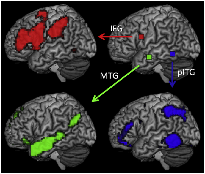
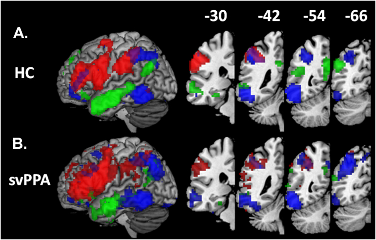
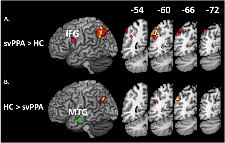
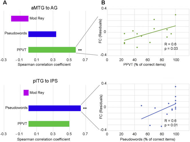
The semantic variant of primary progressive aphasia (svPPA) is a clinical syndrome characterized by semantic memory deficits with relatively preserved motor speech, syntax, and phonology. There is consistent evidence linking focal neurodegeneration of the anterior temporal lobes (ATL) to the semantic deficits observed in svPPA. Less is known about large-scale functional connectivity changes in this syndrome, particularly regarding the interplay between affected and spared language networks that leads to the unique cognitive dissociations typical of svPPA. Using whole-brain, seed-based connectivity on task-free Magnetic Resonance Imaging (MRI) data, we studied connectivity of networks anchored to three left-hemisphere regions crucially involved in svPPA symptomatology: ATL just posterior to the main atrophic area, opercular inferior frontal gyrus, and posterior inferior temporal lobe. First, in 32 healthy controls, these seeds isolated three networks: a ventral semantic network involving anterior middle temporal and angular gyri, a dorsal articulatory-phonological system involving inferior frontal and supramarginal regions, and a third functional connection between posterior inferior temporal and intraparietal regions likely involved in linking visual and linguistic processes. We then compared connectivity strength of these three networks between 16 svPPA patients and the 32 controls. In svPPA, decreased functional connectivity in the ventral semantic network correlated with weak semantic skills, while connectivity of the network seeded from the posterior inferior temporal lobe, though not significantly different between the two groups, correlated with pseudoword reading skills. Increased connectivity between the inferior frontal gyrus and the superior portion of the angular gyrus suggested possible adaptive changes. Our findings have two main implications. First, they support a functional subdivision of the left IPL based on its connectivity to specific language-related regions. Second, the unique neuroanatomical and linguistic profile observed in svPPA provides a compelling model for the functional interplay of these networks, being either up- or down- regulated in response to disease.
Keywords: Functional connectivity; Language; Parietal lobe; Primary progressive aphasia; Resting-state connectivity.
Copyright © 2019 The Authors. Published by Elsevier Inc. All rights reserved.
Figures
References
-
- Agosta F., Galantucci S., Valsasina P., Canu E., Meani A., Marcone A. Disrupted brain connectome in semantic variant of primary progressive aphasia. Neurobiol. Aging. 2014;35(11):2646–2655. - PubMed
-
- Ashburner J., Friston K.J. Unified segmentation. Neuroimage. 2005;26(3):839–851. - PubMed
-
- Beeson P.M.R. 2010. K. Arizona Battery for Reading and Spelling (ABRS) (cited; Available from:)
Publication types
MeSH terms
Grants and funding
- P50 AG023501/AG/NIA NIH HHS/United States
- R01 AG038791/AG/NIA NIH HHS/United States
- U01 AG052943/AG/NIA NIH HHS/United States
- T32 AG023481/AG/NIA NIH HHS/United States
- R01 DC013270/DC/NIDCD NIH HHS/United States
- F31 DC009145/DC/NIDCD NIH HHS/United States
- U54 NS092089/NS/NINDS NIH HHS/United States
- K24 DC015544/DC/NIDCD NIH HHS/United States
- R01 DC016345/DC/NIDCD NIH HHS/United States
- R03 DC013403/DC/NIDCD NIH HHS/United States
- P01 AG019724/AG/NIA NIH HHS/United States
- R01 DC016291/DC/NIDCD NIH HHS/United States
- R03 DC010878/DC/NIDCD NIH HHS/United States
- R01 NS100440/NS/NINDS NIH HHS/United States
- R56 NS050915/NS/NINDS NIH HHS/United States
- R01 NS050915/NS/NINDS NIH HHS/United States
