

Novel Peptide-Based Inhibitors for Microtubule Polymerization in Phytophthora capsici
- PMID: 31146360
- PMCID: PMC6600545
- DOI: 10.3390/ijms20112641
Novel Peptide-Based Inhibitors for Microtubule Polymerization in Phytophthora capsici
Abstract
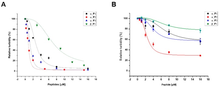
The plant disease Phytophthora blight, caused by the oomycete pathogen Phytophthora capsici, is responsible for major economic losses in pepper production. Microtubules have been an attractive target for many antifungal agents as they are involved in key cellular events such as cell proliferation, signaling, and migration in eukaryotic cells. In order to design a novel biocompatible inhibitor, we screened and identified inhibitory peptides against alpha- and beta-tubulin of P. capsici using a phage display method. The identified peptides displayed a higher binding affinity (nanomolar range) and improved specificity toward P. capsici alpha- and beta-tubulin in comparison to Homo sapiens tubulin as evaluated by fluorometric analysis. One peptide demonstrated the high inhibitory effect on microtubule formation with a nanomolar range of IC50 values, which were much lower than a well-known chemical inhibitor-benomyl (IC50 = 500 µM). Based on these results, this peptide can be employed to further develop promising candidates for novel antifungal agents against Phytophthora blight.
Keywords: microtubules; peptide; phage display; phytophthora blight; phytophthora capsici.
Conflict of interest statement
The authors declare no conflicts of interest.
Figures


Similar articles
-
Screening for peptides binding on Phytophthora capsici extracts by phage display.J Microbiol Methods. 2009 Jul;78(1):54-8. doi: 10.1016/j.mimet.2009.04.006. Epub 2009 Apr 21. J Microbiol Methods. 2009. PMID: 19389430
-
Alpha- and beta-tubulin from Phytophthora capsici KACC 40483: molecular cloning, biochemical characterization, and antimicrotubule screening.Appl Microbiol Biotechnol. 2009 Mar;82(3):513-24. doi: 10.1007/s00253-008-1821-7. Epub 2008 Dec 20. Appl Microbiol Biotechnol. 2009. PMID: 19099300
-
Antifungal activity of zedoary turmeric oil against Phytophthora capsici through damaging cell membrane.Pestic Biochem Physiol. 2019 Sep;159:59-67. doi: 10.1016/j.pestbp.2019.05.014. Epub 2019 May 24. Pestic Biochem Physiol. 2019. PMID: 31400785
-
Structural comparison of the interaction of tubulin with various ligands affecting microtubule dynamics.Curr Cancer Drug Targets. 2012 Jul;12(6):658-66. doi: 10.2174/156800912801784893. Curr Cancer Drug Targets. 2012. PMID: 22385515 Review.
-
Recent developments in tubulin polymerization inhibitors: An overview.Eur J Med Chem. 2014 Nov 24;87:89-124. doi: 10.1016/j.ejmech.2014.09.051. Epub 2014 Sep 16. Eur J Med Chem. 2014. PMID: 25240869 Review.
Cited by
-
Development of a Low-Molecular-Weight Aβ42 Detection System Using a Enzyme-Linked Peptide Assay.Biomolecules. 2021 Dec 2;11(12):1818. doi: 10.3390/biom11121818. Biomolecules. 2021. PMID: 34944462 Free PMC article.
-
4'-O-Methylbroussochalcone B as a novel tubulin polymerization inhibitor suppressed the proliferation and migration of acute myeloid leukaemia cells.BMC Cancer. 2021 Jan 22;21(1):91. doi: 10.1186/s12885-020-07759-4. BMC Cancer. 2021. PMID: 33482772 Free PMC article.
-
Design, Synthesis, and Antifungal/Anti-Oomycete Activities of Novel 1,2,4-Triazole Derivatives Containing Carboxamide Fragments.J Fungi (Basel). 2024 Feb 19;10(2):160. doi: 10.3390/jof10020160. J Fungi (Basel). 2024. PMID: 38392832 Free PMC article.
References
-
- Sanogo S., Ji P. Integrated management of Phytophthora capsici on solanaceous and cucurbitaceous crops: current status, gaps in knowledge and research needs. Can. J. Plant Pathol. 2012;34:479–492. doi: 10.1080/07060661.2012.732117. - DOI
-
- Oh B.T., Hur H., Lee K.J., Shanthi K., Soh B.Y., Lee W.J., Myung H., Kannan S.K. Suppression of Phytophthora blight on pepper (Capsicum annuum L) by bacilli isolated from brackish environment. Biocontrol Sci. Technol. 2011;21:1297–1311. doi: 10.1080/09583157.2011.618264. - DOI
-
- Zhang S., White T.L., Martinez M.C., McInroy J.A., Kloepper J.W., Klassen W. Evaluation of plant growth-promoting rhizobacteria for control of Phytophthora blight on squash under greenhouse conditions. Biol. Control. 2010;53:129–135. doi: 10.1016/j.biocontrol.2009.10.015. - DOI
-
- Lan C.Z., Liu P.Q., Li B.J., Chen Q.H., Weng Q.Y. Development of a specific PCR assay for the rapid and sensitive detection of Phytophthora capsica. Aust. Plant Pathol. 2012;42:379–384. doi: 10.1007/s13313-012-0185-8. - DOI
MeSH terms
Substances
Grants and funding
LinkOut - more resources
Full Text Sources
