. 2019 Jun 4;73(21):2705-2718.
doi: 10.1016/j.jacc.2019.02.074.

Osteopontin Promotes Left Ventricular Diastolic Dysfunction Through a Mitochondrial Pathway

Affiliations

Osteopontin Promotes Left Ventricular Diastolic Dysfunction Through a Mitochondrial Pathway

Keyvan Yousefi et al. J Am Coll Cardiol. .

Abstract

Background: Patients with chronic kidney disease (CKD) and coincident heart failure with preserved ejection fraction (HFpEF) may constitute a distinct HFpEF phenotype. Osteopontin (OPN) is a biomarker of HFpEF and predictive of disease outcome. We recently reported that OPN blockade reversed hypertension, mitochondrial dysfunction, and kidney failure in Col4a3-/- mice, a model of human Alport syndrome.

Objectives: The purpose of this study was to identify potential OPN targets in biopsies of HF patients, healthy control subjects, and human induced pluripotent stem cell-derived cardiomyocytes (hiPS-CMs), and to characterize the cardiac phenotype of Col4a3-/- mice, relate this to HFpEF, and investigate possible causative roles for OPN in driving the cardiomyopathy.

Methods: OGDHL mRNA and protein were quantified in myocardial samples from patients with HFpEF, heart failure with reduced ejection fraction, and donor control subjects. OGDHL expression was quantified in hiPS-CMs treated with or without anti-OPN antibody. Cardiac parameters were evaluated in Col4a3-/- mice with and without global OPN knockout or AAV9-mediated delivery of 2-oxoglutarate dehydrogenase-like (Ogdhl) to the heart.

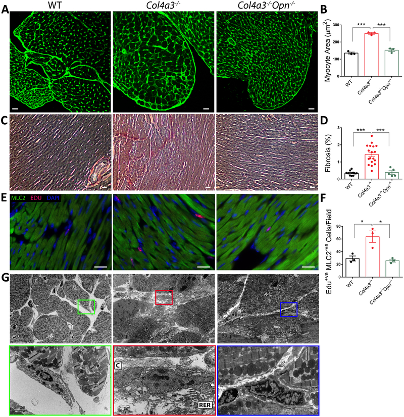
Results: OGDHL mRNA and protein displayed abnormal abundances in cardiac biopsies of HFpEF (n = 17) compared with donor control subjects (n = 12; p < 0.01) or heart failure with reduced ejection fraction patients (n = 12; p < 0.05). Blockade of OPN in hiPS-CMs conferred increased OGDHL expression. Col4a3-/- mice demonstrated cardiomyopathy with similarities to HFpEF, including diastolic dysfunction, cardiac hypertrophy and fibrosis, pulmonary edema, and impaired mitochondrial function. The cardiomyopathy was ameliorated by Opn-/- coincident with improved renal function and increased expression of Ogdhl. Heart-specific overexpression of Ogdhl in Col4a3-/- mice also improved cardiac function and cardiomyocyte energy state.

Conclusions: Col4a3-/- mice present a model of HFpEF secondary to CKD wherein OPN and OGDHL are intermediates, and possibly therapeutic targets.

Keywords: Alport syndrome; HFpEF; OGDHL; hiPS-CM; mitochondria; osteopontin.


Figures

Central Illustration:
Central Illustration:. Osteopontin and Mitochondrial OGDHL in Diastolic Dysfunction.
The phenotype of a subset of patients with HFpEF including a possible central and causative role for Osteopontin (OPN) is mimicked in Alport (Col4a3−/−) mice. Our studies show that OPN regulates mitochondrial 2-Oxoglutarate Dyhydrogenase (OGDHL) and myocardial bioenergetics in a preclinical model, and this at least partially drives the HFpEF phenotype. The results were validated in human induced pluripotent-derived cardiomyocytes (hiPS-CMs), and cardiac biopsies of HFpEF patients.
Figure 1:
Figure 1:. OPN deficiency ameliorates cardiac dysfunction in Col4a3−/− mice.
A-D. Echocardiography of 2-month Col4a3−/− mixed background mice shows diastolic dysfunction as indicated by a prolonged IVRT, increased E/E’, reduced E/A, and elevated MPI. E-F. GLS and GCS were impaired in the Col4a3−/− mice indicating subclinical systolic dysfunction. G Col4a3−/− mice show preserved EF. H. Pulmonary edema is suggested by increased body weight-normalized lung weight as well as by elevated Lung wet-to-dry (W/D) ratio, indicating failing hearts in Col4a3−/− mice. Hetero-/homo-zygous deletion of OPN in Col4a3−/− mice improves cardiac function and prevents pulmonary edema. Complete echocardiographic and morphometric measurements are presented in Online Tables 1 and 2. Data are mean±SEM. N=10–37 mice per group. *p<0.05, **p<0.01, ***p<0.001 using student’s t-test for two groups (panel I) comparisons or one-way ANOVA with Tukey’s post hoc test for multiple groups (all other panels).
Figure 2:
Figure 2:. OPN deficiency ameliorates cardiac hypertrophy and fibrosis in Col4a3−/− mice.
A-B. WGA staining of Col4a3−/− hearts shows cardiac hypertrophy in Col4a3−/− mice as increased myocyte cross-sectional area that is reduced with OPN knockout. Scale bar: 20μm. N=3 mice per group. C-D. Picrosirius Red staining of cardiac cross-sections indicated increased Collagen deposition in Col4a3−/− heart that is significantly reduced by OPN deficiency. Scale bar: 100μm. N=5–15 mice per group. E-F. The number of Edu positive interstitial cells (MLC2 negative) is elevated in Col4a3−/− hearts but normalized with OPN deficiency Scale bar: 20μm. N=3 per mice group. G. OPN deficiency markedly decreased myocardial fibrosis in Col4a3−/− mice as shown by EM images of reduced collagen fibers and interstitial “activated” fibroblasts. C: Collagen fibers, RER: Rough Endoplasmic Reticulum. Data are mean±SEM. *p<0.05, **p<0.01, ***p<0.001 using one-way ANOVA with Tukey’s post hoc test.
Figure 3:
Figure 3:. OGDHL is dysregulated in Col4a3−/− hearts and normalized by OPN deficiency.
A. Microarray heatmap shows genes that are altered in the Col4a3−/− hearts and upregulated with OPN deficiency. B. The downregulation of selected genes in the Col4a3−/− hearts is validated by qPCR. Target validation in whole heart tissue (C) or mitochondrial fraction (D) shows reduced OGDHL protein and suppressed OGDH activity (E). F. OGDHL protein levels are upregulated with OPN knockout in the Col4a3−/− hearts. Data are mean±SEM. N=3–5 hearts per group. *p<0.05 and **p<0.01 using unpaired Student’s t-test.
Figure 4:
Figure 4:. OPN negatively regulates OGDHL in hiPS-CMs and respiration in nCMs.
A-C. Treatment of hiPS-CMs with OPN mAb significantly increases OGDHL protein levels by immunostaining and western blotting. N= 3 wells per group. D-E. Mouse neonatal cardiomyocytes treated with 20 μg/ml mouse recombinant OPN for 48 hours show significantly reduced ATP-linked oxygen consumption rate. N=9–16 wells per group. Data are representative of 3 independent experiments. Data are mean±SEM. *p<0.05, **p<0.01, ***p<0.001 using unpaired Student’s t-test.
Figure 5:
Figure 5:. Cardiac dysfunction and impaired mitochondrial function in Col4a3−/− mice are rescued by heart-specific Ogdhl overexpression.
A-G Cardiac function of Col4a3−/− 129J mice was evaluated by echocardiography and strain analysis four weeks after AAV9-cTnt-Ogdhl or AAV9-cTnt-Luciferase injections showing improved MPI (B), improved strain (F,G) and reduced weight loss (H). Complete echocardiographic and morphometric measurements are presented in Online Tables 4 and 5. I-J. Extracellular flux assay confirmed suppressed mitochondrial respiration in adult cardiomyocytes isolated from Col4a3−/− hearts. Four weeks post AAV9-cTnt-Ogdhl injections, the adult myocytes show significantly improved oxygen consumption rate (OCR). Data are mean±SEM. N=15–29 mice per group. *p<0.05, **p<0.01, ***p<0.001 using one-way ANOVA with Tukey’s post hoc test.
Figure 6:
Figure 6:. Pressure Volume Analysis of Col4a3−/− 129J mice.
Invasive hemodynamic measurements demonstrated that 2-month Col4a3−/− 129J background mice develop diastolic dysfunction. Complete invasive hemodynamic measurements are presented in Online Table 6. Data are mean±SEM. N=13–18 mice per group. *p<0.05, **p<0.01, ***p<0.001 using unpaired Student’s t-test.
Figure 7:
Figure 7:. OGDHL expression is dysregulated in cardiac biopsies of HFpEF patients.
Ogdhl mRNA (A) and protein (B) levels are upregulated in HFpEF patients as shown by qPCR and western blotting, respectively. n.c.u.: Normalized Delta C, n.d.u.: Normalized Densitometry Unit. Data are mean±SEM. N=12–18 per group. *p<0.05, and **p<0.01, using Kruskal-Wallis nonparametric test with Dunn’s multiple comparisons test for qPCR and one-way ANOVA with Holm-Sidak’s post hoc test for western blots.

Comment in

References

    1. Shah SJ, Kitzman DW, Borlaug BA et al. Phenotype-specific treatment of heart failure with preserved ejection fraction: a multiorgan roadmap. Circulation 2016;134:73–90. - PMC - PubMed
    1. Lam CSP, Donal E, Kraigher-Krainer E, Vasan RS. Epidemiology and clinical course of heart failure with preserved ejection fraction. Eur J Heart Fail 2011;13:18–28. - PMC - PubMed
    1. Bhatia RS, Tu JV, Lee DS et al. Outcome of heart failure with preserved ejection fraction in a population-based study. N Engl J Med 2006;355:260–269. - PubMed
    1. Owan TE, Hodge DO, Herges RM, Jacobsen SJ, Roger VL, Redfield MM. Trends in Prevalence and Outcome of Heart Failure with Preserved Ejection Fraction. N Engl J Med 2006;355:251–259. - PubMed
    1. Shah SJ, Gheorghiade M. Heart failure with preserved ejection fraction: treat now by treating comorbidities. JAMA 2008;300:431–433. - PubMed

Publication types

MeSH terms