. 2019 Sep:102:1-11.
doi: 10.1016/j.msec.2019.04.026. Epub 2019 Apr 11.

Enhanced cell functions on graphene oxide incorporated 3D printed polycaprolactone scaffolds

Affiliations

Enhanced cell functions on graphene oxide incorporated 3D printed polycaprolactone scaffolds

Janitha M Unagolla et al. Mater Sci Eng C Mater Biol Appl. 2019 Sep.

Abstract

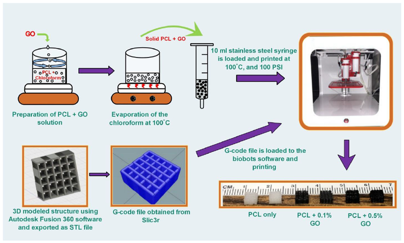
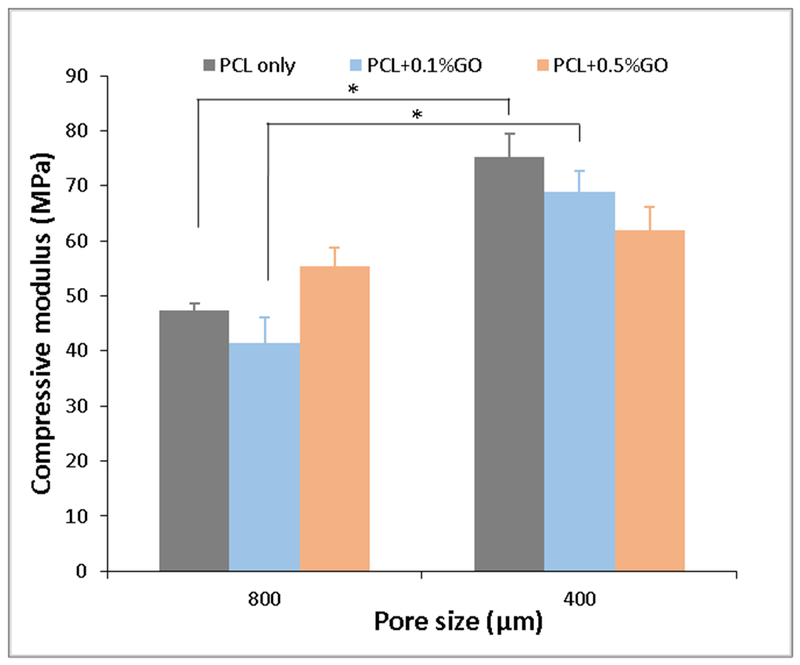
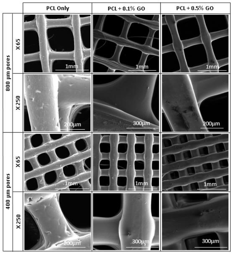
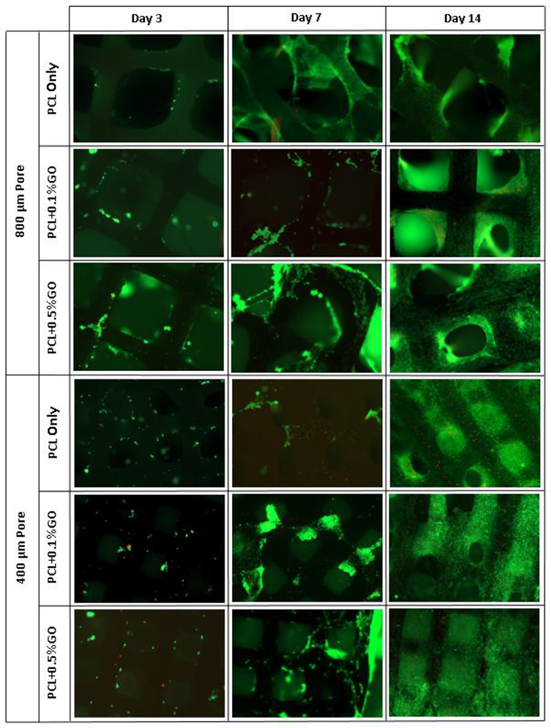
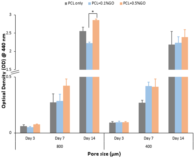
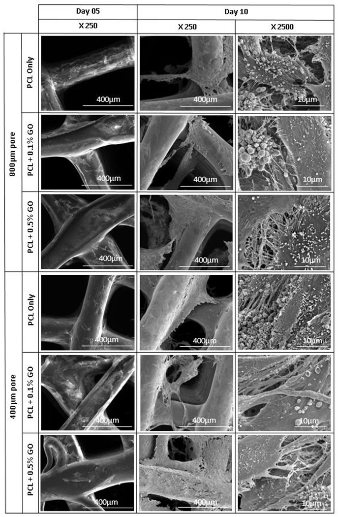
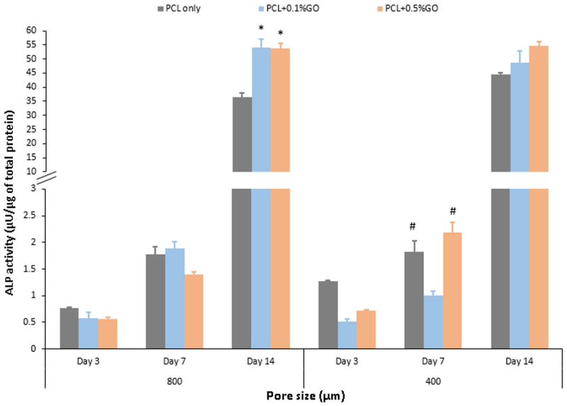
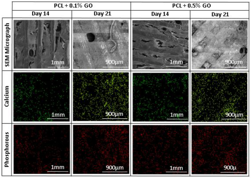
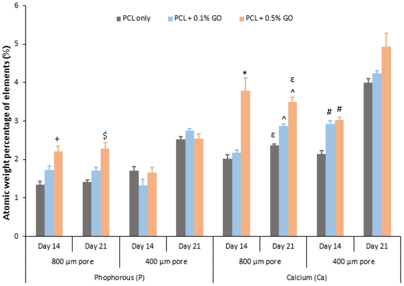
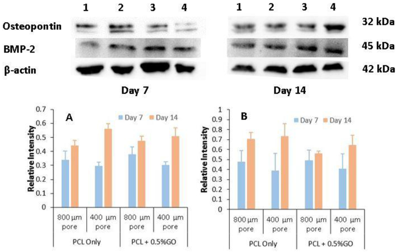
For tissue engineering applications, a porous scaffold with an interconnected network is essential to facilitate the cell attachment and proliferation in a three dimensional (3D) structure. This study aimed to fabricate the scaffolds by an extrusion-based 3D printer using a blend of polycaprolactone (PCL), and graphene oxide (GO) as a favorable platform for bone tissue engineering. The mechanical properties, morphology, biocompatibility, and biological activities such as cell proliferation and differentiation were studied concerning the two different pore sizes; 400 μm, and 800 μm, and also with two different GO content; 0.1% (w/w) and 0.5% (w/w). The compressive strength of the scaffolds was not significantly changed due to the small amount of GO, but, as expected scaffolds with 400 μm pores showed a higher compressive modulus in comparison to the scaffolds with 800 μm pores. The data indicated that the cell attachment and proliferation were increased by adding a small amount of GO. According to the results, pore size did not play a significant role in cell proliferation and differentiation. Alkaline Phosphate (ALP) activity assay further confirmed that the GO increase the ALP activity and further Elemental analysis of Calcium and Phosphorous showed that the GO increased the mineralization compared to PCL only scaffolds. Western blot analysis showed the porous structure facilitate the secretion of bone morphogenic protein-2 (BMP-2) and osteopontin at both day 7 and 14 which galvanizes the osteogenic capability of PCL and PCL + GO scaffolds.

Keywords: Biocompatibility; Cell proliferation; Differentiation; Graphene oxide; Polycaprolactone.


Figures

Figure 1:
Figure 1:
Schematic representation of preparation of scaffolds using 3D printer
Figure 2:
Figure 2:
Compressive modulus of scaffolds at two different pore sizes; * indicates p<0.05; n=7
Figure 3:
Figure 3:
SEM micrographs of scaffolds; X indicates the magnification
Figure 4:
Figure 4:
Live and dead cell assay fluorescence images at days 3, 7, and 14; green- live cells, read- dead cells; scale 1 mm
Figure 5:
Figure 5:
WST-1 assay optical density values at 440 nm for three different time points; * indicates the p<0.05; n=3
Figure 6:
Figure 6:
SEM micrograph of osteoblast cell attachment at days 5 and 10; X indicates the magnification
Figure 7:
Figure 7:
ALP activity of the scaffolds at three different time points; * indicates the significance of p<0.05 with respect to (wrt) PCL only scaffold at day 14; # indicates the significance of p<0.05 wrt PCL +0.1% GO scaffold at day 7; n=3
Figure 8:
Figure 8:
Elemental mapping images and SEM micrographs of selected scaffolds after osteogenic differentiation at days 14 and 21
Figure 9:
Figure 9:
Quantitative analysis of calcium and phosphorous elements using EDX method; + indicates the significance p<0.05 wrt PCL only; $ indicates the significance p<0.05 wrt PCL only; * indicates the significance p<0.05 wrt to both PCL only and PCL + 0.1% GO; ε indicates the significance p<0.05 wrt PCL + 0.1% GO; ^ indicates the significance p<0.05 wrt PCL only; # indicates the significance p<0.05 wrt PCL only
Figure 10:
Figure 10:
Western blot analysis of preosteoblasts following the induction of osteogenic differentiation at day 7 and day 14 for different samples, 1- PCL only scaffolds with 800 μm pores, 2- PCL only scaffolds with 400 μm pores, 3- PCL + 0.5% GO scaffolds with 800 μm pores, and 4- PCL + 0.5% GO scaffolds with 400 μm pores; graph A: Bone Morphogenic Protein-2 (BMP-2), graph B: Osteopontin (OPN) (relative intensity wrt β-actin)

References

    1. Woodfield TBF, Malda J, De Wijn J, Péters F, Riesle J, Van Blitterswijk CA, Design of porous scaffolds for cartilage tissue engineering using a three-dimensional fiber-deposition technique, Biomaterials. 25 (2004) 4149–4161. doi: 10.1016/j.biomaterials.2003.10.056. - DOI - PubMed
    1. Hutmacher DW, Schantz T, Zein I, Ng KW, Teoh SH, Tan KC, Mechanical properties and cell cultural response of polycaprolactone scaffolds designed and fabricated via fused deposition modeling, J. Biomed. Mater. Res 55 (2001) 203–216. doi: 10.1002/1097-4636(200105)55:2<203::AID-JBM1007>3.0.CO;2-7. - DOI - PubMed
    1. Park SA, Lee SH, Kim WD, Fabrication of porous polycaprolactone/hydroxyapatite (PCL/HA) blend scaffolds using a 3D plotting system for bone tissue engineering, Bioprocess Biosyst. Eng 34 (2011) 505–513. doi: 10.1007/s00449-010-0499-2. - DOI - PubMed
    1. Trachtenberg JE, Placone JK, Smith BT, Piard CM, Santoro M, Scott DW, Fisher JP, Mikos AG, Extrusion-Based 3D Printing of Polypropylene fumarate) in a Full-Factorial Design, ACS Biomater. Sci. Eng 2 (2016) 1771–1780. doi:10.1021/acsbiomaterials.6b00026. - DOI - PubMed
    1. Tarafder S, Bose S, Polycaprolactone-coated 3D printed tricalcium phosphate scaffolds for bone tissue engineering: In vitro alendronate release behavior and local delivery effect on in vivo osteogenesis, ACS Appl. Mater. Interfaces 6 (2014) 9955–9965. doi:10.1021/am501048n. - DOI - PMC - PubMed