

Diagnostic accuracy of intracoronary optical coherence tomography-derived fractional flow reserve for assessment of coronary stenosis severity
- PMID: 31147309
- PMCID: PMC8130381
- DOI: 10.4244/EIJ-D-19-00182
Diagnostic accuracy of intracoronary optical coherence tomography-derived fractional flow reserve for assessment of coronary stenosis severity
Abstract
Aims: A novel method for computation of fractional flow reserve (FFR) from optical coherence tomography (OCT) was developed recently. This study aimed to evaluate the diagnostic accuracy of a new OCT-based FFR (OFR) computational approach, using wire-based FFR as the reference standard.
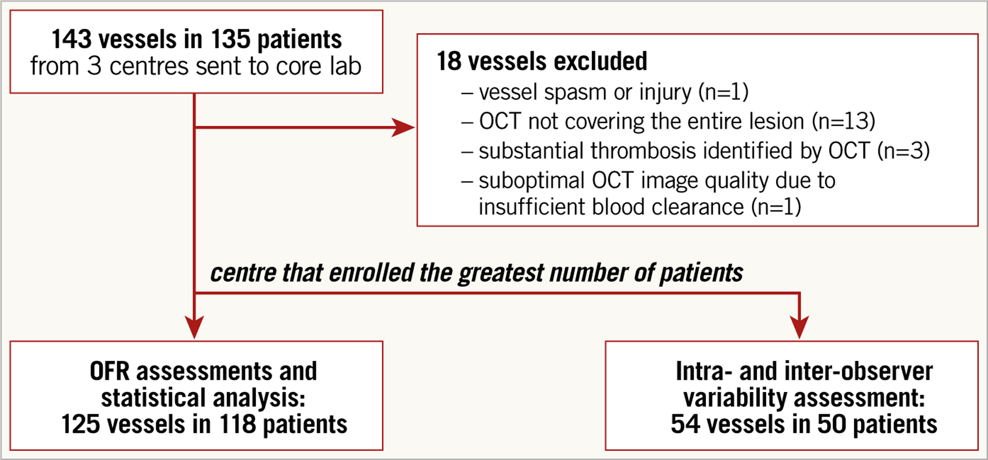
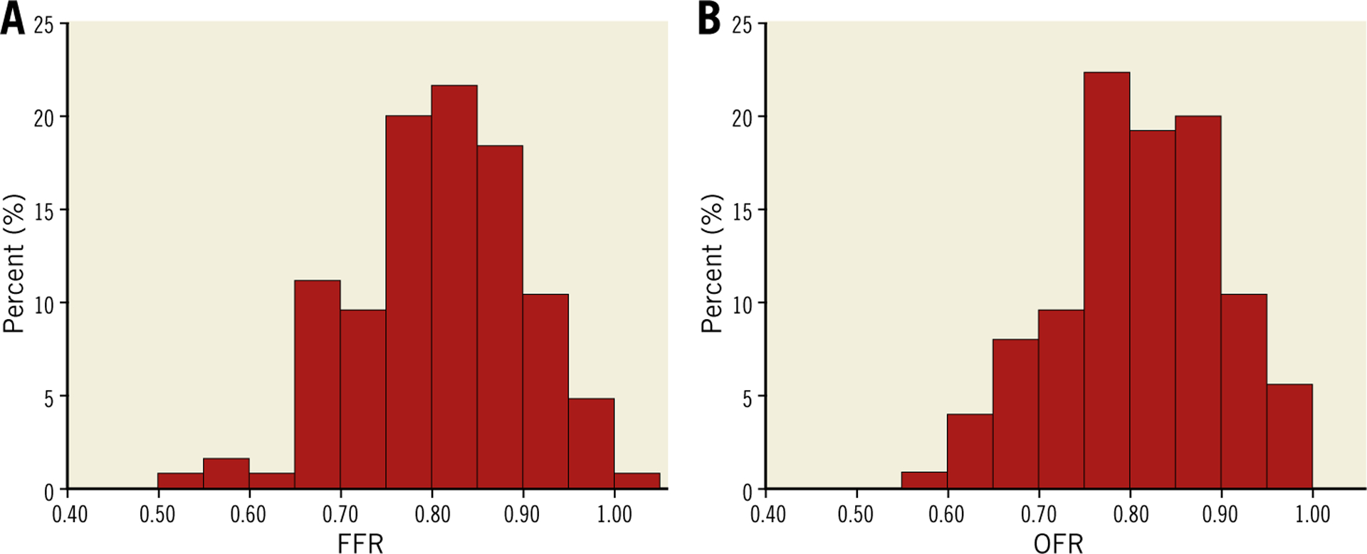
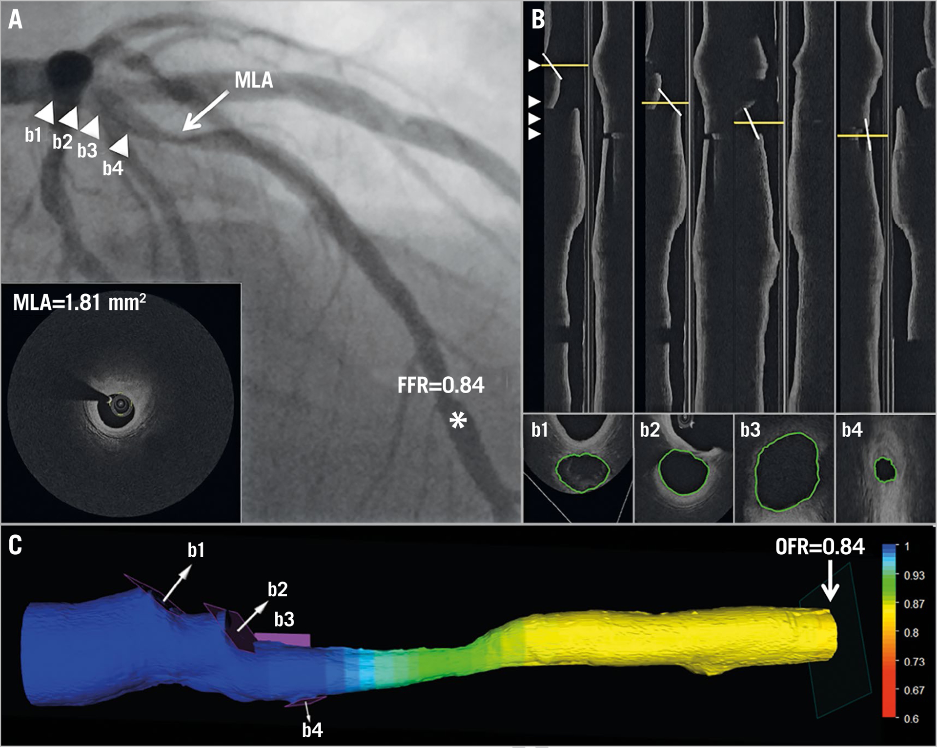
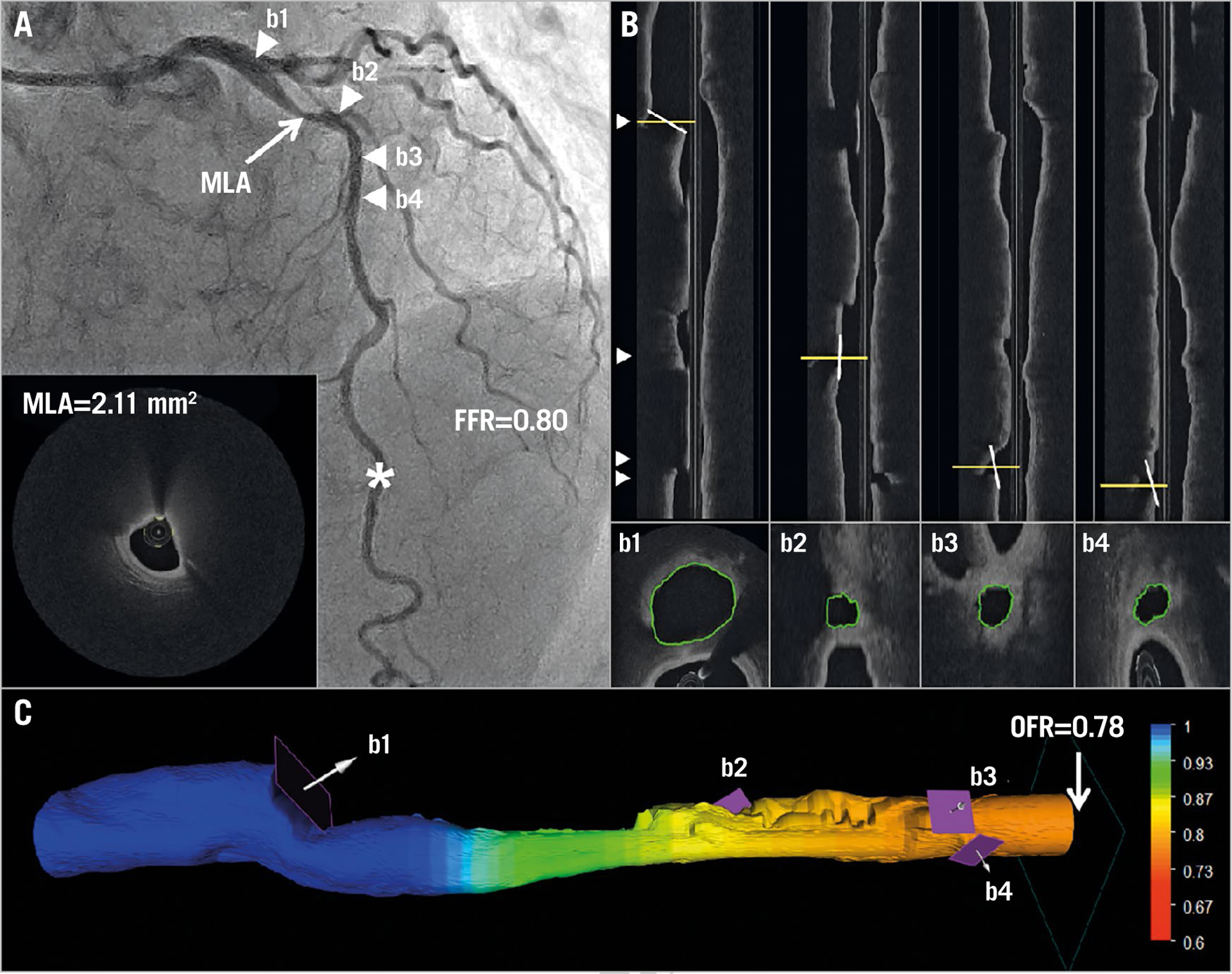
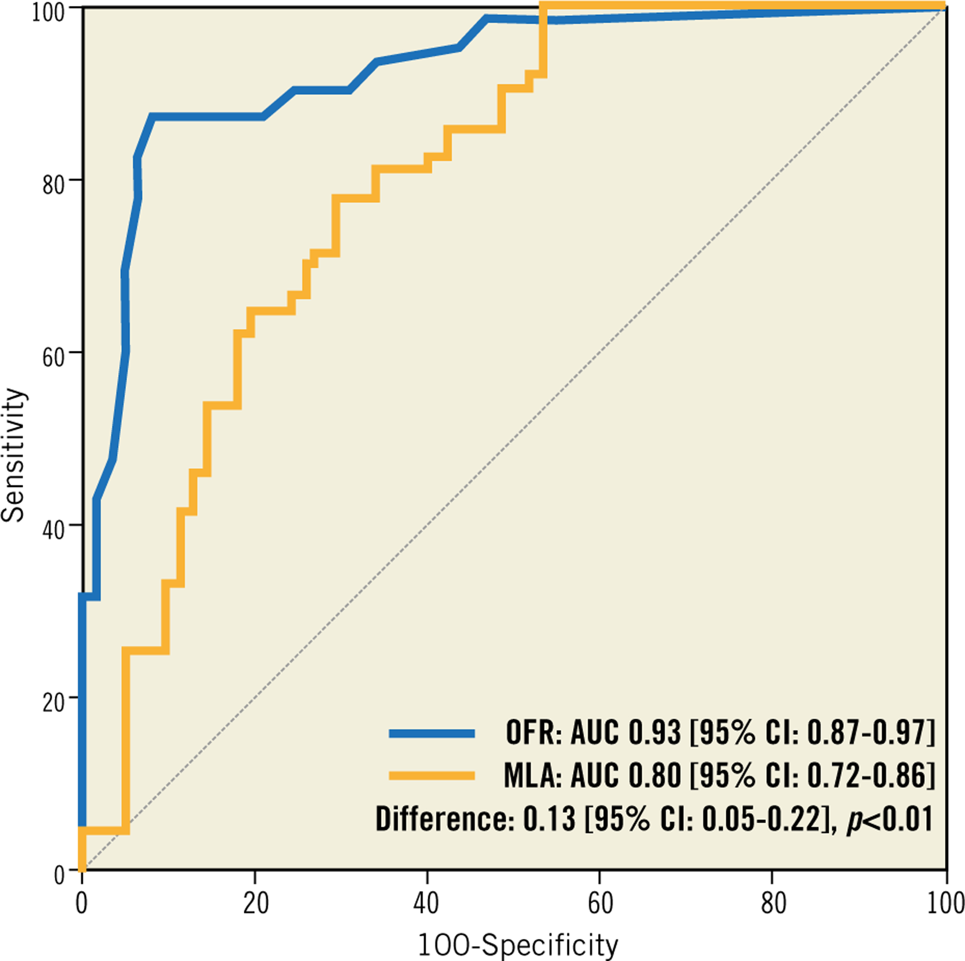
Methods and results: Patients who underwent both OCT and FFR prior to intervention were analysed. The lumen of the interrogated vessel and the ostia of the side branches were automatically delineated and used to compute OFR. Bifurcation fractal laws were applied to correct the change in reference lumen size due to the step-down phenomenon. OFR was compared with FFR, both using a cut-off value of 0.80 to define ischaemia. Computational analysis was performed in 125 vessels from 118 patients. Average FFR was 0.80±0.09. Accuracy, sensitivity, specificity, positive predictive value, and negative predictive value for OFR to identify FFR ≤0.80 was 90% (95% CI: 84-95), 87% (95% CI: 77-94), 92% (95% CI: 82-97), 92% (95% CI: 82-97), and 88% (95% CI: 77-95), respectively. The AUC was higher for OFR than minimal lumen area (0.93 [95% CI: 0.87-0.97] versus 0.80 [95% CI: 0.72-0.86], p=0.002). Average OFR analysis time was 55±23 seconds for each OCT pullback. Intra- and inter-observer variability in OFR analysis was 0.00±0.02 and 0.00±0.03, respectively.
Conclusions: OFR is a novel and fast method allowing assessment of flow-limiting coronary stenosis without pressure wire and induced hyperaemia. The good diagnostic accuracy and low observer variability bear the potential of improved integration of intracoronary imaging and physiological assessment.
Figures

References
-
- Tonino PA, De Bruyne B, Pijls NHJ, Siebert U, Ikeno F, van `t Veer M, Klauss V, Manoharan G, Engstrøm T, Oldroyd KG, Ver Lee PN, MacCarthy PA, Fearon WF; FAME Study Investigators. Fractional flow reserve versus angiography for guiding percutaneous coronary intervention. N Engl J Med. 2009;360:213–24. - PubMed
-
- Xaplanteris P, Fournier S, Pijls NHJ, Fearon WF, Barbato E, Tonino PAL, Engstrøm T, Kääb S, Dambrink JH, Rioufol G, Toth GG, Piroth Z, Witt N, Fröbert O, Kala P, Linke A, Jagic N, Mates M, Mavromatis K, Samady H, Irimpen A, Oldroyd K, Campo G, Rothenbühler M, Jüni P, De Bruyne B; FAME 2 Investigators. Five-Year Outcomes with PCI Guided by Fractional Flow Reserve. N Engl J Med. 2018;379:250–9. - PubMed
-
- Götberg M, Cook CM, Sen S, Nijjer S, Escaned J, Davies JE. The Evolving Future of Instantaneous Wave-Free Ratio and Fractional Flow Reserve. J Am Coll Cardiol. 2017;70:1379–402. - PubMed
-
- Tu S, Westra J, Yang J, von Birgelen C, Ferrara A, Pellicano M, Nef H, Tebaldi M, Murasato Y, Lansky A, Barbato E, van der Heijden LC, Reiber JHC, Holm NR, Wijns W; FAVOR Pilot Trial Study Group. Diagnostic Accuracy of Fast Computational Approaches to Derive Fractional Flow Reserve From Diagnostic Coronary Angiography: The International Multicenter FAVOR Pilot Study. JACC Cardiovasc Interv. 2016;9:2024–35. - PubMed
-
- Xu B, Tu S, Qiao S, Qu X, Chen Y, Yang J, Guo L, Sun Z, Li Z, Tian F, Fang W, Chen J, Li W, Guan C, Holm NR, Wijns W, Hu S. Diagnostic Accuracy of Angiography-Based Quantitative Flow Ratio Measurements for Online Assessment of Coronary Stenosis. J Am Coll Cardiol. 2017;70:3077–87. - PubMed
MeSH terms
Grants and funding
LinkOut - more resources
Full Text Sources
Medical