

Ensemble of Convolutional Neural Networks Improves Automated Segmentation of Acute Ischemic Lesions Using Multiparametric Diffusion-Weighted MRI
- PMID: 31147354
- PMCID: PMC6715290
- DOI: 10.3174/ajnr.A6077
Ensemble of Convolutional Neural Networks Improves Automated Segmentation of Acute Ischemic Lesions Using Multiparametric Diffusion-Weighted MRI
Abstract
Background and purpose: Accurate automated infarct segmentation is needed for acute ischemic stroke studies relying on infarct volumes as an imaging phenotype or biomarker that require large numbers of subjects. This study investigated whether an ensemble of convolutional neural networks trained on multiparametric DWI maps outperforms single networks trained on solo DWI parametric maps.
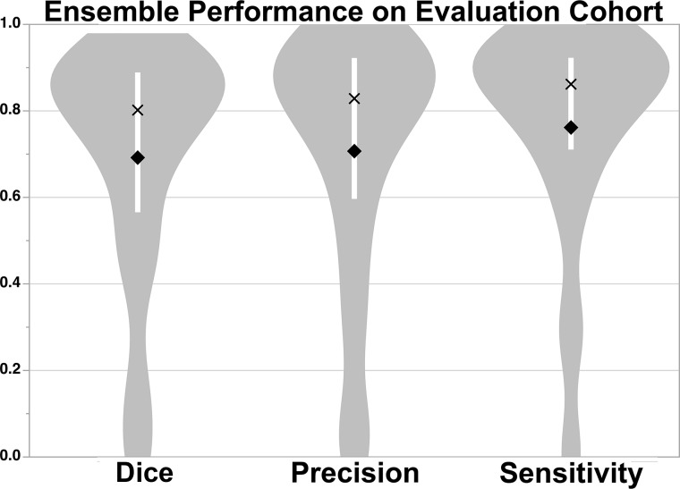
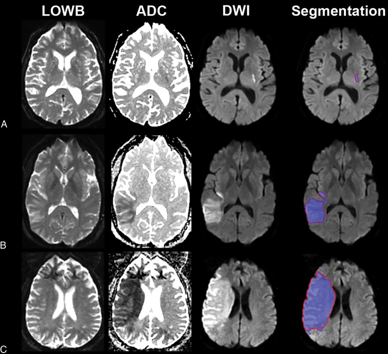
Materials and methods: Convolutional neural networks were trained on combinations of DWI, ADC, and low b-value-weighted images from 116 subjects. The performances of the networks (measured by the Dice score, sensitivity, and precision) were compared with one another and with ensembles of 5 networks. To assess the generalizability of the approach, we applied the best-performing model to an independent Evaluation Cohort of 151 subjects. Agreement between manual and automated segmentations for identifying patients with large lesion volumes was calculated across multiple thresholds (21, 31, 51, and 70 cm3).
Results: An ensemble of convolutional neural networks trained on DWI, ADC, and low b-value-weighted images produced the most accurate acute infarct segmentation over individual networks (P < .001). Automated volumes correlated with manually measured volumes (Spearman ρ = 0.91, P < .001) for the independent cohort. For the task of identifying patients with large lesion volumes, agreement between manual outlines and automated outlines was high (Cohen κ, 0.86-0.90; P < .001).
Conclusions: Acute infarcts are more accurately segmented using ensembles of convolutional neural networks trained with multiparametric maps than by using a single model trained with a solo map. Automated lesion segmentation has high agreement with manual techniques for identifying patients with large lesion volumes.
© 2019 by American Journal of Neuroradiology.
Figures
References
-
- Hevia-Montiel N, Jimenez-Alaniz JR, Medina-Banuelos V, et al. Robust nonparametric segmentation of infarct lesion from diffusion-weighted MR images. Conf Proc IEEE Eng Med Biol Soc 2007;2007:2102–05 - PubMed
Publication types
MeSH terms
Grants and funding
- R01 NS082285/NS/NINDS NIH HHS/United States
- S10 RR019307/RR/NCRR NIH HHS/United States
- R01 NS086905/NS/NINDS NIH HHS/United States
- U01 NS069208/NS/NINDS NIH HHS/United States
- R01 NS051412/NS/NINDS NIH HHS/United States
- R21 NS077442/NS/NINDS NIH HHS/United States
- R01 DC012584/DC/NIDCD NIH HHS/United States
- R21 NS085574/NS/NINDS NIH HHS/United States
- U10 NS086729/NS/NINDS NIH HHS/United States
- R01 NS038477/NS/NINDS NIH HHS/United States
- P50 NS051343/NS/NINDS NIH HHS/United States
- R01 NS105875/NS/NINDS NIH HHS/United States
- U01 NS095869/NS/NINDS NIH HHS/United States
- R01 NS063925/NS/NINDS NIH HHS/United States
- P41 EB015896/EB/NIBIB NIH HHS/United States
- R01 NS059775/NS/NINDS NIH HHS/United States
LinkOut - more resources
Full Text Sources
Other Literature Sources