. 2019 Jul 1;218(7):2136-2149.
doi: 10.1083/jcb.201901116. Epub 2019 May 30.

Repair of nuclear ruptures requires barrier-to-autointegration factor

Affiliations

Repair of nuclear ruptures requires barrier-to-autointegration factor

Charles T Halfmann et al. J Cell Biol. .

Abstract

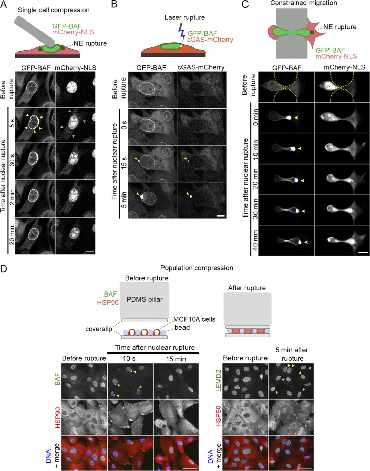
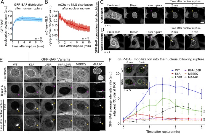
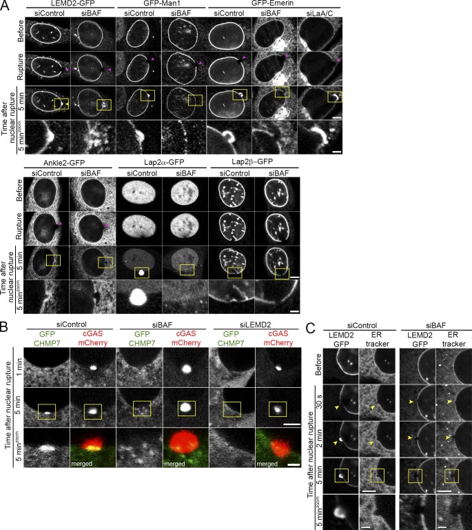
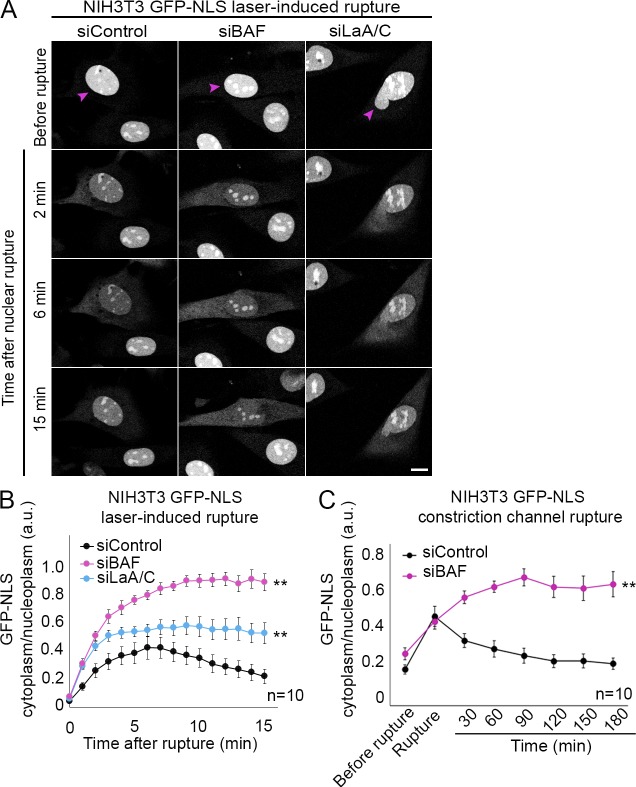
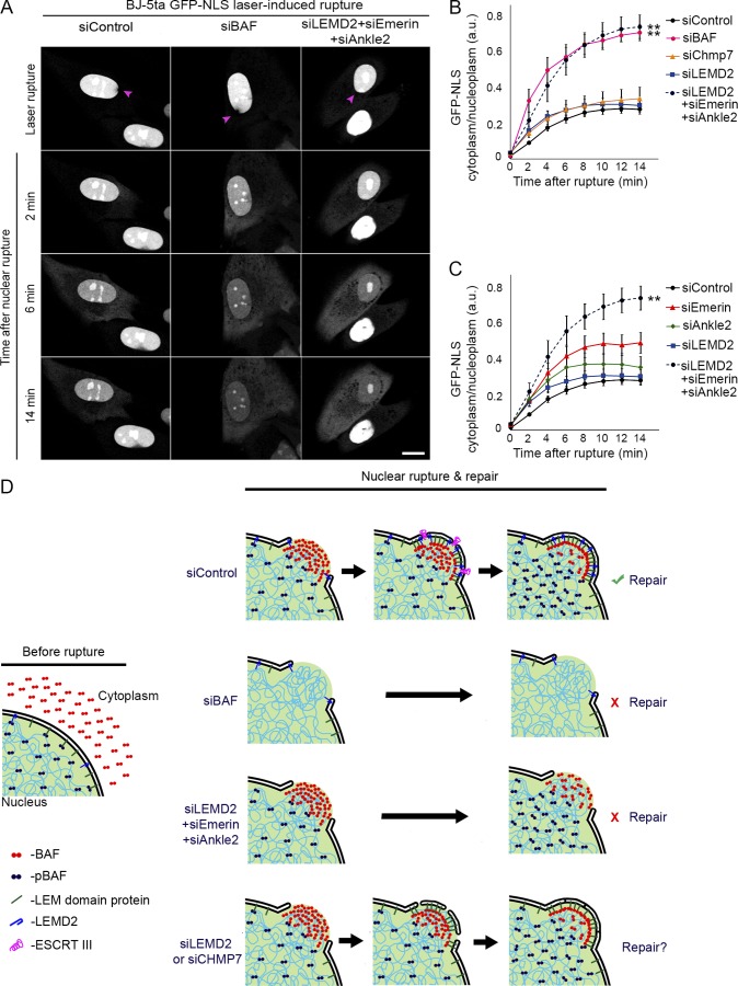
Cell nuclei rupture following exposure to mechanical force and/or upon weakening of nuclear integrity, but nuclear ruptures are repairable. Barrier-to-autointegration factor (BAF), a small DNA-binding protein, rapidly localizes to nuclear ruptures; however, its role at these rupture sites is unknown. Here, we show that it is predominantly a nonphosphorylated cytoplasmic population of BAF that binds nuclear DNA to rapidly and transiently localize to the sites of nuclear rupture, resulting in BAF accumulation in the nucleus. BAF subsequently recruits transmembrane LEM-domain proteins, causing their accumulation at rupture sites. Loss of BAF impairs recruitment of LEM-domain proteins and nuclear envelope membranes to nuclear rupture sites and prevents nuclear envelope barrier function restoration. Simultaneous depletion of multiple LEM-domain proteins similarly inhibits rupture repair. LEMD2 is required for recruitment of the ESCRT-III membrane repair machinery to ruptures; however, neither LEMD2 nor ESCRT-III is required to repair ruptures. These results reveal a new role for BAF in the response to and repair of nuclear ruptures.


Figures

Figure 1.
Figure 1.
BAF rapidly but transiently localizes to sites of nuclear rupture. (A) Sequential images of a representative NIH3T3 cell expressing GFP-BAF after nuclear rupture via compression by a blunted microcapillary. GFP-BAF localizes to sites of nuclear rupture on the nuclear rim (yellow arrowheads) and resolves within minutes. Coexpression of mCherry-NLS serves as a nuclear rupture marker (blue arrowheads). (B) Sequential images of a representative NIH3T3 coexpressing GFP-BAF and cGAS-mCherry undergoing laser-induced NE rupture (purple arrowheads). Sites of nuclear rupture are indicated by cGAS accumulation (yellow arrowheads). (C) Sequential images of a representative NIH3T3 cell coexpressing GFP-BAF and mCherry-NLS undergoing nuclear rupture during constricted migration through a channel (height, 3 µm) in a microfluidic device. Sites of nuclear rupture are indicated by BAF accumulation (yellow arrowheads). (D) Populations of MCF10A cells that were compressed, fixed at indicated time points, and labeled with anti-BAF or LEMD2 (green) and HSP90 (red). Hoechst dye was used to label DNA (blue). Cells with ruptured nuclei are apparent at 10 s in BAF-labeled cells and 5 min in LEMD2-labeled cells (yellow arrowheads). Bars: (A–C) 10 µm; (D) 50 µm.
Figure 2.
Figure 2.
Cytoplasmic BAF predominantly localizes to nuclear ruptures, and its behavior is primarily driven by association with DNA. (A) The nucleoplasmic/cytoplasmic GFP-BAF intensity was measured in live NIH3T3 cells following nuclear rupture with a blunted microcapillary. (B) The ratio of cytoplasmic/nucleoplasmic mCherry-NLS intensity was measured to monitor the repair of the ruptures. The shaded areas represent SEM (n = 5 cells). (C and D) Sequential images of NIH3T3 cells expressing GFP-BAF for which the GFP-BAF signal was photobleached in either the cytoplasm (n = 4 cells; C) or nucleoplasm (n = 4 cells; D), followed by laser-induced NE rupture (purple arrowheads). Yellow arrowheads indicate accumulation of GFP-BAF from each representative compartment at the site of nuclear rupture. (E) NIH3T3 cells expressing GFP-tagged BAF mutants, reduced DNA/histone-binding affinity (K6A), reduced LEM-domain protein-binding capability (L58R), double mutant (K6A, L58R), phosphomimetic (MEEEQ), or nonphosphorylatable (MAAAQ) underwent nuclear bleach then were ruptured using laser ablation. Migration of mutated BAF into the nucleus was monitored and compared with the WT BAF nuclear migration. (F) The average intensity of BAF was measured in regions of interest (ROIs) located in the nucleoplasm either distal (green circle) or proximal (orange circle) to the rupture site. The proximal-to-distal average intensity ratio for each cell was calculated for the first 3 min after nuclear rupture. The graph represents mean values ± SEM (n = 5 cells for each; **, P < 0.0001 from WT by a mixed-effects model with Tukey’s post hoc comparison test). Bars: 10 µm.
Figure 3.
Figure 3.
Recruitment of membranes and membrane proteins to nuclear ruptures is impaired by the loss of BAF. (A) NIH3T3 cells expressing GFP-tagged LEM-domain proteins LEMD2, Man1, Emerin, Ankle2, Lap2α, or Lap2β were transfected with siBAF, siControl, or siLaA/C (for GFP-Emerin–expressing cells) for 72 h before laser-induced nuclear rupture (purple arrowheads). Accumulation, or the lack thereof, of each LEM-domain protein at rupture sites (yellow arrowheads) is monitored over 5 min. (B) NIH3T3 cells coexpressing GFP-Chmp7 and cGAS-mCherry were transfected with siBAF, siControl, or siLEMD2 for 72 h before laser-induced nuclear rupture, and GFP-Chmp7 accumulation was monitored over 5 min. Merged channels in zoomed-in images in the bottom row show GFP-Chmp7 (green) and cGAS-mCherry (red). (C) LEMD2-GFP expressing NIH3T3 cells were transfected with either siBAF or siControl siRNAs for 96 h, followed by incubation with ER-tracker red before laser-induced NE rupture. Cells were imaged for 5 min to monitor GFP-LEMD2 and ER-tracker red accumulation at nuclear ruptures (yellow arrowheads). Bars, 5 µm. Yellow boxes indicate the area of zoomed images. Bar, 1 µm.
Figure 4.
Figure 4.
BAF is required to functionally repair the ruptured NE. (A) NIH3T3 cells expressing GFP-NLS were transfected with siControl, siBAF, or siLaA/C for 96 h before laser-induced NE rupture (purple arrowheads). Bar, 10 µm. (B) The cytoplasmic/nucleoplasmic ratios of GFP-NLS were measured over 15 min after laser-induced NE rupture. The graph represents mean values ± SEM (n = 10 cells for each; **, P < 0.0001 from siControl by a mixed-effects model with Tukey’s post hoc comparison test). (C) Quantification of cytoplasmic/nucleoplasmic ratios of GFP-NLS from NIH3T3 cells transfected with either siControl of siBAF siRNAs before rupture via constricted migration in constrained channels (height, 3 µm). The graph represents mean values ± SEM (n = 10 cells for each; **, P < 0.0001 from siControl by a mixed-effects model).
Figure 5.
Figure 5.
BAF is required to functionally repair the ruptured NE via recruitment of multiple LEM-domain proteins. (A) Representative images of BJ-5ta human fibroblast cells expressing GFP-NLS transfected with siControl, siBAF, or a combination of siLEMD2, siEmerin, and siANKLE2 for 96 h before laser-induced NE rupture (purple arrowheads). Bar, 10 µm. (B) Quantification of the cytoplasmic/nucleoplasmic ratio of GFP-NLS following laser-induced nuclear rupture in BJ-5ta cells transfected with siControl, siBAF, siLEMD2, siChmp7, or a combination of siLEMD2, siEmerin, and siANKLE2 for 96 h. The graph represents mean values ± SEM (n = 17, 10, 12, 12, 11 cells, respectively; **, P < 0.0001 from siControl by a mixed-effects model with Tukey’s post hoc comparison). (C) Quantification of cytoplasmic/nucleoplasmic ratio of GFP-NLS following laser-induced nuclear rupture in BJ-5ta cells transfected with siControl, siEmerin, siANKLE2, siLEMD2, or a combination of siLEMD2, siEmerin, and siANKLE2 for 96 h. The graph represents mean values ± SEM (n = 17, 14, 16, 12, 11 cells, respectively; **, P < 0.0001 from siControl by a mixed-effects model with Tukey’s post hoc comparison). (D) A model for NE rupture repair. In control cells (siControl), cytosolic BAF is recruited to the nuclear rupture via DNA binding, followed by the subsequent mobilization of LEM-domain proteins, ESCRT-III, and membranes to the rupture. Loss of BAF (siBAF) results in failure to functionally recruit ESCRT-III, LEM-domain proteins, and membranes, resulting in a failure to repair the NE rupture. The combinatorial loss of NE- and ER-resident LEM-domain proteins (siLEMD2 + siEmerin + siAnkle2), also results in a failure to actively repair the NE rupture. The loss of either LEMD2 or CHMP7 (siLEMD2 or siCHMP7) does not significantly impair NE rupture repair, suggesting that the ESCRT-III complex may facilitate, but is not required for, rupture resealing.

Comment in

References

    1. Asencio C., Davidson I.F., Santarella-Mellwig R., Ly-Hartig T.B., Mall M., Wallenfang M.R., Mattaj I.W., and Gorjánácz M.. 2012. Coordination of kinase and phosphatase activities by Lem4 enables nuclear envelope reassembly during mitosis. Cell. 150:122–135. 10.1016/j.cell.2012.04.043 - DOI - PubMed
    1. Birendra K.C., May D.G., Benson B.V., Kim D.I., Shivega W.G., Ali M.H., Faustino R.S., Campos A.R., and Roux K.J.. 2017. VRK2A is an A-type lamin-dependent nuclear envelope kinase that phosphorylates BAF. Mol. Biol. Cell. 28:2241–2250. 10.1091/mbc.e17-03-0138 - DOI - PMC - PubMed
    1. Cabanillas R., Cadiñanos J., Villameytide J.A., Pérez M., Longo J., Richard J.M., Alvarez R., Durán N.S., Illán R., González D.J., and López-Otín C.. 2011. Néstor-Guillermo progeria syndrome: a novel premature aging condition with early onset and chronic development caused by BANF1 mutations. Am. J. Med. Genet. A. 155:2617–2625. 10.1002/ajmg.a.34249 - DOI - PubMed
    1. Cai M., Huang Y., Ghirlando R., Wilson K.L., Craigie R., and Clore G.M.. 2001. Solution structure of the constant region of nuclear envelope protein LAP2 reveals two LEM-domain structures: one binds BAF and the other binds DNA. EMBO J. 20:4399–4407. 10.1093/emboj/20.16.4399 - DOI - PMC - PubMed
    1. Capanni C., Cenni V., Haraguchi T., Squarzoni S., Schüchner S., Ogris E., Novelli G., Maraldi N., and Lattanzi G.. 2010. Lamin A precursor induces barrier-to-autointegration factor nuclear localization. Cell Cycle. 9:2600–2610. 10.4161/cc.9.13.12080 - DOI - PubMed

Publication types