. 2019 Jul 1;218(7):2277-2293.
doi: 10.1083/jcb.201804201. Epub 2019 May 30.

Scribble, Erbin, and Lano redundantly regulate epithelial polarity and apical adhesion complex

Affiliations

Scribble, Erbin, and Lano redundantly regulate epithelial polarity and apical adhesion complex

Jongho Choi et al. J Cell Biol. .

Abstract

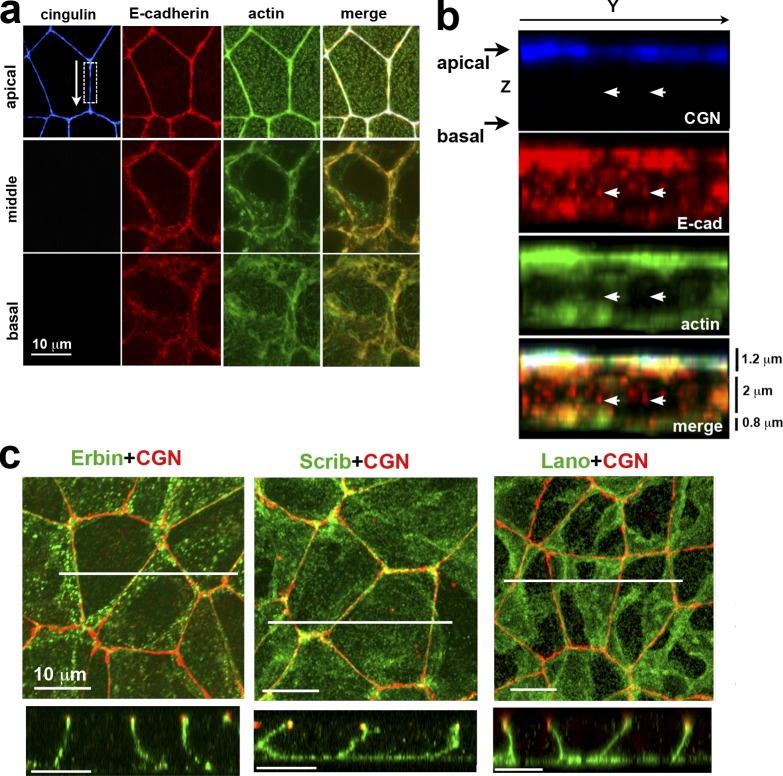
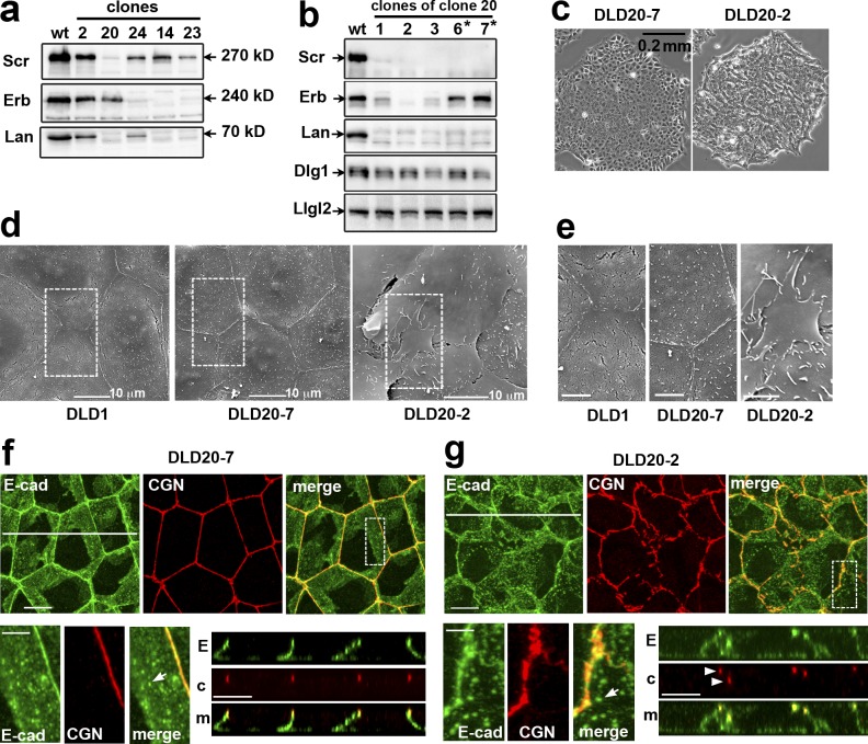
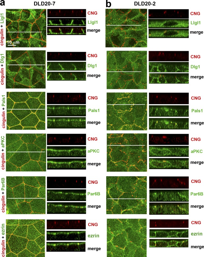
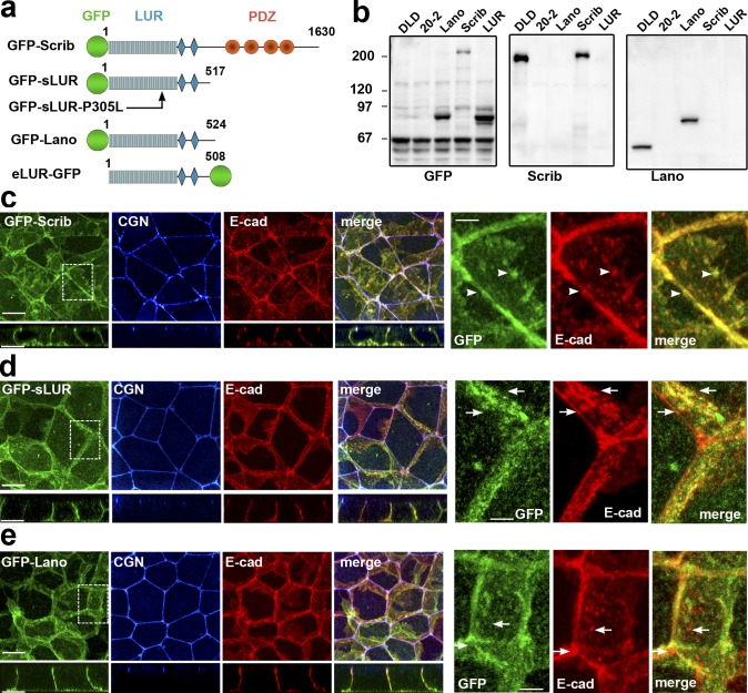
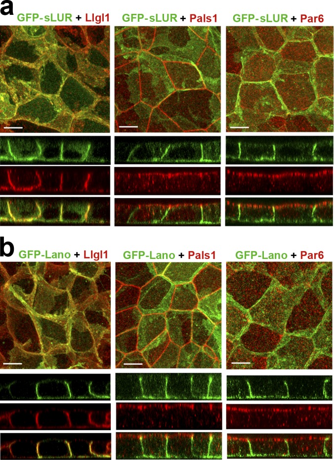
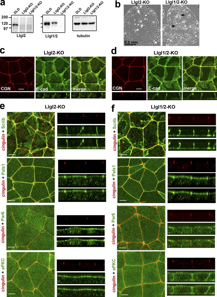
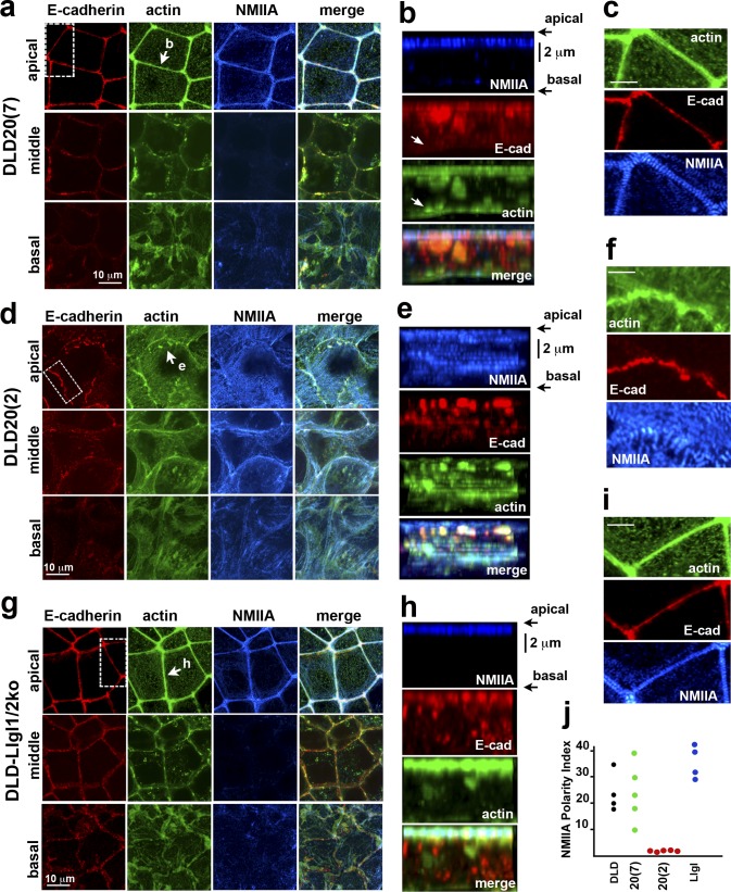
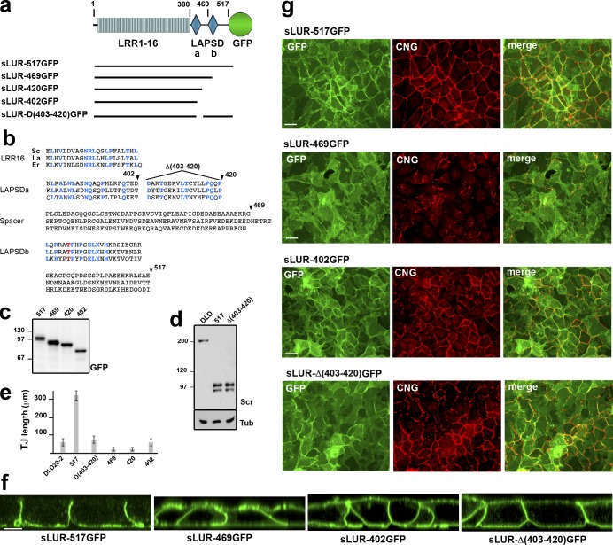
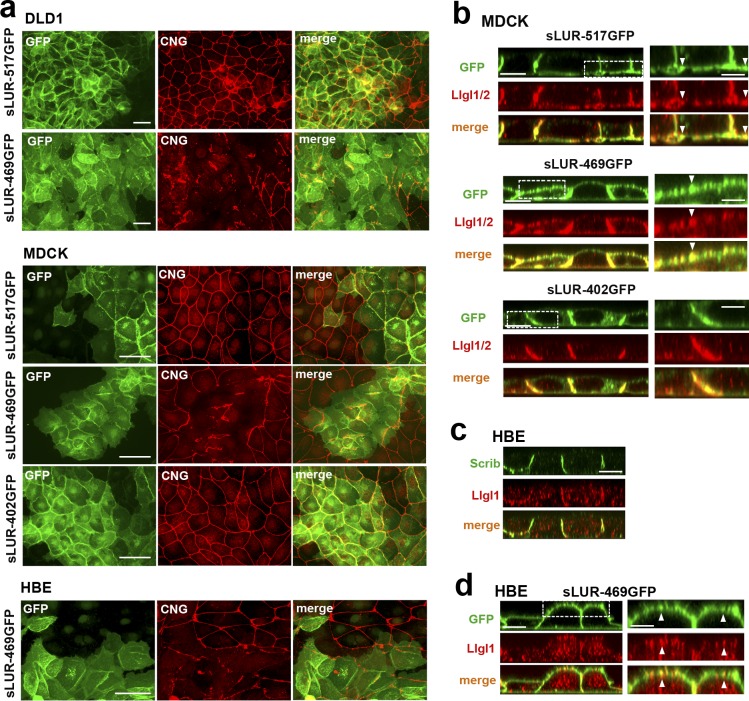
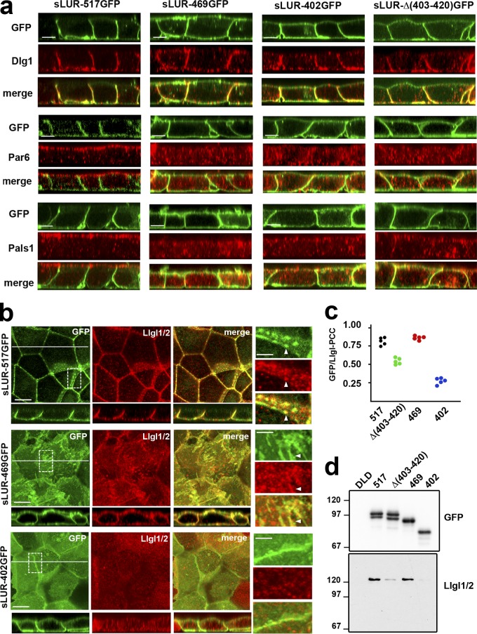
The basolateral protein Scribble (Scrib), a member of the LAP protein family, is essential for epithelial apicobasal polarity (ABP) in Drosophila However, a conserved function for this protein in mammals is unclear. Here we show that the crucial role for Scrib in ABP has remained obscure due to the compensatory function of two other LAP proteins, Erbin and Lano. A combined Scrib/Erbin/Lano knockout disorganizes the cell-cell junctions and the cytoskeleton. It also results in mislocalization of several apical (Par6, aPKC, and Pals1) and basolateral (Llgl1 and Llgl2) identity proteins. These defects can be rescued by the conserved "LU" region of these LAP proteins. Structure-function analysis of this region determined that the so-called LAPSDb domain is essential for basolateral targeting of these proteins, while the LAPSDa domain is essential for supporting the membrane basolateral identity and binding to Llgl. In contrast to the key role in Drosophila, mislocalization of Llgl proteins does not appear to be critical in the scrib ABP phenotype.


Figures

Figure 1.
Figure 1.
ABP of DLD1 cells. (a) Projections of optical z slices spanning apical, middle, and basal portions of the cells. The apical region is reconstructed by a projection of three optical z slices spanning the apical 1.2 µm of cells, the middle region is a projection of five optical z slices spanning 2 µm of the central cell portion, and the basal region is a projection of the two optical z slices spanning 0.8 µm of the basal cell region. (b) Full-face y-z view of the lateral membrane indicated by the dashed box in a. Arrows in b point to the slAJs in which E-cadherin (E-cad, red) does not colocalaze with TJ marker cingulin (CGN, blue) and actin (green). (c) Projections of all x-y optical slices of the cells stained for CGN (red) and Erbin, hScrib, or Lano (green). Only merged images are shown. Confocal cross-sections (along the white lines) of the projections are presented at the bottom. Note that all three LAPPs are located exclusively along the basolateral membranes.
Figure 2.
Figure 2.
DLD20-2 cells deficient for all three LAPPs, but not Erbin-expressing DLD20-7 counterparts, exhibit abnormal cell–cell contacts. (a) Western blot analysis for LAPPs of total cell lysates of DLDcas9 cells (wt) and some of their clones (their numbers are indicated above the lanes) obtained after transfection with the combination of oligos targeting hScrib (Scr), Erbin (Erb), and Lano (Lan) genes. Note that clone 20 lost hScrib and Lano, while clones 14 and 23 lost Erbin and Lano. (b) Total cell lysates of subclones of clone 20 obtained after a second round of transfection with Erbin gene-specific oligonucleotide. Asterisks mark the subclones exhibiting the parental DLD1 phenotype. The lysates were probed for LAPP expression and for Dlg1 and Llgl2. Note that two subclones, DLD20-2 and DLD20-3 (2 and 3 correspondingly), show no expression of all three LAPPs. The expression level of two other Scrib module proteins Llgl2 and Dlg1 is unchanged. (c) Phase contrast of the LAPP-deficient DLD20-2 cells and Erbin only expressing DLD20-7 cells. (d) Scanning EM of the control DLD1 cells and their DLD20-7 and DLD20-2 progenitors. Bar, 10 µm. The dashed line–selected areas are zoomed in (e). Note numerous overlapping apical lamellae in LAPP-deficient DLD20-2 cells. Bar, 5 µm. (f and g) Projections of all x-y optical slices of DLD20-7 and DLD20-2 cells stained for E-cad (green) and CGN (red). Bar, 10 µm. The zoomed areas (dashed boxes) are presented at the bottom left. Bar, 5 µm. The optical z–cross-sections along the white lines are shown at the bottom right. E, E-cad; c, CGN; m, merge. Bar, 10 µm. Arrowheads point to atypical TJs placed behind one another. Arrows show that both cell lines preserve slAJs.
Figure 3.
Figure 3.
Polarity proteins in the control DLD20-7 and LAPP-deficient DLD20-2 cells. The confluent 3-d-old cultures of DLD20-7 (a) and DLD20-2 (b) cells were stained with anti-cingulin antibody (CNG, red) and a panel of antibodies against different polarity proteins (green). Among them, two are against Scrib module proteins, Llgl1 and Dlg1, one is against Crb module protein, Pals1, two are against Par6/aPKC complex proteins, Par6B and aPKCζ (aPKC), and one is against the general apical membrane marker, ezrin. A magnification in both the x-y projections and x-z optical sections is the same and is indicated at the top left image. In all images, the central white line indicates position of the Z stacks.
Figure 4.
Figure 4.
GFP-tagged hScrib, hScrib-LUR, and Lano are localized at the basolateral membrane and rescue the integrity of AJC. (a) Schematic representation of the recombinant full-length hScrib (GFP-Scrib), its PDZ domain–deficient mutant (GFP-sLUR), its point mutant bearing P305L substitution (GFP-SLUR-P305L), full-length Lano (GFP-Lano), and a PDZ-deficient mutant of Erbin (eLUR-GFP): GFP (green circle); LUR (gray) consisting of 16 LRRs (16 rectangles) and two LAPS domains (diamonds) and PDZ region (red circles). The sizes of the proteins are indicated by the C-terminal amino acid numbers. (b) Western blot of the total cell lysates of the parental DLD1 cells (DLD), the LAPP-deficient DLD20-2 cells (20-2), and DLD20-2 cells expressing GFP-Lano (Lano), GFP-Scrib (Scrib), or GFP-sLUR (LUR). The blots were probed for GFP-tagged proteins using anti-GFP (GFP), hScrib (Scrib), and Lano (Lano). Note that anti-hScrib antibody recognizing C-terminal epitope does not react with GFP-sLUR. Molecular weight markers (in kD) are shown on the left. (c–e) Projections of all x-y optical slices of DLD20-2 cells expressing GFP-Scrib (c), GFP-LUR (d), and GFP-Lano (e). The optical z-sections through approximately the middle of each image are shown at the bottom. Cells were stained for CGN (blue) and E-cad (red) and also imaged for GFP fluorescence (green). Bars, 10 µm. The zoomed areas marked by the dashed line are presented on right panel. Bars, 3.5 µm. Note that majority of AJs associate with GFP-Scrib clusters (arrowheads), but not with GFP-LUR and GFP-Lano (arrows).
Figure 5.
Figure 5.
Both PDZ domain-deficient GFP-sLUR and GFP-Lano rescue defects in polarity protein localization. (a and b) Projections of all optical slices of DLD20-2 cells expressing GFP-sLUR (a) and GFP-Lano (b). Cells were imaged for GFP fluorescence (green) in conjunction with anti-Llgl1/2, Pals1, or Par6B staining (all red). The corresponding x-z sections (with the same magnification) through the middle of each image are shown at the bottom. Bars, 10 µm.
Figure 6.
Figure 6.
DLD1 cells deficient for Llgl1/2 exhibit only mild polarity defects. (a) Western blotting of total cell lysates of DLDcas9 cells (DLD), their Llgl2-deficient clone (Llgl2-KO), and the clone of the latter clone obtained after Llgl1 knockout (Llgl1/2-KO). The lysates were probed with antibodies indicated at the bottom. Note that the anti-Lgl1 antibody used in our study (ab18302) cross-reacts with Llgl2. (b) Phase contrast of the Llgl2- and Llgl1/2-deficient colonies (Llgl2-KO and Llgl1/2-KO). Arrows indicate the sites of clear multilayered organization. (c and d) Projections of all x-y optical slices of Llgl2- and Llgl1/2-deficient cells stained for E-cad (green) and CGN (red). The optical z–cross-sections of these images at the same magnification are shown at the bottom. Bars, 10 µm. (e and f) The confluent 3-d-old cultures of Llgl2-KO (e) and Llgl1/2-KO (f) cells were stained with anti-cingulin antibody (cingulin, red) and with antibodies against polarity proteins (green): hScrib (Scrib), Pals1, Par6B (Par6), and aPKCζ (aPKC). Bars, 10 µm. Separate and merged Z stacks for each antibody staining at the same magnification are provided in the right panel.
Figure 7.
Figure 7.
The changes in the actin cytoskeleton architecture upon LAPP knockout. The Erbin-only expressing DLD20(7) cells (a–c); the LAPP-deficient DLD20(2) cells (d–f), and the Llgl1/2-deficient cells (g–i) were stained for E-cadherin (red), actin (green), and NMIIA (blue). (a, d, and g) x-y projections of optical z slices of these cells spanning 1.2 µm of their apical (apical), 2 µm of the middle (middle), and 0.8 µm of the basal (basal) portions. Projections of each region were collected as described for Fig. 1 a. (b, e, and h) Full-face view of the lateral membrane indicated by the arrow in a, d, and g. Images were reconstructed using all available confocal z-slices. Arrows in b point to the slAJs, which does not recruit actin. Bars (vertical), 2 µm. (c, f, and i) The areas indicated by the dashed boxed in a, d, and g are zoomed to show the organization of the apical actin bundles. Bars, 5 µm. (j) NMIIA-AI of the selected lateral membranes of the control DLD1 cells (DLD) and their progenitors, DLD20(7) (20[7]), DLD20(2) (20[2]), and DLD-Llgl1/2ko (Llgl) cells. Five lateral membranes from different images were taken for each cell line.
Figure 8.
Figure 8.
Structural and functional characterization of hScrib LUR. (a) Schematic representation of the mutant sLUR-517GFP containing the full-length LUR of hScrib. The representations of all domains are as in Fig. 4. Numbers indicate the C-terminal amino acids of LRR (380) and the LAPSDa/LAPSDb spacer (469). Solid lines show the portions of the protein present in the corresponding mutants. (b) Comparison of the C-terminal LUR portions of hScrib (Sc), Lano (La), and Erbin (Er) that include the last LRR (LRR16), LAPSDa, LAPSDa/LAPSDb spacer (Spacer), LAPSDb, and the short post LUR present in the sLUR-517GFP. Numbers and arrowheads show structure of the deletion mutants: sLUR-469GFP, sLUR-420GFP, sLUR-402GFP (terminated at aa 469, 420, and 402, respectively), and sLUR-Δ(403–420)GFP bearing corresponding internal deletion. The identical residues are highlighted in blue. (c) GFP-probed Western blot of total cell lysates of DLD20-2 cells expressing sLUR-517GFP (517), sLUR-469GFP (469), sLUR-420GFP (420), and sLUR-402GFP (402). Molecular weight markers are indicated as in Fig. 4. (d) Western blot of DLD1 cells (DLD) and DLD20-2 clones expressing sLUR-517GFP and sLUR-Δ(403–420)GFP probed for hScrib (Scr) and tubulin (Tub). Note that the recombinant proteins and endogenous hScrib are expressed at the comparable levels. (e and f) 3-d-old cultures of DLD20-2 cells and their descendants (marked as above) were stained for cingulin. (e) The continuous length of TJs in these cultures assessed as described in Materials and methods. The error bars represent SEs (n = 10). (g) Representative widefield images of these cultures showing GFP (green) and cingulin (red) distribution. Note that LAPSDb-deficient mutant sLUR469GFP results in dramatic TJ disintegration. Bars, 20 µm. (f) Representative confocal optical sections of these cultures. Note that all mutants, except sLUR-517GFP, are localized at both apical and basolateral membranes. Bar, 10 µm.
Figure 9.
Figure 9.
Dominant-negative effect of the LAPSDb-deficient mutant. (a) Representative widefield images of DLD1, MDCK, and HBE cells expressing different sLUR mutants (indicated on the left) imaged for GFP (green) and CNG (red). The cells were cultured 48 h before staining. Note that the cells expressing the LAPSDb-deficient mutant, sLUR-469GFP, but not other mutants exhibit dramatic disintegration of TJs. Bars, 30 µm. (b) Representative optical z-sections of MDCK cells expressing the full-length sLUR (sLUR-517GFP), and its mutants sLUR-469 GFP and sLUR-402GFP stained for Llgl1/2 (red). Note apical localization of Llgl1/2 in cells expressing sLUR-469GFP. Bars, 10 µm. The boxed regions are zoomed on the right panel. Bars, 3 µm. Note partial colocalization of the mutant and Llgl1/2 (arrowhead). (c) WT HBE cells stained for hScrib (Scrib) and Llgl1. Note the absence of both at the apical cortex. (d) HBE cells expressing sLUR-469GFP imaged for GFP and Llgl1. Note apical enrichment of both proteins. Bar, 10 µm. The boxed area is zoomed on the right. Bar, 5 µm.
Figure 10.
Figure 10.
Polarity markers in DLD20-2 cells expressing different sLUR mutants. (a) Representative z-sections of DLD20-2 cells expressing sLUR-517GFP, sLUR-469GFP, sLUR-402GFP, and sLUR-Δ(403–420). The cells were imaged for GFP fluorescence (green) in conjunction with immunostaining for Dlg1, Par6B, and Pals1 (all red). Bars, 10 µm. Note that neither of the sLUR-517GFP mutants rescues ABP of DLD20-2 cells. (b) The cells were imaged for GFP (green) and for Llgl1/2 (red). x-y projections of three optical z slices spanning only 1.2 µm of the apical cell regions are shown. The entire optical cross-sections of these cells along the white lines are at the bottom. Bars, 10 µm. Dashed boxes show regions of higher magnifications (right). Note that some GFP and Llgl1/2 clusters are colocalized (arrowheads). (c) PCC values between green (GFP) and red (Llgl1/2) fluorescence of the random optical z slices of the images shown in b. (d) Western blot of the anti-GFP precipitates obtained from the confluent cultures of the control DLD cells and DLD20-2 cells expressing sLUR-517GFP mutants (marked as in Fig. 8, c and d) probed for GFP and Llgl1/2.

References

    1. Abe K., and Takeichi M.. 2008. EPLIN mediates linkage of the cadherin catenin complex to F-actin and stabilizes the circumferential actin belt. Proc. Natl. Acad. Sci. USA. 105:13–19. 10.1073/pnas.0710504105 - DOI - PMC - PubMed
    1. Albertson R., Chabu C., Sheehan A., and Doe C.Q.. 2004. Scribble protein domain mapping reveals a multistep localization mechanism and domains necessary for establishing cortical polarity. J. Cell Sci. 117:6061–6070. 10.1242/jcs.01525 - DOI - PubMed
    1. Barros C.S., Phelps C.B., and Brand A.H.. 2003. Drosophila nonmuscle myosin II promotes the asymmetric segregation of cell fate determinants by cortical exclusion rather than active transport. Dev. Cell. 5:829–840. 10.1016/S1534-5807(03)00359-9 - DOI - PubMed
    1. Bilder D. 2004. Epithelial polarity and proliferation control: links from the Drosophila neoplastic tumor suppressors. Genes Dev. 18:1909–1925. 10.1101/gad.1211604 - DOI - PubMed
    1. Bilder D., and Perrimon N.. 2000. Localization of apical epithelial determinants by the basolateral PDZ protein Scribble. Nature. 403:676–680. 10.1038/35001108 - DOI - PubMed

Publication types

MeSH terms