. 2019 Jul 5;294(27):10663-10673.
doi: 10.1074/jbc.RA119.008577. Epub 2019 May 30.

Structure of the super-elongation complex subunit AFF4 C-terminal homology domain reveals requirements for AFF homo- and heterodimerization

Affiliations

Structure of the super-elongation complex subunit AFF4 C-terminal homology domain reveals requirements for AFF homo- and heterodimerization

Ying Chen et al. J Biol Chem. .

Abstract

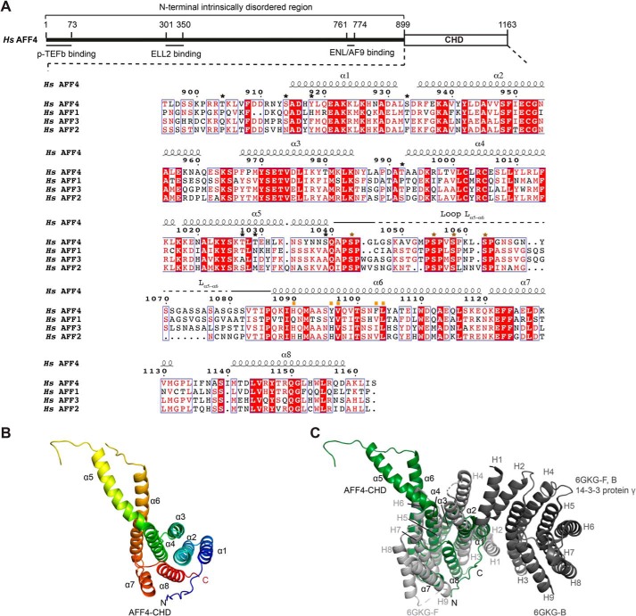
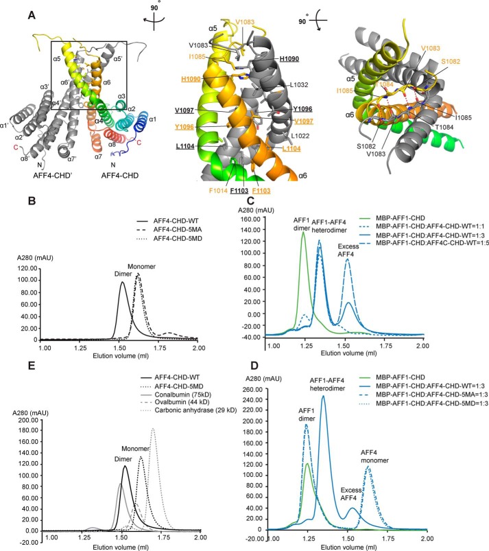
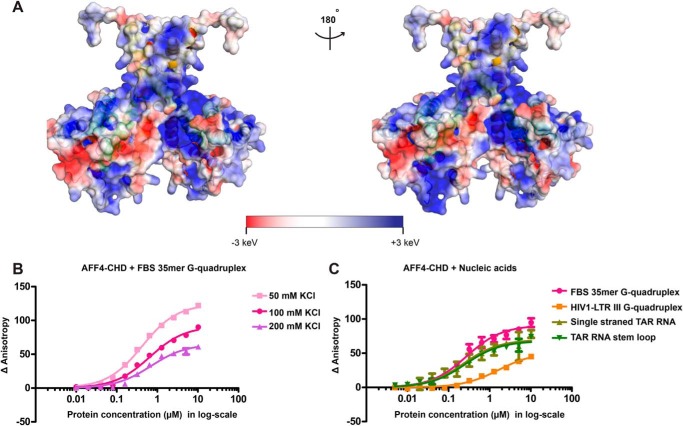
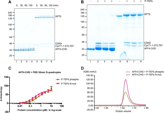
AF4/FMR2 family member 4 (AFF4) is the scaffold protein of the multisubunit super-elongation complex, which plays key roles in the release of RNA polymerase II from promoter-proximal pausing and in the transactivation of HIV-1 transcription. AFF4 consists of an intrinsically disordered N-terminal region that interacts with other super-elongation complex subunits and a C-terminal homology domain (CHD) that is conserved among AF4/FMR2 family proteins, including AFF1, AFF2, AFF3, and AFF4. Here, we solved the X-ray crystal structure of the CHD in human AFF4 (AFF4-CHD) to 2.2 Å resolution and characterized its biochemical properties. The structure disclosed that AFF4-CHD folds into a novel domain that consists of eight helices and is distantly related to tetratrico peptide repeat motifs. Our analyses further revealed that AFF4-CHD mediates the formation of an AFF4 homodimer or an AFF1-AFF4 heterodimer. Results from fluorescence anisotropy experiments suggested that AFF4-CHD interacts with both RNA and DNA in vitro Furthermore, we identified a surface loop region in AFF4-CHD as a substrate for the P-TEFb kinase cyclin-dependent kinase 9, which triggers release of polymerase II from promoter-proximal pausing sites. In conclusion, the AFF-CHD structure and biochemical analyses reported here reveal the molecular basis for the homo- and heterodimerization of AFF proteins and implicate the AFF4-CHD in nucleic acid interactions. The high conservation of the CHD among several other proteins suggests that our results are also relevant for understanding other CHD-containing proteins and their dimerization behavior.

Keywords: AF4/FMR2 family member 4 (AFF4); C-terminal homology domain (CHD); RNA polymerase II; X-ray crystallography; dimerization; gene regulation; phosphorylation; super elongation complex; transcription elongation factor.


Conflict of interest statement

The authors declare that they have no conflicts of interest with the contents of this article

Figures

Figure 1.
Figure 1.
Crystal structure of the AFF4-CHD. A, scheme of the domain organization of AFF4 and the sequence alignment of human AFF4 (NP_055238.1), AFF1 (NP_001160165.1), AFF3 (NP_002276.2), and AFF2 (NP_001162594.1). The interaction regions of AFF4 with P-TEFb, ELL2, and AF9/ENL are shown as lines under the human AFF4 scheme. The alignment was performed using full-length AFF1, AFF2, AFF3, and AFF4 proteins from human, mouse, chicken, and zebrafish and Lilliputian protein from Drosophila using Jalview (muscle with default) (65). The C-terminal sequences of the human AFF proteins are shown here. The C-terminal sequences of the AFF proteins from all above species are shown in Fig. S1. The secondary structure on top of the alignment was assigned based on the crystal structure of AFF4-CHD to the multisequence alignment using ESPript 3 (66). Stars above the sequences indicate residues that are phosphorylated by P-TEFb identified by MS in this study. Black stars show the residues that are not conserved among AFFs. Brown stars show the phosphorylation sites that are conserved among AFFs. The conserved phosphorylation region Lα5–α6 is marked by solid and dashed lines above the sequence. A dashed line indicates the invisible residues in the crystal structure. Mutated residues on the dimerization interface are marked by orange squares. B, crystal structure of AFF4-CHD in one asymmetry unit. The model is colored from N to C using rainbow colors from blue to red. C, superposition of AFF4-CHD monomer with homodimer of 14-3-3 protein γ (Protein Data Bank code 6GKG-F, B (31)). 14-3-3 protein γ is colored in gray, and AFF4-CHD is colored in dark green as figures above. The α-helices of AFF4-CHD are labeled as α1–α8, and the α-helices of 14-3-3 protein γ are labeled as H1–H9. H1, H3, and H4 of the two 14-3-3 protein γ monomers (in light and dark gray) mediate the homodimerization of 14-3-3 protein γ.
Figure 2.
Figure 2.
AFF4-CHD forms homodimer and can form heterodimer with AFF1-CHD in solution. A, dimerization interface of AFF4-CHD homodimer. The left panel shows the overview of the homodimerization interface. In the middle panel, the model is rotated ∼90° clockwise around the vertical axis relative to the overview in the left panel. In the right panel, the model is rotated ∼90° toward the reader around the horizontal axis relative to the view in the middle panel. The two monomers in the dimer are colored gray (AFF4-CHD′) and rainbow (AFF4-CHD), respectively. Residues that are involved in the interface are labeled in orange (AFF4-CHD) and gray (AFF4-CHD′). The side chains (middle panel) or main chains (right panel) are shown as sticks. Underlined letters and numbers indicate residues that are mutated to alanine or aspartate in the 5MA or 5MD constructs for biochemical studies in panels B–D. In the right panel, red dashed lines indicate hydrogen bonds. B, disruption of the dimerization interface abolished the homodimerization of AFF4-CHD. The state of WT and mutants (5MA and 5MD) of AFF4-CHD are analyzed by analytical gel filtration. The elution profiles of WT and mutants of AFF4-CHD are shown as smoothed lines. The raw data were exported from UNICORN and plotted as smoothed lines in Excel. AFF4-CHD–5MA contains the following mutations: H1090A, Y1096A, V1097A, F1103A, and L1104A. AFF4-CHD–5MD contains mutations H1090D, Y1096D, V1097D, F1103D, and L1104D. The WT, 5MA, and 5MD elution profiles are shown in solid, dashed, and dotted black lines, respectively. C, WT AFF4-CHD forms a heterodimer with MBP–AFF1-CHD. The elution profiles of MBP–AFF1-CHD and premixed MBP–AFF1-CHD/AFF4-CHD are shown as smoothed lines. MBP–AFF1-CHD alone (in green) serves as a control. To form the AFF4-CHD/AFF1-CHD complex, AFF4-CHD and MBP–AFF1-CHD were mixed in different ratios (1:1, 1:3, and 1:5 molar ratios) and incubated overnight at 4 °C. The protein mixtures were sequentially injected onto analytical gel filtration column, and the profiles of the preincubated MBP–AFF1/AFF4 protein at different ratios are presented as solid (1:3) and dashed blue lines (1:1 and 1:5) as indicated in the figure. D, the AFF4-CHD mutants failed to form heterodimers with MBP–AFF1-CHD. The MBP–AFF1-CHD are premixed with AFF4-CHD–WT or AFF4-CHD mutants (5MA and 5MD) in 1:3 ratio and incubated overnight prior injection to analytical gel filtrations. The MBP–AFF1-CHD alone (in green) serves as a control. Solid, dashed, and dotted blue lines indicate the elution profile of MBP–AFF1-CHD premixed WT, 5MA, and 5MD, respectively. E, comparison of the molecular mass of AFF4-CHD–WT and 5MD with size exclusion standards. The elution profiles of AFF4-CHD–WT, AFF4-CHD–5MD, and size exclusion standards including conalbumin, ovalbumin, and carbonic anhydrase are shown as a black solid line, a black dashed line, a gray solid line, and a gray dashed line, respectively.
Figure 3.
Figure 3.
AFF4-CHD harbors large positively charged surface and associates with nucleic acids. A, electrostatic surface potential generated with APBS software (67, 68). Blue, red, and white represent positive, negative, and neutral residues, respectively, with electrostatic potentials showing from −3 to +3 keV. The right panel shows the back side of the molecule, with 180° rotation relative to the left panel. B, binding of AFF4-CHD to 10 nm fluorescence labeled FBS 35-mer G-quadruplex in buffer containing 50, 100, and 200 mm KCl. C, binding of AFF4-CHD to 10 nm fluorescence labeled FBS 35-mer G-quadruplex, structured and unstructured TAR RNA, and HIV-1 LTR-III G-quadruplex DNA in buffer containing 100 mm KCl. The experiments in B were repeated three times, each time with two pipetting replicates using 10 nm 5′-FAM–labeled RNA and protein concentration from 0.01 to 10 μm. The experiments in C were repeated three times using 10 nm 5′-FAM–labeled RNA and protein concentration from 0.005 to 10 μm. The standard deviation is shown as error bars on the figure. The curves were fitted with quadratic single-site binding equation (see “Experimental procedures”).
Figure 4.
Figure 4.
AFF4-CHD is a substrate of P-TEFb kinase. A, time course of the phosphorylation of AFF4-CHD by P-TEFb. AFF4-CHD and SPT4/SPT5 were incubated with P-TEFb over a time course of 2 h, and samples were taken at 0, 30, 60, and 120 min. SPT5 serves as a control to monitor the activity of P-TEFb and demonstrate the shift of protein bands after incubation with P-TEFb in the presence of ATP. B, final products of the phosphorylation assay triplicates for the identification of phosphorylation sites using MS. Lanes 1 and 5 are apo proteins before adding P-TEFb. Lanes 2–4 and 6–8 are the triplicates of the phosphorylation assay after 2 h of incubation. C, binding of AFF4-CHD to 10 nm fluorescence labeled FBS 35-mer G-quadruplex in apo and phosphorylated states. The binding assay was performed after incubating AFF4-CHD with WT P-TEFb kinase or catalytical mutant of P-TEFb kinase in the presence of ATP. The experiments were repeated three times, each time with two pipetting repeats as in Fig. 3C. D, the final products of the three phosphorylation triplicates of the AFF4-CHD incubated with P-TEFb and P-TEFb–N-mut were injected onto the gel filtration column (Superdex 200 increase 3.2/300; GE Healthcare), respectively.

References

    1. Hahn S., and Young E. T. (2011) Transcriptional regulation in Saccharomyces cerevisiae: transcription factor regulation and function, mechanisms of initiation, and roles of activators and coactivators. Genetics 189, 705–736 10.1534/genetics.111.127019 - DOI - PMC - PubMed
    1. Li J., and Gilmour D. S. (2011) Promoter proximal pausing and the control of gene expression. Curr. Opin. Genet. Dev. 21, 231–235 10.1016/j.gde.2011.01.010 - DOI - PMC - PubMed
    1. Sainsbury S., Bernecky C., and Cramer P. (2015) Structural basis of transcription initiation by RNA polymerase II. Nat. Rev. Mol. Cell Biol. 16, 129–143 10.1038/nrm3952 - DOI - PubMed
    1. Jonkers I., and Lis J. T. (2015) Getting up to speed with transcription elongation by RNA polymerase II. Nat. Rev. Mol. Cell Biol. 16, 167–177 10.1038/nrm3953 - DOI - PMC - PubMed
    1. Vos S. M., Farnung L., Boehning M., Wigge C., Linden A., Urlaub H., and Cramer P. (2018) Structure of activated transcription complex Pol II–DSIF–PAF–SPT6. Nature 560, 607–612 10.1038/s41586-018-0440-4 - DOI - PubMed

Publication types

MeSH terms

Associated data

LinkOut - more resources