. 2019 Jun 18;116(25):12400-12409.
doi: 10.1073/pnas.1901237116. Epub 2019 May 30.

Building bridges to move recombination complexes

Affiliations

Building bridges to move recombination complexes

Emeline Dubois et al. Proc Natl Acad Sci U S A. .

Abstract

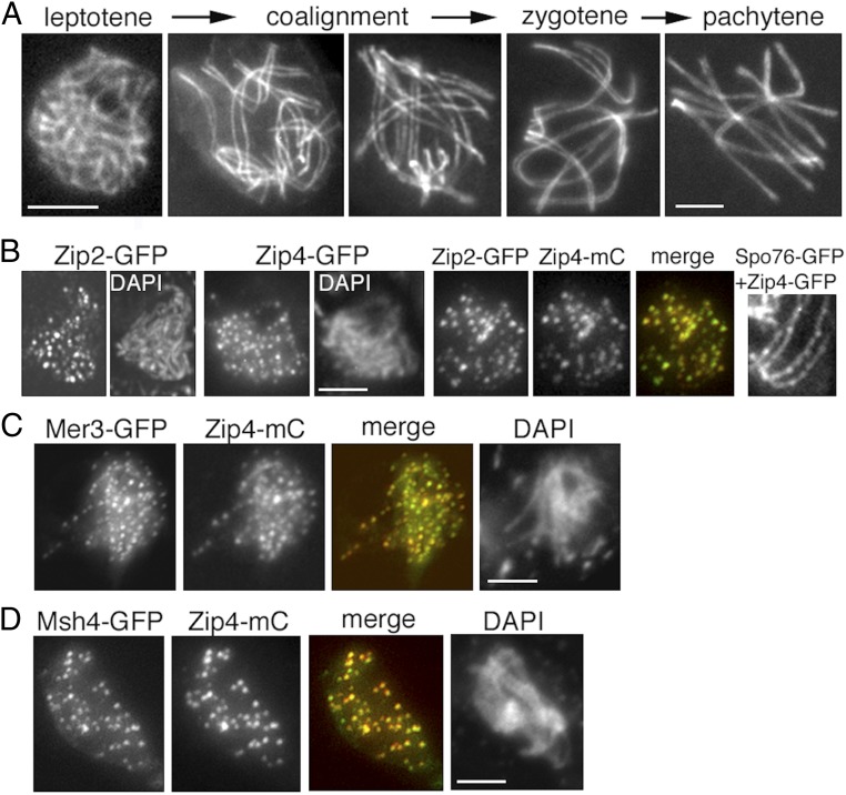
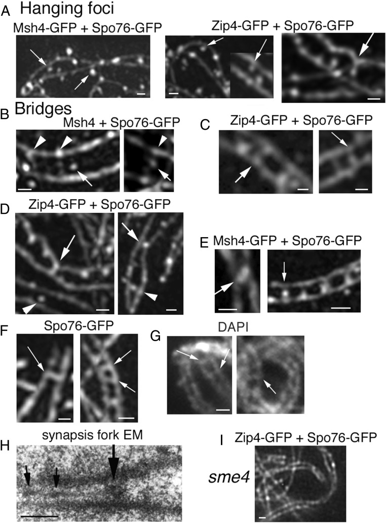
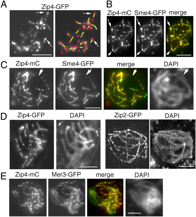
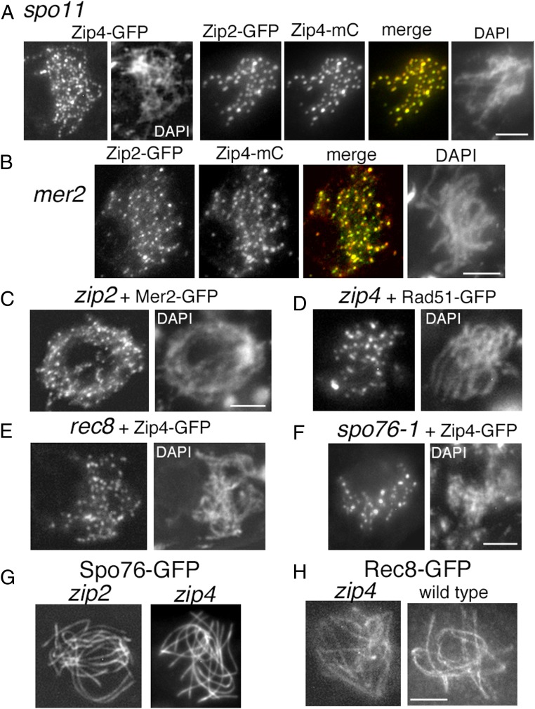
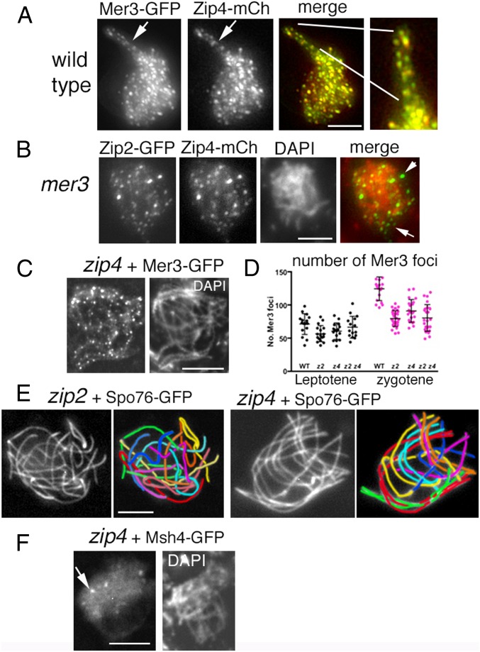
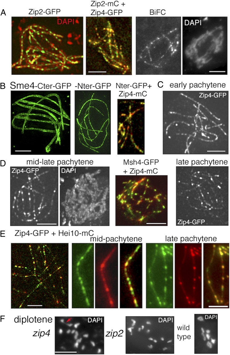
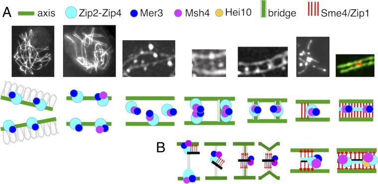
A central feature of meiosis is pairing of homologous chromosomes, which occurs in two stages: coalignment of axes followed by installation of the synaptonemal complex (SC). Concomitantly, recombination complexes reposition from on-axis association to the SC central region. We show here that, in the fungus Sordaria macrospora, this critical transition is mediated by robust interaxis bridges that contain an axis component (Spo76/Pds5), DNA, plus colocalizing Mer3/Msh4 recombination proteins and the Zip2-Zip4 mediator complex. Mer3-Msh4-Zip2-Zip4 colocalizing foci are first released from their tight axis association, dependent on the SC transverse-filament protein Sme4/Zip1, before moving to bridges and thus to a between-axis position. Ensuing shortening of bridges and accompanying juxtaposition of axes to 100 nm enables installation of SC central elements at sites of between-axis Mer3-Msh4-Zip2-Zip4 complexes. We show also that the Zip2-Zip4 complex has an intrinsic affinity for chromosome axes at early leptotene, where it localizes independently of recombination, but is dependent on Mer3. Then, later, Zip2-Zip4 has an intrinsic affinity for the SC central element, where it ultimately localizes to sites of crossover complexes at the end of pachytene. These and other findings suggest that the fundamental role of Zip2-Zip4 is to mediate the recombination/structure interface at all post-double-strand break stages. We propose that Zip2-Zip4 directly mediates a molecular handoff of Mer3-Msh4 complexes, from association with axis components to association with SC central components, at the bridge stage, and then directly mediates central region installation during SC nucleation.

Keywords: Zip2-Zip4; chromosome structure; interaxis bridges; meiotic recombination; synaptonemal complex.


Conflict of interest statement

The authors declare no conflict of interest.

Figures

Fig. 1.
Fig. 1.
Zip2 and Zip4 localizations at leptotene. (A) WT Sordaria prophase. From left to right: early leptotene, coalignment at 400 and 200 nm, partial synapsis at zygotene, and complete synapsis at pachytene. Chromosome axes are marked by Spo76/Pds5-GFP. (B) Early leptotene. (B, Left) Zip2 and Zip4 foci are regularly spaced along chromosomes with similar numbers of foci for the two molecules from early to late leptotene [respectively, for Zip2: 60 ± 9 and 73 ± 3 (n = 33 nuclei); and for Zip4: 59 ± 5 and 82 ± 11 (n = 32)]. (B, Middle) Perfect colocalization of the two proteins. (B, Right) Costaining with Spo76-GFP shows that Zip4 foci are localized on axes. (C and D) Colocalization of Zip4 foci with Mer3 foci (C) and Msh4 foci (D). (Scale bars: 2 μm.)
Fig. 2.
Fig. 2.
Interaxis bridges. (AE) 3D-SIM pictures with costaining of Spo76-GFP (axes) and Zip4-GFP or Msh4-GFP. (A) First step: Msh4 foci (Left; arrows) and Zip4 foci (Middle; arrows) partially “detach” from their coaligned axes. (A, Right) Axis indentation with attached focus located in-between axes (arrow). (B) Examples of early bridges when axes are at 400-nm distance with Msh4 foci located in-between axes (arrows) sometimes (Right) linked by bridge-like structures (arrowheads) or nearby such structures (Left). (C) At the 200-nm coalignment stage, Zip4 foci are located either at matching sites (arrow, Left) or on bridges (arrow, Right). (D) Two examples of single Zip4-GFP foci located in-between axis. At 100-nm distance (arrows), axes are “constricted” by foci that are either fusing (Left) or are single (Right). At 200-nm distance, foci are in the middle between two straight axes (arrowheads). (E, Left) Two fusing Msh4 foci (arrow) with close axes. (E, Right) Row of four bridges with single Zip4 foci (arrow). (F) Spo76-GFP is visible on bridges (arrows) when axes are at 200-nm (Left) and almost 100-nm (Right) distance (3D-SIM). (G) Examples of DAPI bridges visible when homolog axes are at 200-nm (arrows) distance (classical fluorescent microscope). (H) EM section of a pairing fork with a bridge-like structure (large arrow). Note that SC initiates at one side of this structure and exhibits two small recombination nodules (small arrows). (I) In the absence of Sme4, homologs coalign but Zip4 foci remain on axes. (Scale bars: 200 nm.)
Fig. 3.
Fig. 3.
SC nucleation. (A) At early zygotene, Zip2-Zip4 occur either as single foci or as foci with small lines extending in one direction from a focus (arrows) or as lines with no obvious focus. (Right) Corresponding drawing with indications of lines (yellow) and foci (red). (B) Zip4 foci and foci with lines (arrows), colocalize perfectly with corresponding Sme4/Zip1 morphologies (arrows). (C) Zip4 and Sme4/Zip1 morphologies colocalize also throughout zygotene (arrow points to an unsynapsed region, clearly visible in DAPI; Right). (D, Left) At late-zygotene, Zip4-GFP elongates further. (Right) At early pachytene, Zip2-GFP makes continuous foci/lines along all homologs. (E) Colocalization of Mer3 foci with Zip4 foci during zygotene. (Scale bars: 2 μm.)
Fig. 4.
Fig. 4.
Localization of Zip2-Zip4 in the absence of DSBs and cohesins, plus Mer2, Rad51, Spo76, and Rec8 localizations in the absence of Zip2 and/or Zip4. (A) In spo11-null mutant, Zip4 makes regularly spaced foci along all chromosomes (Left) that colocalize with Zip2 foci (Right). (B) Zip2 and Zip4 foci colocalize also in the absence of Mer2. (C and D) WT-like loading of Mer2-GFP (Left) and Rad51-GFP (Right) in zip2 and zip4 null mutants. (E) Zip4 loads normally on axes in the absence of Rec8. (F) Reduced number of Zip4 foci in the nonnull spo76-1 mutant, likely due to the mutant’s defective sister cohesion and/or abnormal chromatin diffuseness (as seen in the corresponding DAPI picture, compared with the DAPI of rec8). (G and H) Spo76 (G) and Rec8 (H) loading is WT-like along all axes in the absence of Zip2 or Zip4. Note that Rec8 lines (Right) are more irregular than the Spo76 lines (Left) in both mutant and WT backgrounds (WT Spo76-GFP in Fig. 1A). (Scale bars: 2 μm.)
Fig. 5.
Fig. 5.
Zip2-Zip4, Mer3, and Msh4 localizations and zip2-zip4 pairing defects. (A) Zip4 localizes to the matching pairs of Mer3 foci in WT. (A, Right) Enlarged segment of the coaligned region. (B) Zip2 and Zip4 colocalize in the absence of Mer3, but foci are no longer exclusively located on chromosomes, as shown by their merge with DAPI (Right; arrows point to foci which do not overlap with DAPI staining). (C) Mer3 foci are regularly spaced along all zip4 chromosomes. (D) Number of Mer3 foci in single and double zip2 zip4 mutants (z2, z4, z2z4) compared with WT at leptotene (Left, black dots) and at early zygotene (Right, pink dots) nuclei (determined by ascus sizes, which grow from 20 μm at early leptotene to 60 μm at zygotene and 100 μm at pachytene, in mutants and WT). (E) zip2 and zip4 mutant nuclei show either no coalignment at all (Left) or partial coalignment (Right). The seven homologs are distinguishable by their lengths and color in the corresponding drawings. In the zip4 nucleus (Right), red, green, cyan, and orange pairs are coaligned while purple pair is partially coaligned and yellow plus blue pairs are not coaligned. Chromosome axes are marked by Spo76-GFP. (F) Only few Msh4 foci (arrow) are visible in the absence of Zip4 (compare with Mer3 foci in C). (Scale bars: 2 μm.)
Fig. 6.
Fig. 6.
Pachytene localization of Zip2-Zip4 versus Sme4/Zip1 and Hei10. (A) All nuclei are at Pachytene. (A, Left) Costaining of Zip2-GFP with DAPI (red). (A, Middle) Colocalization of Zip2-mCherry and Zip4-GFP. (A, Right) Reconstitution of a single GFP signal by Zip2-Zip4 BiFC analysis. (B) 3D-SIM pictures of Sme4 C terminus–GFP define two lines (Left), while the N terminus–GFP is seen as a single line (second picture from left). (B, Middle) Colocalization of Sme4 N terminus–GFP with Zip4-mCherry. (C) Early pachytene nucleus with regularly spaced Zip4 foci (as Zip2 foci in A). (D) During mid-late pachytene, some Zip4 foci increase in volume and brightness (Left, compare with C). They colocalize with Msh4 foci (Middle). (D, Right) At late pachytene, the nonbright Zip4 foci are even more dim. (E) Costaining of Zip4-GFP and Hei10-mCherry during mid and late pachytene. (E, Left) 3D-SIM picture of a late pachytene nucleus. (E, Middle and Right) Foci along one homolog. At mid-pachytene (Middle), Zip4 foci (green) are more numerous and thus only partially colocalize with Hei10 foci (red), while at late pachytene (Right), the two types of foci colocalize mostly. (F) Diplotene nuclei. (F, Left) zip4 with 1 chiasma (red arrow) and zip2 with 14 univalents (Middle) versus WT nucleus with 7 chiasmate bivalents (Right). (Scale bars: 2 μm.)
Fig. 7.
Fig. 7.
From early leptotene to SC formation. (A, Upper) From left to right: pictures corresponding to the different stages described in the paper. (A, Lower) Cartoon of the experimentally observed proteins (indicated above) involved in the corresponding transitions. (B) Proposed model for Zip2-Zip4-mediated coalignment-to-synapsis transition. From left to right: Zip2-Zip4-Mer3-Msh4–mediated recruitment of SC central component Sme4/Zip1 (red arrows) and SC central components (black line). The resulting ensemble, bound to axes components, is released from the axes through bridge formation before relocalization halfway between axes. A signal for SC nucleation then occurs which disassembles the bridges and brings axes closer together, allowing SC initiation. During this process, the Zip2-Zip4-Mer3-Msh4 ensemble now gets associated only with SC central components. SC is thus nucleated by centrally located Zip2-Zip4-Mer3-Msh4, and SC initiation occurs at sites of recombination specifically because of their association with Zip2-Zip4. Recombination complexes are thereby automatically localized on SC central elements. Instead of recombination complexes being “moved” to the SC, the SC comes to the recombination complex.

References

    1. Lam I, Keeney S (2014) Mechanism and regulation of meiotic recombination initiation. Cold Spring Harb Perspect Biol 7:a016634. - PMC - PubMed
    1. Hunter N. (2015) Meiotic recombination: The essence of heredity. Cold Spring Harb Perspect Biol 7:a016618. - PMC - PubMed
    1. Page SL, Hawley RS (2004) The genetics and molecular biology of the synaptonemal complex. Annu Rev Cell Dev Biol 20:525–558. - PubMed
    1. Fraune J, et al. (2016) Evolutionary history of the mammalian synaptonemal complex. Chromosoma 125:355–360. - PubMed
    1. Gao J, Colaiácovo MP (2018) Zipping and unzipping: Protein modifications regulating synaptonemal complex dynamics. Trends Genet 34:232–245. - PMC - PubMed

Publication types

Substances

LinkOut - more resources