. 2019 Jun 11;116(24):11900-11905.
doi: 10.1073/pnas.1901841116. Epub 2019 May 30.

Insights into IgM-mediated complement activation based on in situ structures of IgM-C1-C4b

Affiliations

Insights into IgM-mediated complement activation based on in situ structures of IgM-C1-C4b

Thomas H Sharp et al. Proc Natl Acad Sci U S A. .

Abstract

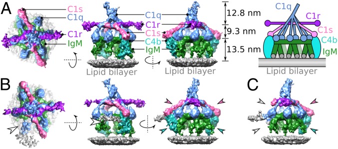
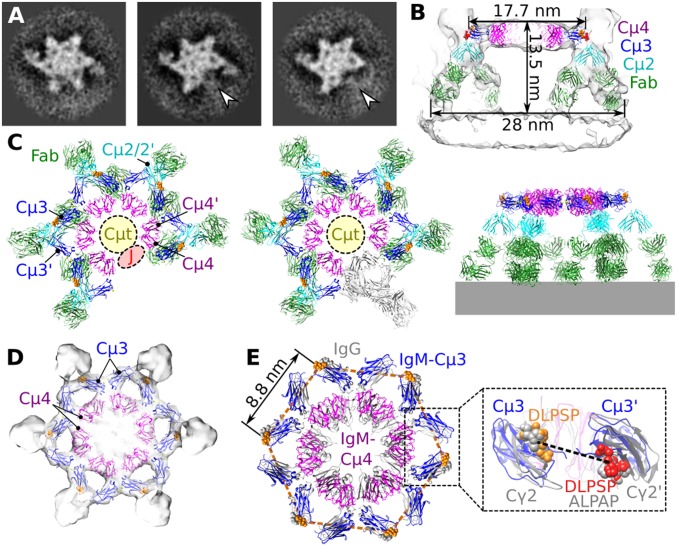
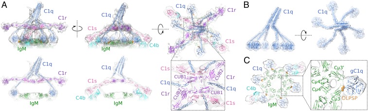
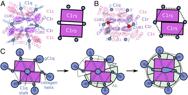
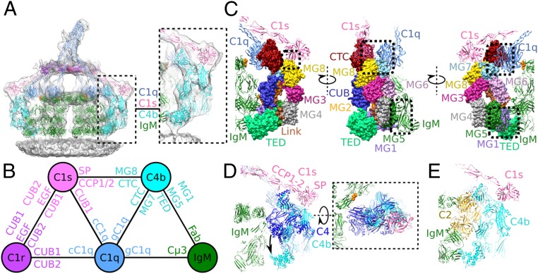
Antigen binding by serum Ig-M (IgM) protects against microbial infections and helps to prevent autoimmunity, but causes life-threatening diseases when mistargeted. How antigen-bound IgM activates complement-immune responses remains unclear. We present cryoelectron tomography structures of IgM, C1, and C4b complexes formed on antigen-bearing lipid membranes by normal human serum at 4 °C. The IgM-C1-C4b complexes revealed C4b product release as the temperature-limiting step in complement activation. Both IgM hexamers and pentamers adopted hexagonal, dome-shaped structures with Fab pairs, dimerized by hinge domains, bound to surface antigens that support a platform of Fc regions. C1 binds IgM through widely spread C1q-collagen helices, with C1r proteases pointing outward and C1s bending downward and interacting with surface-attached C4b, which further interacts with the adjacent IgM-Fab2 and globular C1q-recognition unit. Based on these data, we present mechanistic models for antibody-mediated, C1q-transmitted activation of C1 and for C4b deposition, while further conformational rearrangements are required to form C3 convertases.

Keywords: C1; IgM; complement; cryoelectron tomography; subtomogram averaging.


Conflict of interest statement

The authors declare no conflict of interest.

Figures

Fig. 1.
Fig. 1.
IgM-C1-C4b complexes imaged by phase-plate cryoelectron tomography. (A) Top and two side views of the hexameric IgM-C1-C4b2 density map. Shown are assemblies containing C1q (blue), two molecules of C1r (purple), two molecules of C1s (pink), hexameric IgM (green), and two molecules of C4b (cyan) formed on liposome bilayers (gray). (Right) Relative heights of components are indicated along with a simplified schematic of the complex. (B) Density map of the pentameric IgM-C1-C4b2 complex indicating two molecules of C4b (cyan arrowheads) and two C1s arms (pink arrowheads). Weak density is observed for the C1q leg above empty space and noisy density of pentameric IgM (white arrowhead). (C) Density map of the pentameric IgM-C1-C4b1 complex, showing a single molecule of C4b (cyan arrowhead) and the associated C1s CCP1/2-SP domains (pink arrowhead). On the opposite side disordered noisy density (gray) and increased flexibility of C1s due to the absent C4b molecule are apparent as missing density (white arrowheads).
Fig. 2.
Fig. 2.
Pentameric and hexameric IgM complexes form dome-shape structures. (A) Tomographic slices through the Fc platforms of hexameric IgM-C1-C4b2 (Left), pentameric IgM-C1-C4b2 (Middle), and pentameric IgM-C1-C4b1 (Right) showing 60° spacing of protomers. The site of the missing IgM protomer in the pentameric Fc platforms is indicated (white arrowheads). (B) Side view of the hexameric IgM complex fitted into the density, showing binding of both Fab2 arms (green) to the antigenic surface, dimeric Cµ2/2′-hinge domains (cyan), and the Fc (Cµ3–4/3′-4′) platform (Cµ3 and Cµ4 domains are colored blue and purple, respectively). DLPSP residues (432–436) are colored orange (top face) and red (bottom face). (C) Models of pentameric IgM (Left) and hexameric IgM from the side (Middle) and top (Right). The region for the 18 Cµt peptide extensions is indicated by yellow circles, the positions of the DLPSP residues mediating C1q binding are shown in orange, and the putative site for the J-chain is shown as a red ellipse in the pentameric complex. A single protomer in hexameric IgM is colored gray. (D) Fc platform comprising Cµ3 and Cµ4 domains fitted into the C6-symmetrised density map. (E) Top view of the hexameric IgM model (colored) overlaying the hexameric IgG Fc platform (gray). The distance between adjacent DLPSP motifs (orange) is 8.8 nm (orange hexagon). (Inset) A side view of a Fc region indicating the 31-Å Cα distance between Pro434-Pro434’ (dashed line) in IgM (blue) with DLPSP (orange and red, as described above) and hexamerized IgG1 (gray) aligned to the homologous ALPAP residues (gray spheres).
Fig. 3.
Fig. 3.
C1 binding to hexameric IgM platforms. (A) Model of C1qr2s2 in density (truncated below the IgM Fc platform) bound to hexameric IgM showing the orientations of C1r and C1s. (Below) Central slices through the model. The Inset displays the CUB1 and CUB2 domains of the two antiparallel C1r molecules binding to C1q-collagen helices. (B) C1q has six collagen legs spiraling down to hexagonally arranged globular head domains. (C) Binding of heterotrimeric gC1q units to DLPSP residues (orange) of the FG loop of Fc Cµ3 (SI Appendix, Fig. S6).
Fig. 4.
Fig. 4.
Comparing C1r2s2 in the IgM-C1-C4b complex and the isolated crystal structure suggests a mechanism for C1 activation. (A) Interactions of C1q-collagen helices with C1r and C1s observed in IgM-C1; structures of CUB1-EGF-CUB2 domains in C1r and C1s with positions of C1q indicated by blue circles with specific binding to collagen-binding sites indicated by connecting black lines. Schematic shows the C1rs cores with positions of collagen arms. (B) Crystal structure (Protein Data Bank code 6F1C) (33) and schematic representations of C1rs heterodimers with projected collagen locations added (blue circles), as deduced from C1-IgM; the two obscured collagen positions are indicated by red circles and lines. (C) Schematic representation of C1 conformational rearrangement upon binding to antibody-Fc platforms; with C1q (blue), C1rs heterodimers (purple), and antibodies (green). Empty sites for antibody binding are indicated in gray; C1q positions at the height of the proteases by small blue circles; gC1q headpieces by large blue circles, connecting C1q collagen helices by blue bars; and upright and tilted C1q stalks as a blue circle or ovoid. Dotted lines highlight the hexagonal arrangements. The sliding motion of two halves of the complex is indicated by the colored arrow. See also Movie S1.
Fig. 5.
Fig. 5.
C4b binding to IgM-C1 complexes. (A) The full model in density of the hexameric IgM-C1-C4b2 complex, colored as in Fig. 1. (Inset) The C4b density fit. (B) Protein interaction map showing the domains mediating binding between the protein complexes. (C) Interactions of C4b (surface model) with IgM, C1q, and C1s (ribbon models) are indicated by dashed boxes. C4b colored as in SI Appendix, Fig. S1. (D) Hypothetical IgM-C1-C4 model. The TED domain is structurally unhindered and free to rearrange upon cleavage to C4b. (E) Hypothetical IgM-C1-C4bC2 model showing steric clashes with both the Fc and Fab regions of IgM.

References

    1. Boes M., Role of natural and immune IgM antibodies in immune responses. Mol. Immunol. 37, 1141–1149 (2000). - PubMed
    1. Frank M. M., Humphrey J. H., The subunits in rabbit anti-Forssman IgM antibody. J. Exp. Med. 127, 967–982 (1968). - PMC - PubMed
    1. Mäkelä O., Ruoslahti E., Seppälä I. J., Affinity of IgM and IgG antibodies. Immunochemistry 7, 917–932 (1970). - PubMed
    1. Zhou Z. H., et al. , Polyreactive antibodies plus complement enhance the phagocytosis of cells made apoptotic by UV-light or HIV. Sci. Rep. 3, 2271 (2013). - PMC - PubMed
    1. Quartier P., Potter P. K., Ehrenstein M. R., Walport M. J., Botto M., Predominant role of IgM-dependent activation of the classical pathway in the clearance of dying cells by murine bone marrow-derived macrophages in vitro. Eur. J. Immunol. 35, 252–260 (2005). - PubMed

Publication types

LinkOut - more resources