

Lithium Chloride-Releasing 3D Printed Scaffold for Enhanced Cartilage Regeneration
- PMID: 31147532
- PMCID: PMC6559007
- DOI: 10.12659/MSM.916918
Lithium Chloride-Releasing 3D Printed Scaffold for Enhanced Cartilage Regeneration
Abstract
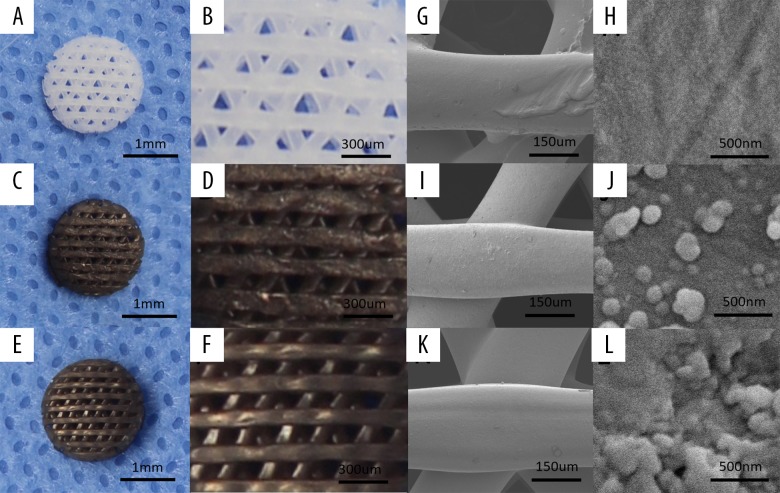
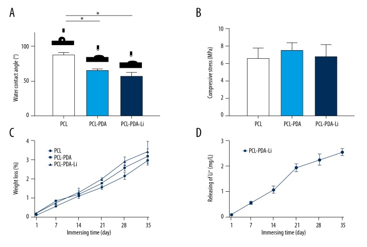
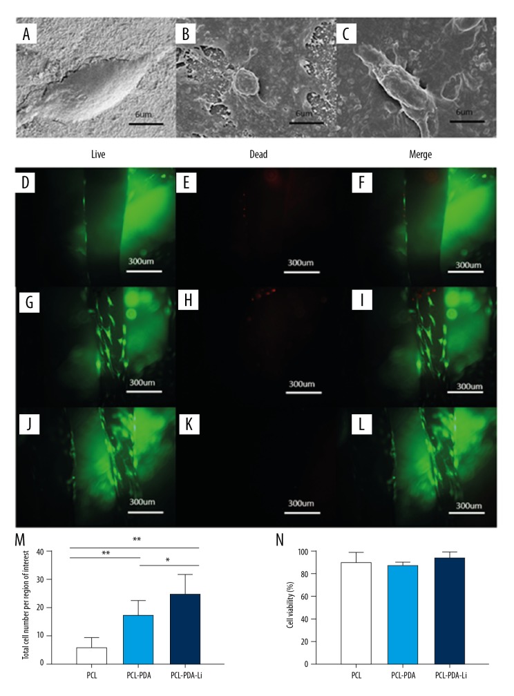
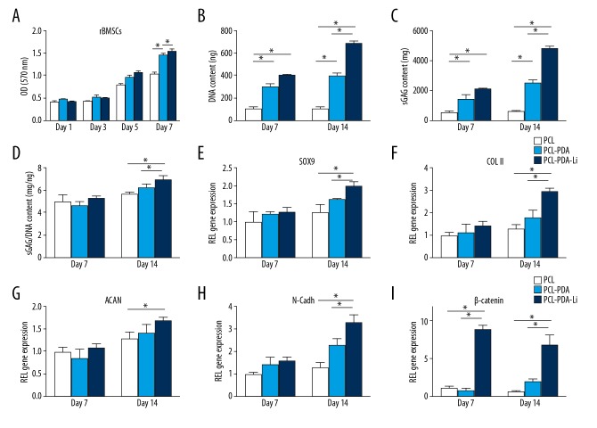
BACKGROUND We synthetized a 3D printed poly-ε-caprolactone (PCL) scaffold with polydopamine (PDA) coating and lithium chloride (LiCl) deposition for cartilage tissue engineering and analyzed its effect on promoting rabbit bone marrow mesenchymal stem cells (rBMSC) chondrogenesis in vitro. MATERIAL AND METHODS PCL scaffolds were prepared by 3D printing with a well-designed CAD digital model, then modified by PDA coating to produce PCL-PDA scaffolds. Finally, LiCl was deposited on the PDA coating to produce PCL-PDA-Li scaffolds. The physicochemical properties, bioactivity, and biocompatibility of PCL-PDA-Li scaffolds were accessed by comparing them with PCL scaffolds and PCL-PDA scaffolds. RESULTS 3D PCL scaffolds exhibited excellent mechanical integrity as designed. PDA coating and LiCl deposition improved surface hydrophilicity without sacrificing mechanical strength. Li⁺ release was durable and ion concentration did not reach the cytotoxicity level. This in vitro study showed that, compared to PCL scaffolds, PCL-PDA and PCL-PDA-Li scaffolds significantly increased glycosaminoglycan (GAG) formation and chondrogenic marker gene expression, while PCL-PDA-Li scaffolds showed far higher rBMSC viability and chondrogenesis. CONCLUSIONS 3D printed PCL-PDA-Li scaffolds promoted chondrogenesis in vitro and may provide a good method for lithium administration and be a potential candidate for cartilage tissue engineering.
Figures
References
-
- Armiento AR, Stoddart MJ, Alin M, et al. Biomaterials for articular cartilage tissue engineering: Learning from biology. Acta Biomater. 2018;65:1–20. - PubMed
-
- Wei B, Yao Q, Guo Y, et al. Three-dimensional polycaprolactone-hydroxyapatite scaffolds combined with bone marrow cells for cartilage tissue engineering. J Biomater Appl. 2015;30:160–70. - PubMed
-
- Noreikait A, Antanaviciute I, Mikalayeva V, et al. Scaffold design for artificial tissue with bone marrow stem cells. Medicina (Kaunas) 2017;53:203–10. - PubMed
MeSH terms
Substances
LinkOut - more resources
Full Text Sources
Miscellaneous
