Review
. 2019 May 30;5(1):38.
doi: 10.1038/s41572-019-0087-y.

Paediatric and adult-onset male hypogonadism

Affiliations
Review

Paediatric and adult-onset male hypogonadism

Andrea Salonia et al. Nat Rev Dis Primers. .

Abstract

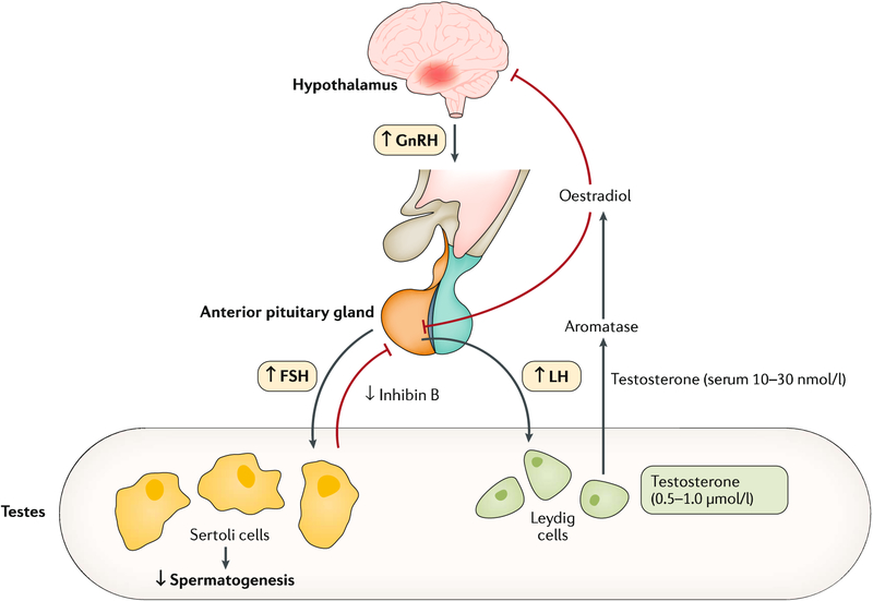
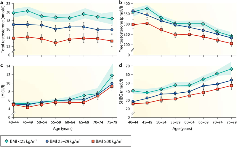
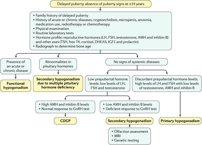
The hypothalamic-pituitary-gonadal axis is of relevance in many processes related to the development, maturation and ageing of the male. Through this axis, a cascade of coordinated activities is carried out leading to sustained testicular endocrine function, with gonadal testosterone production, as well as exocrine function, with spermatogenesis. Conditions impairing the hypothalamic-pituitary-gonadal axis during paediatric or pubertal life may result in delayed puberty. Late-onset hypogonadism is a clinical condition in the ageing male combining low concentrations of circulating testosterone and specific symptoms associated with impaired hormone production. Testosterone therapy for congenital forms of hypogonadism must be lifelong, whereas testosterone treatment of late-onset hypogonadism remains a matter of debate because of unclear indications for replacement, uncertain efficacy and potential risks. This Primer focuses on a reappraisal of the physiological role of testosterone, with emphasis on the critical interpretation of the hypogonadal conditions throughout the lifespan of the male individual, with the exception of hypogonadal states resulting from congenital disorders of sex development.


Conflict of interest statement

Competing interests

The authors declare no competing interests.

Figures

Fig. 1 |
Fig. 1 |. The hypothalamic–pituitary–gonadal axis.
Both testosterone synthesis and male fertility result from the delicate coordination throughout the hypothalamic–pituitary–gonadal axis, thereby ensuring normal testicular function. Gonadotropin-releasing hormone (GnRH) stimulates the release of luteinizing hormone (LH) from the pituitary gland. This triggers the Leydig cells within the testes to respond by producing adequate levels of testosterone, which, in turn, exerts negative feedback control on the hypothalamus and pituitary gland. Likewise, GnRH stimulates the release of follicle-stimulating hormone (FSH) from the pituitary gland. This triggers and sustains the spermatogenesis within the exocrine part of the testes. The testes contribute >95% of total circulating testosterone in the postpubertal male; testosterone is secreted into the circulation down a concentration gradient, where it equilibrates between protein-bound (98%) and free hormone (1–2%) fractions. Circulating testosterone and other sex hormones are bound either to low-affinity, high-availability proteins (primarily albumin) or to the high-affinity glycoprotein sex hormone-binding globulin (SHBG). These binding proteins play an important part in regulating the transport, distribution, metabolism and biological activity of the sex hormones,. Conditions that alter SHBG levels (for instance, ageing, obesity, insulin resistance and liver disease) influence free testosterone levels. The free hormone fraction is postulated to be the biologically active form of testosterone,,. Testosterone secretion varies throughout the day and is usually the highest in the morning. Hence, samples to determine testosterone levels need to be taken in the morning. Figure adapted from REF., Springer Nature Limited.
Fig. 2 |
Fig. 2 |. Anatomical changes and serum hormone levels associated with male sex determination and maturation.
In the fetal period, testicular hormones begin to be secreted independently of fetal pituitary gonadotropins in the first trimester of fetal life and drive fetal differentiation of the genitalia. In the second and third trimesters, growth of the genitalia and testicular descent are stimulated by androgen secretion dependent on fetal luteinizing hormone (LH). In the postnatal period, testicular volume increases during childhood owing essentially to Sertoli proliferation. After the postnatal activation in the 0–6-month period (usually called ‘mini-puberty’), serum levels of gonadotropins and testosterone (T) decline, but those of the Sertoli cell markers anti-Mullerian hormone (AMH) and inhibin B persist at clearly detectable levels. During puberty, testicular volume increases dramatically owing to spermatogenic development, secondary to gonadotropin and T action. Sertoli cell markers show opposite profiles: AMH is inhibited by T whereas inhibin B is upregulated by follicle-stimulating hormone (FSH) and germ cells. INSL3, insulin-like factor 3; O, testicular volume measured by comparison to Prader’s orchidometer; US, testicular volume measured by ultrasonography.
Fig. 3 |
Fig. 3 |. Pathophysiology of hypogonadism.
Hypogonadism may be caused by a primary testicular pathology (primary hypogonadism, otherwise known as hypergonadotropic hypogonadism, which is defined as low testicular hormones, with high gonadotropins) resulting from malfunction at the level of the testes due to a genetic cause, injury, inflammation or infection (panel a). Conversely, central defects of the hypothalamus or the pituitary gland lead to secondary hypogonadism (also called central hypogonadism or hypogonadotropic hypogonadism, which is defined as low testicular hormones, with low or normal gonadotropins), which is most often caused by genetic defects, neoplasm or infiltrative disorders (panel b). FSH, follicle-stimulating hormone; GnRFI, gonadotropin-releasing hormone; LH, luteinizing hormone. Adapted with permission from REF., Oxford University Press.
Fig. 4 |
Fig. 4 |. Pathophysiology of congenital secondary hypogonadism.
Genes associated with congenital secondary hypogonadism. GnRH, gonadotropin-releasing hormone.
Fig. 5 |
Fig. 5 |. Relationship between age, BMI and reproductive hormones.
The graphs present mean levels of total and calculated free testosterone, luteinizing hormone (LH) and sex hormone-binding globulin (SHBC). a | Total testosterone is reduced in overweight and obese men compared with nonobese men at all ages. b | Free testosterone, similar to total testosterone, is reduced in overweight and obese men compared with nonobese men at all ages. c | LH increases with age but is not associated with body mass index (BMI). d | SHBC increases with age. For total testosterone and SHBC, no interaction between BMI and age were found, whereas free testosterone showed an interaction between BMI and age. The data were derived from a cohort of 3,220 men aged 40–79 years recruited in the European Male Ageing Study (EMAS) study. Shaded areas and vertical lines represent the 95% CI. Adapted with permission from REF., Oxford University Press.
Fig. 6 |
Fig. 6 |. Diagnostic algorithm for hypogonadism in pubertal age.
Diagnostic flowchart in a boy presenting with no signs of pubertal development by 14 years of age. Tests mentioned may help to distinguish among and/or confirm diagnoses, but the full battery is not recommended or warranted in all cases and may not lead to a conclusive diagnosis. The main text describes considerations regarding test use. AMH, anti-Müllerian hormone; CDGP, constitutional delay of growth and puberty; DHEAS, dehydroepiandrosterone; FSH, follicle-stimulating hormone; GnRH, gonadotropin-releasing hormone; IGF1, insulin-like growth factor 1; LH, luteinizing hormone; T4, thyroxine; TSH, thyroid-stimulating hormone.

References

    1. Corradi PF, Corradi RB & Greene LW Physiology of the hypothalamic pituitary gonadal axis in the male. Urol. Clin. North Am 43, 151–162 (2016). - PubMed
    2. This manuscript comprehensively describes the complex physiology of the male HPG axis.

    1. Fluck CE et al. Why boys will be boys: two pathways of fetal testicular androgen biosynthesis are needed for male sexual differentiation. Am. J. Hum. Genet 89, 201–218 (2011). - PMC - PubMed
    1. Ross A & Bhasin S Hypogonadism: its prevalence and diagnosis. Urol. Clin. North Am 43, 163–176 (2016). - PubMed
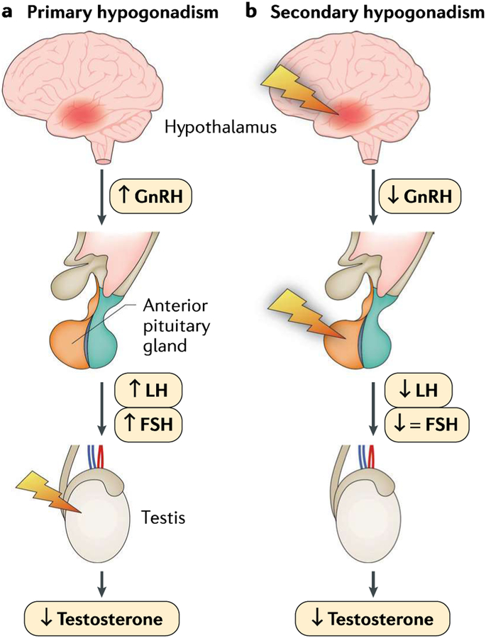
    1. Rey RA et al. Male hypogonadism: an extended classification based on a developmental, endocrine physiology-based approach. Andrology 1,3–16 (2013). - PubMed
    2. This work provides a classification of male hypogonadism, explaining the pathophysiology and specific diagnostic procedures needed according to the age of establishment of the disorder, from fetal life to adulthood.

    1. Rastrelli G, Vignozzi L & Maggi M Different medications for hypogonadotropic hypogonadism. Endocr. Dev 30, 60–78 (2016). - PubMed