. 2019 May 30;9(1):8092.
doi: 10.1038/s41598-019-44250-y.

Retinal Oxygen Delivery, Metabolism and Extraction Fraction and Retinal Thickness Immediately Following an Interval of Ophthalmic Vessel Occlusion in Rats

Affiliations

Retinal Oxygen Delivery, Metabolism and Extraction Fraction and Retinal Thickness Immediately Following an Interval of Ophthalmic Vessel Occlusion in Rats

Norman P Blair et al. Sci Rep. .

Abstract

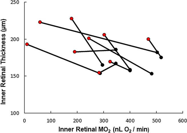
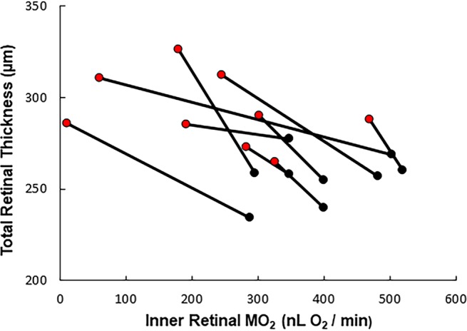
Limited knowledge is currently available about alterations of retinal blood flow (F), oxygen delivery (DO2), oxygen metabolism (MO2), oxygen extraction fraction (OEF), or thickness after the ophthalmic blood vessels have been closed for a substantial interval and then reopened. We ligated the ophthalmic vessels for 120 minutes in one eye of 17 rats, and measured these variables within 20 minutes after release of the ligature in the 10 rats which had immediate reflow. F, DO2 and MO2 were 5.2 ± 3.1 μL/min, 428 ± 271 nL O2/min, and 234 ± 133 nL O2/min, respectively, that is, to 58%, 46% and 60% of values obtained from normal fellow eyes (P < 0.004). OEF was 0.65 ± 0.23, 148% of normal (P = 0.03). Inner and total retinal thicknesses were 195 ± 24 and 293 ± 20 μm, respectively, 117% and 114% of normal, and inversely related to MO2 (P ≤ 0.02). These results reflect how much energy is available to the retina immediately after an interval of nonperfusion for 120 minutes. Thus, they elucidate aspects of the pathophysiology of nonperfusion retinal injury and may improve therapy in patients with retinal artery or ophthalmic artery obstructions.


Conflict of interest statement

The authors declare no competing interests.

Figures

Figure 1
Figure 1
Relationship between inner retinal thickness and inner retinal oxygen metabolism (MO2) in rats with normal fellow and nonperfusion eyes. Data from the normal fellow eyes (black) are connected to the corresponding data obtained from the eyes immediately after 120 minutes of total ocular nonperfusion (red). An inverse linear relationship was found between increases in inner retinal thickness values and nonperfusion-induced reductions in MO2 values (β = −0.093 µm * min * nL O2−1, P = 0.019, N = 9).
Figure 2
Figure 2
Relationship between total retinal thickness and inner retinal oxygen metabolism (MO2) in rats with normal fellow and nonperfusion eyes. Data from the normal fellow eyes (black) are connected to the corresponding data obtained from the eyes immediately after 120 minutes of total ocular nonperfusion (red). An inverse linear relationship was found between increases in total retinal thickness values and nonperfusion-induced reductions in MO2 values (β = −0.12 µm * nL O2−1 * min, P = 0.006, N = 9).

Similar articles

Cited by

References

    1. Bresnick GH, De Venecia G, Myers FL, Harris JA, Davis MD. Retinal ischemia in diabetic retinopathy. Archives of ophthalmology. 1975;93:1300–1310. doi: 10.1001/archopht.1975.01010020934002. - DOI - PubMed
    1. Shimizu K, Kobayashi Y, Muraoka K. Midperipheral fundus involvement in diabetic retinopathy. Ophthalmology. 1981;88:601–612. doi: 10.1016/S0161-6420(81)34983-5. - DOI - PubMed
    1. Silva PS, et al. Diabetic Retinopathy Severity and Peripheral Lesions Are Associated with Nonperfusion on Ultrawide Field Angiography. Ophthalmology. 2015;122:2465–2472. doi: 10.1016/j.ophtha.2015.07.034. - DOI - PubMed
    1. Stefansson E. Oxygen and diabetic eye disease. Graefe’s archive for clinical and experimental ophthalmology=Albrecht von Graefes Archiv fur klinische und experimentelle Ophthalmologie. 1990;228:120–123. doi: 10.1007/BF02764304. - DOI - PubMed
    1. Do BK, Rodger DC. Sickle cell disease and the eye. Curr Opin Ophthalmol. 2017;28:623–628. doi: 10.1097/ICU.0000000000000423. - DOI - PubMed

Publication types