. 2019 Jun;36(6):1251-1261.
doi: 10.1007/s10815-019-01424-x. Epub 2019 May 30.

Femtosecond laser is effective tool for zona pellucida engraving and tagging of preimplantation mammalian embryos

Affiliations

Femtosecond laser is effective tool for zona pellucida engraving and tagging of preimplantation mammalian embryos

Inna V Ilina et al. J Assist Reprod Genet. 2019 Jun.

Abstract

Purpose: Our purpose was to study whether application of femtosecond laser pulses for alphanumeric code marking in the volume of zona pellucida (ZP) could be effective and reliable approach for direct tagging of preimplantation embryos.

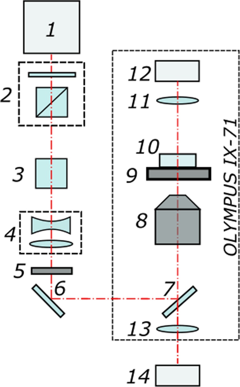
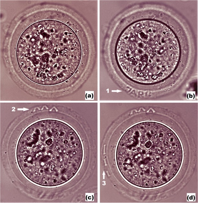
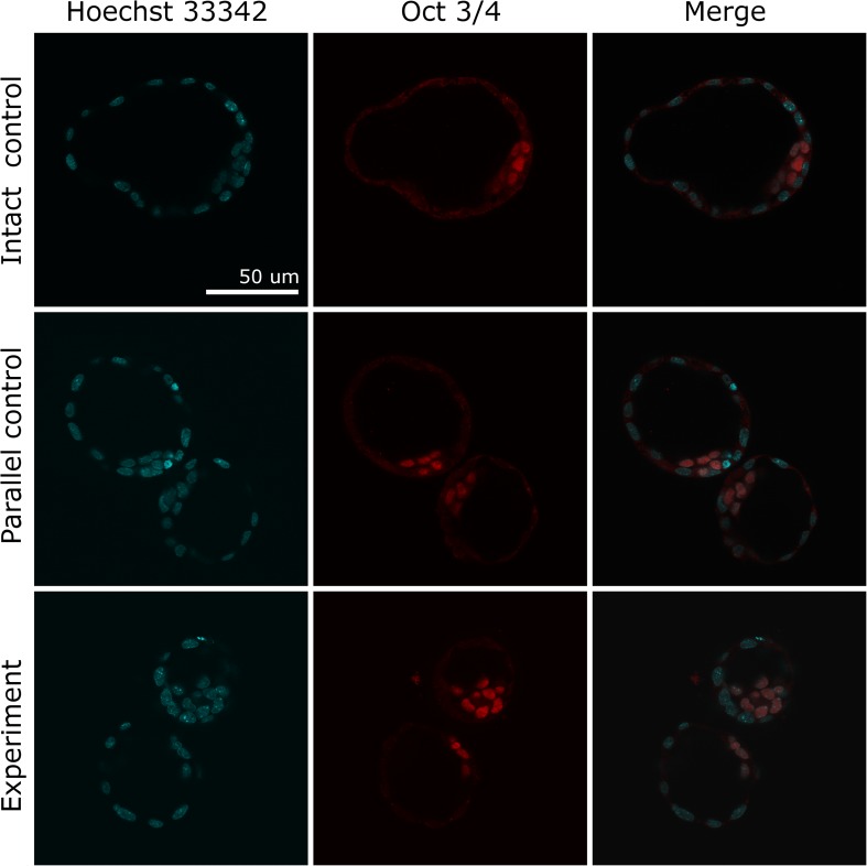
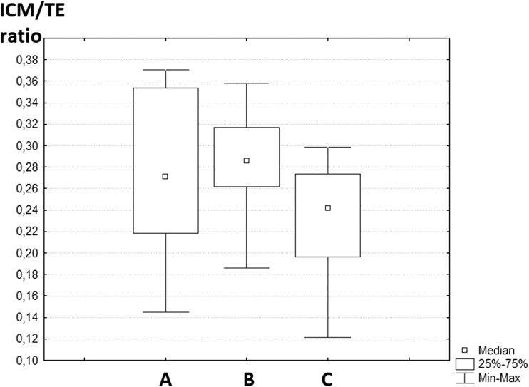
Methods: Femtosecond laser pulses (wavelength of 514 nm, pulse duration of 280 fs, repetition rate of 2.5 kHz, pulse energy of 20 nJ) were applied for precise alphanumeric code engraving on the ZP of mouse embryos at the zygote stage for individual embryo marking and their accurate identification. Embryo quality assessment every 24 h post laser-assisted marking as well as immunofluorescence staining (for ICM/TE cell number ratio calculation) were performed.

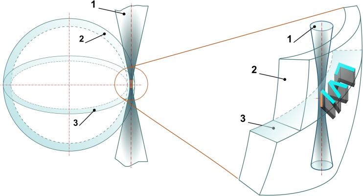
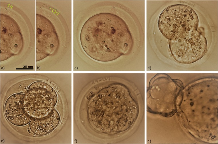
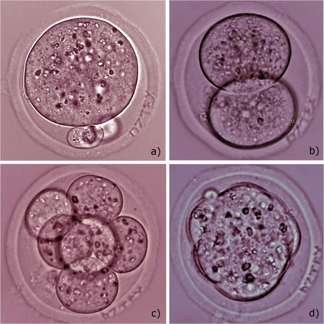
Results: Initial experiments have started with embryo marking in a single equatorial plane. The codes engraved could be clearly recognized until the thinning of the ZP prior to hatching. Since embryo may change its orientation during the ART cycle, multi-plane code engraving seems to be more practical for simplifying the process of code searching and embryo identification. We have marked the ZP in three planes, and no decrease in developmental rates as well as no morphological changes of embryos post laser-assisted engraving have been observed as compared to control group embryos.

Conclusions: Our results demonstrate the suitability of femtosecond laser as a novel tool for noninvasive embryo tagging, enabling embryo identification from day 0.5 post coitum to at least early blastocyst stage. Thus, the versatility and the potential use of femtosecond lasers in the field of developmental biology and assisted reproduction have been shown.

Keywords: Embryo identification; Embryo tagging; Femtosecond laser; Laser microsurgery; Laser-assisted embryo marking; Zona pellucida.


Conflict of interest statement

The authors declare that they have no conflict of interest.

Figures

Fig. 1
Fig. 1
Schematic diagram of the experimental set-up. (1) Femtosecond ytterbium laser. (2) Beam attenuator. (3) Second-harmonic generator. (4) Telescope. (5) Electro-mechanical shutter. (6) Mirror. (7) Dichroic mirror. (8) Microscope objective. (9) X-Y motorized stage. (10) Petri dish. (11) Condenser lens. (12) Microscope lamp. (13) Tube lens. (14) CMOS camera
Fig. 2
Fig. 2
Schematic illustration of laser-assisted engraving of “LVI” code in the volume of the ZP. (1) Laser beam. (2) The zona pellucida. (3) The equatorial plane
Fig. 3
Fig. 3
Femtosecond laser-assisted tagging of preimplantation mouse embryos. a Drawing of numeric characters “1” and “4” in the software. b Laser engraving of numeric characters “1” and “4” on the zona pellucida and drawing of alphabetic characters “L, V, I” in the software. c Alphanumeric code engraved on the zygote’s zona pellucida. (df) Development of embryo with the engraved code on the 1.5, 2.5, 3.5 dpc. (g) Embryo hatching on the 4.5 dpc
Fig. 4
Fig. 4
Femtosecond laser-assisted engraving of code “07TEX” on the zona pellucida of preimplantation mouse embryos. a Embryo right after the code engraving (0.5 dpc). b Embryo on the following day (1.5 dpc) with clearly visible code. cd Embryo at 2.5 dpc and 3.5 dpc with codes “07TEX” slightly shifted outwards but still recognizable
Fig. 5
Fig. 5
Femtosecond laser-assisted engraving of three codes in various planes. a Embryo prior to laser-assisted code engraving. b Laser-assisted engraving of code “JARG”. c, d laser-assisted engraving of codes “V V V” and “I I I” correspondingly
Fig. 6
Fig. 6
Differential staining of inner cell mass and trophectoderm cells of mouse embryos. Row 1 is fully hatched blastocyst from intact control group, rows 2 and 3 are 8-shaped hatching blastocysts from parallel and experimental groups. All nuclei were stained with Hoechst 33342 (blue color), and ICM cells were stained with anti-Oct3/4 antibody conjugated with Alexa Fluor 568 (red color)
Fig. 7
Fig. 7
The box-and-whisker plot illustrating ICM/TE ratio in blastocysts from three different groups. (A) Experimental group (three-plane code engraving, samples n = 7). (B) Parallel control (n = 7). (C) Intact control (n = 7)

References

    1. Bedient C, Khanna P, Desai N. Laser pulse application in IVF. In: Jakubczak K, editor. Lasers - applications in science and industry: InTech; 2011. p. 193–214.
    1. Karu TI. Lasers in infertility treatment: irradiation of oocytes and spermatozoa. Photomed Laser Surg. 2012;30:239–241. doi: 10.1089/pho.2012.9888. - DOI - PMC - PubMed
    1. Abdel-Salam Z, Harith MA. Laser researches on livestock semen and oocytes: a brief review. J Adv Res. 2015;6:311–317. doi: 10.1016/j.jare.2014.11.006. - DOI - PMC - PubMed
    1. Montag M, Rink K, Delacretaz G, van der Ven H. Laser induced immobilization and plasma membrane permeabilization in human spermatozoa. Hum Reprod. 2000;15:846–852. doi: 10.1093/humrep/15.4.846. - DOI - PubMed
    1. Sato H, Landthaler M, Haina D, Schill WB. The effects of laser light on sperm motility and velocity in vitro. Andrologia. 1984;16:23–25. doi: 10.1111/j.1439-0272.1984.tb00229.x. - DOI - PubMed