. 2019 Jul;8(14):e1900411.
doi: 10.1002/adhm.201900411. Epub 2019 May 30.

Hyaluronic Acid Hydrogel Integrated with Mesenchymal Stem Cell-Secretome to Treat Endometrial Injury in a Rat Model of Asherman's Syndrome

Affiliations

Hyaluronic Acid Hydrogel Integrated with Mesenchymal Stem Cell-Secretome to Treat Endometrial Injury in a Rat Model of Asherman's Syndrome

Feiran Liu et al. Adv Healthc Mater. 2019 Jul.

Abstract

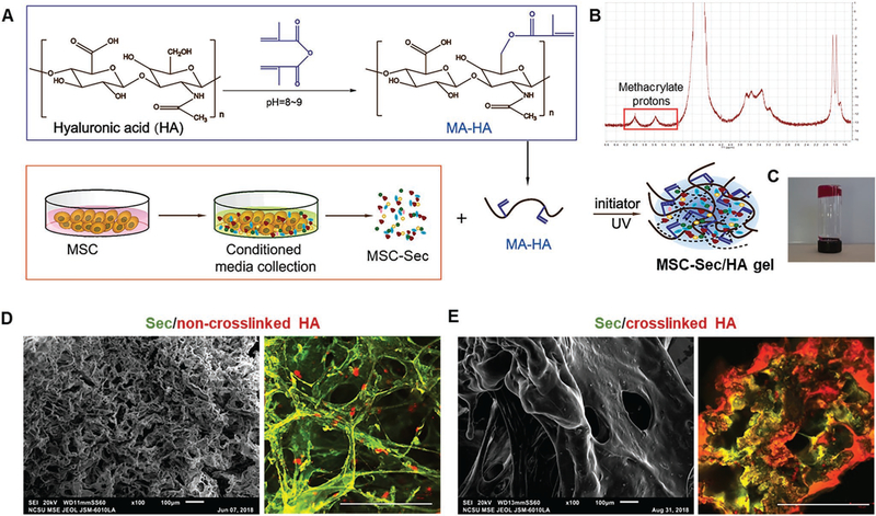
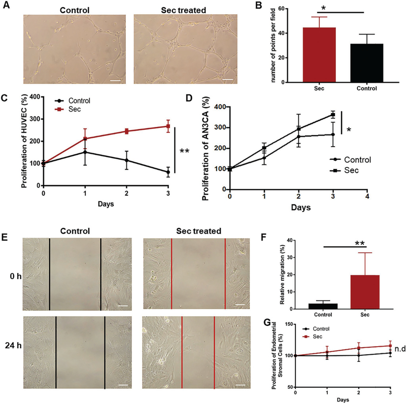
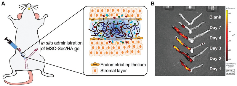
Stem cell therapies have made strides toward the efficacious treatment of injured endometrium and the prevention of intrauterine adhesions, or Asherman's syndrome (AS). Despite this progress, they are limited by their risk of tumor formation, low engraftment rates, as well as storage and transportation logistics. While attempts have been made to curb these issues, there remains a need for simple and effective solutions. A growing body of evidence supports the theory that delivering media, conditioned with mesenchymal stem cells, might be a promising alternative to live cell therapy. Mesenchymal stem cell-secretome (MSC-Sec) has a superior safety profile and can be stored without losing its regenerative properties. It is versatile enough to be added to a number of delivery vehicles that improve engraftment and control the release of the therapeutic. Thus, it holds great potential for the treatment of AS. Here, a new strategy for loading crosslinked hyaluronic acid gel (HA gel) with MSC-Sec is reported. The HA gel/MSC-Sec treatment paradigm creates a sustained release system that repairs endometrial injury in rats and promotes viable pregnancy.

Keywords: Asherman's syndrome; endometrial injury; hyaluronic acid; mesenchymal stem cells; secretome.


Conflict of interest statement

Conflict of Interest

The authors declare no conflict of interest.

Figures

Figure 1.
Figure 1.
Characterization of MSCs. Flow cytometry analysis of common MSC markers such as A) CD105 and B) CD90. MSCs are negative for C) CD31, D) CD34, and E) CD117. F) Summary of positive MSC cells with different markers.
Figure 2.
Figure 2.
Human cytokine arrays. A,B) Human cytokine array analysis (AAH-CYT-6 and AAH-CYT-7) of MSC-Sec and densitometric analysis of proteins associated with tissue regeneration. Graph shows the mean value. The proteins in graph are pointed out in red.
Figure 3.
Figure 3.
Fabrication and characterization of MSC-Sec-loaded, crosslinked HA gel. A) Schematic showing the synthesis of MSC-Sec-loaded, crosslinked HA gel. B) MA-HA: 1H NMR (D2O, 300 MHz, δ ppm): 1.85–1.96 (m, 3H, CH2 = C(CH3)CO), 1.99 (s, 3H, NHCOCH3), 5.74 (s, 1H, CH1H2 = C(CH3) CO), 6.17 (s, 1H, CH1H2 = C(CH3)CO). C) State of HA gel at room temperature when the bottle is upside down (gelation). D) Representative SEM (scale bar: 100 μm) and color-depth projection confocal images of MSC-Sec/non-crosslinked HA; green represents MSC-Sec, red represents HA. Scale bar in confocal image: 100 μm. E) Representative SEM (scale bar: 100 μm) and color-depth projection confocal images of MSC-Sec/crosslinked HA; green represents MSC-Sec, red represents HA. Scale bar in confocal image: 100 μm.
Figure 4.
Figure 4.
In vitro cell-based assays. A) HUVEC tubing images of MSC-Sec-treated group and control group. Scale bar: 20 μm. B) Graph comparing the number of tube points between MSC-Sec-treated group and control group, n = 3. C) Proliferation of HUVEC with IMDM (control group) or MSC-Sec group. D) Proliferation of AN3CA treated with IMDM or MSC-Sec. * indicates P < 0.05 when compared to the control group. E) The image of cell migration of rat endometrial epithelial cells (EEC) cultured with IMDM (control group) or MSC-Sec. Scale bar: 20 μm. F) Analysis of the migration rate of EEC of the control group and MSC-Sec group. G) Proliferation of rat endometrial stromal cells cultured with IMDM or MSC-Sec. n = 6 for each group if no further description, * indicates P < 0.05, ** indicates P < 0.01.
Figure 5.
Figure 5.
MSC-Sec/HA gel injection and rodent model of endometrium injury. A) Schematic showing intrauterine injection of MSC-Sec/HA gel. B) Ex vivo fluorescent imaging of rat uteri at days 1, 2, 3, 4, and 7 after injection.
Figure 6.
Figure 6.
The electrocoagulation-induced animal model of AS. A) Pictures showing the procedure of inducing injury and delivering the treatment. I: abdominal skin incision. II: incision of rectus abdominis and uterus exposure. III: electrocoagulation injury in both sides of uterus. IV: electrocoagulation machine. V: intrauterine injection. Right side is treatment side. Left side is control side. VI: closure of uterus incision. VII: closure of abdominis. VIII: closure of abdominal skin incision. B) Representative H&E and Masson’s trichrome-stained uterus section 14 days after injury (Scale bar: 70 μm). C) Quantitative data comparing the number of glands of endometrium of injured group and control group. D) Quantitative data comparing the thickness of endometrium of injured group and control group. n = 5, ** indicates P < 0.01 when compared to the other group.
Figure 7.
Figure 7.
The effects of MSC-Sec HA gel on fetal development. A) Schematic showing the animal study design. Both sides of the uterus are injured. One side gets treatment while left side gets PBS as control. And there are three treatment groups: MSC-Sec, crosslinked HA gel group; crosslinked HA group, and MSC-Sec group, n = 5. B) Representative uterus images and quantitative data comparing the numbers of fetuses on both sides (red arrow indicates treated side). * indicates P < 0.05 when compared to the other side.
Figure 8.
Figure 8.
MSC-Sec/HA treatment increases gland numbers and endometrium thickness. A) Representative H&E images of sham, the control side of MSC-Sec/HA group, and the treated side of MSC-Sec/HA group. Scale bar: 70 μm. B) Comparison of endometrium thickness between the control side and the treated side of MSC-Sec/HA group. C) Comparison of number of glands between the control side and the treated side of MSC-Sec/HA group. n = 5, ** indicates P < 0.01, *** indicates P < 0.001 when compared to the other group.

References

    1. Hooker AB, Lemmers M, Thurkow AL, Heymans MW, Opmeer BC, Brolmann HA, Mol BW, Huirne JA, Hum. Reprod. Update 2014, 20, 262. - PubMed
    1. Berman JM, Semin. Reprod. Med. 2008, 26, 349. - PubMed
    1. Chen L, Zhang H, Wang Q, Xie F, Gao S, Song Y, Dong J, Feng H, Xie K, Sui L, J. Minimally Invasive Gynecol. 2017, 24, 299. - PubMed
    1. Capella-Allouc S, Morsad F, Rongieres-Bertrand C, Taylor S, Fernandez H, Hum. Reprod 1999, 14, 1230; - PubMed
    2. Roy KK, Baruah J, Sharma JB, Kumar S, Kachawa G, Singh N, Arch. Gynecol. Obstet 2010, 281, 355. - PubMed
    1. Huberlant S, Fernandez H, Vieille P, Khrouf M, Ulrich D, deTayrac R, Letouzey V, PLoS One 2015, 10, e0125610; - PMC - PubMed
    2. Mais V, Cirronis MG, Peiretti M, Ferrucci G, Cossu E, Melis GB, Eur. J. Obstet. Gynecol. Reprod. Biol 2012, 160, 1. - PubMed

Publication types