. 2019 Apr 13;9(8):2380-2394.
doi: 10.7150/thno.29724. eCollection 2019.

SIRT6, a novel direct transcriptional target of FoxO3a, mediates colon cancer therapy

Affiliations

SIRT6, a novel direct transcriptional target of FoxO3a, mediates colon cancer therapy

Yingjie Zhang et al. Theranostics. .

Abstract

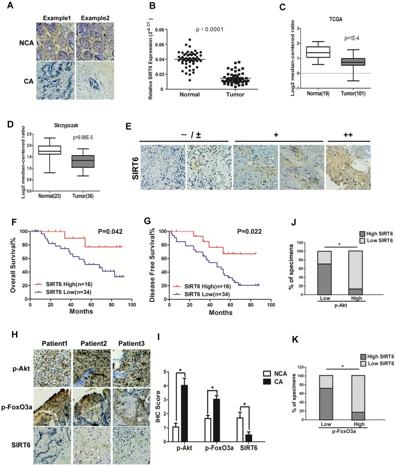
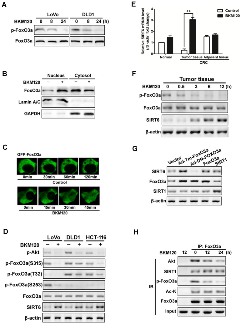
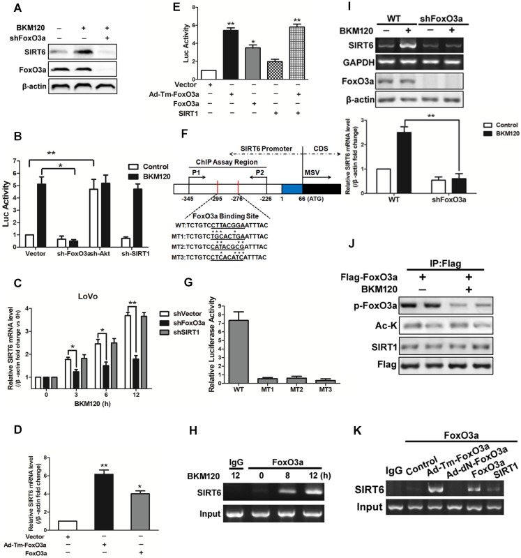
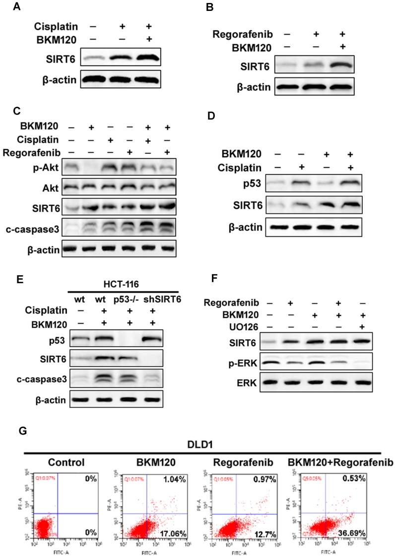
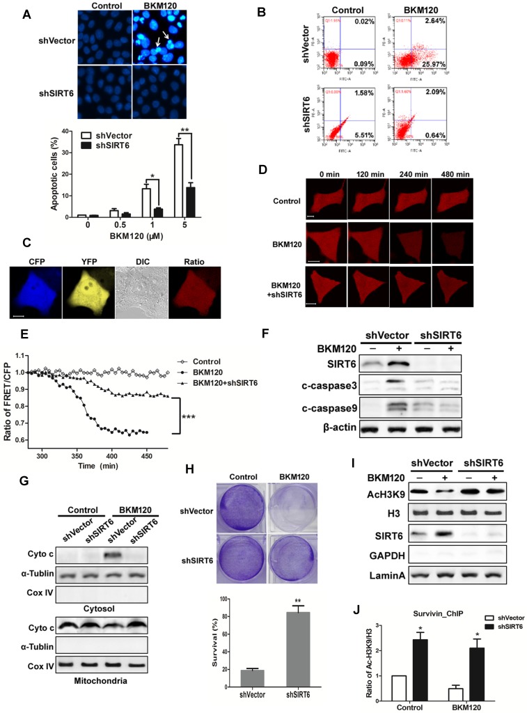
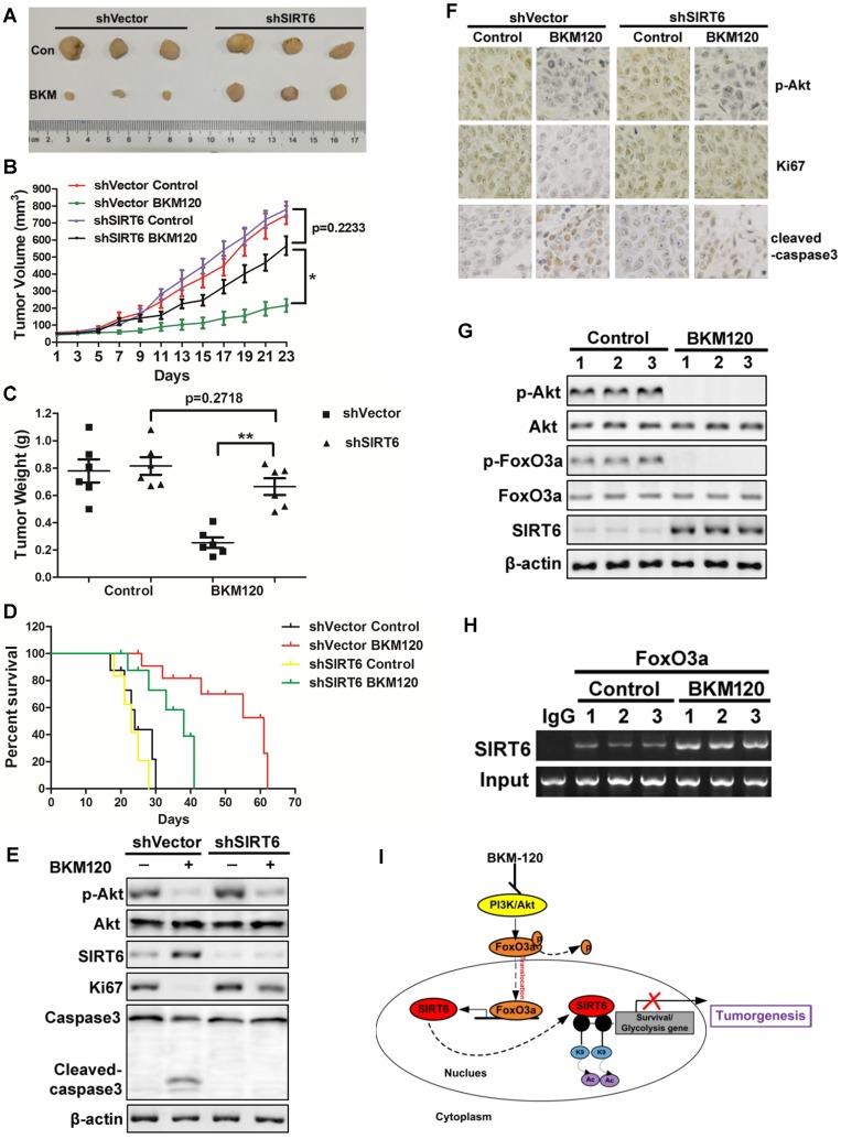
SIRT6, NAD+-dependent deacetylase sirtuin 6, has recently shown to suppress tumor growth in several types of cancer. Colon cancer is a challenging carcinoma associated with high morbidity and death. However, whether SIRT6 play a direct role in colon tumorigenesis and the underlying mechanism are not understood. Methods: To investigate the role of SIRT6 in colon cancer, we firstly analyzed the specimens from 50 colorectal cancer (CRC) patients. We generated shSIRT6 LoVo cells and xenograft mouse to reveal the essential role of SIRT6 in cell apoptosis and tumor growth. To explore the underlying mechanism of SIRT6 regulation, we performed FRET and real-time fluorescence imaging in living cells, real-time PCR, immunoprecipitaion, immunohistochemistry, flow cytometry and luciferase reporter assay. Results: The expression level of SIRT6 in patients' specimens is lower than that of normal controls, and patients with higher SIRT6 level have a better prognosis. Here, we identified that transcriptional factor FoxO3a is a direct up-stream of SIRT6 and positively regulated SIRT6 expression, which in turn, promotes apoptosis by activating Bax and mitochondrial pathway. Functional studies reveal that Akt inactivation increases FoxO3a activity and augment its binding to SIRT6 promoter, leading to elevated SIRT6 expression. Knocking down SIRT6 abolished apoptotic responses and conferred resistance to the treatment of BKM120. Combinational therapies with conventional drugs showed synergistic chemosensitization, which was SIRT6-dependent both in vitro and in vivo. Conclusion: The results uncover SIRT6 as a new potential biomarker for colon cancer, and its unappreciated mechanism about transcription and expression via Akt/FoxO3a pathway.

Keywords: Akt; Apoptosis; Colon cancer; FoxO3a; SIRT6.


Conflict of interest statement

Competing Interests: The authors have declared that no competing interest exists.

Figures

Figure 1
Figure 1
Clinical significance of SIRT6 signaling in human colorectal cancer. (A-D) SIRT6 is down-regulated in colorectal cancer. (A) Immunohistochemical of SIRT6 level in colotectal cancer tissues (CA) and paired non-cancerous tissues (NCA). (100×). (B-D) Down-regulation of SIRT6 in patient samples from our hospital and from Oncomine database (TCGA and Skrzypczak). (B) The logarithmic scale 2-ΔΔCt was carried out to show the relative SIRT6 expression of patient samples. (C and D) Box plot of SIRT6 mRNA Log2 expression levels was evaluated in Oncomine database. (E-G) Low SIRT6 levels in CRC patients correlates with poor prognosis. (E) Representative immunohistochemical staining in colon cancer tissues with different degree of SIRT6 expression. Number of positive cells: (-) <10%; (±) 10-30%; (+) 30-60%; (++)>60%. (F and G) Kaplan-Meier curves of CRC patients with high versus low expression of SIRT6 (n=50; p< 0.05, log-rank test). (H-K) Inverse correlation between SIRT6 and p-Akt or p-FoxO3a expression in human colon cancer. (H) Immunohistochemical stainings of SIRT6, p-Akt and p-FoxO3a in colon cancer tissue. Three representative cases are shown. (I) Total IHC score of SIRT6, p-FoxO3a and p-Akt in colon cancer tissue and non-cancerous tissue (n=50). *P<0.05. (J and K) The percentages of CRC specimens with low or high p-FoxO3a or p-Akt levels and their relationship to SIRT6 expression.
Figure 2
Figure 2
Akt inhibition induced SIRT6 expression and cell apoptosis. (A-D) SIRT6 was induced by BKM120. (A and B) Various colorectal cancer cell lines were stimulated with 24 h of 5μM BKM120. (A) Apoptosis analysis was detected by calculating cells with condensed nucleus after Hoechst 33342 staining. (B) Western blotting analysis of SIRT6 expression. (C and D) LoVo cells with different doses of BKM120 for 24 h. (C) Western blot analysis of SIRT6, p-Akt (Ser473), Akt, c-caspase-3 and full-length caspase-3 expression. (D) Real-time PCR analysis of SIRT6 mRNA induction. *P<0.05, **P<0.01 vs. control. (E-G) SIRT6 was activated by Akt inhibition in LoVo cells. (E and F) Western blot analysis of p-Akt (Ser473), Akt or SIRT6 expression in LoVo cells transfected with control vector or (E) a constitutively active Akt (Myr-Akt) or (F) shAkt#1 or shAkt#2 in the presence/absence of 5μM BKM120. (G) SIRT6 mRNA was detected in LoVo cells transfected with Myr-Akt, WT-Akt, K179M mutant Akt in the presence/absence of BKM120.
Figure 3
Figure 3
Activation of FoxO3a by dephosphorylation mediated SIRT6 induction. (A-D) FoxO3a was dephosphorylated and activated by BKM120 in colon cancer cells. (A) Western blot showing p-FoxO3a (S253) after 8 or 24 h of BKM120 treatment in LoVo or in DLD1 cells. (B and C) FoxO3a translocated from cytosol to nucleus after BKM120 treatment in LoVo cells. (B) FoxO3a expression in both nuclear and cytoplamic fractions. LaminA/C and GAPDH were served as the marker of nucleus and cytosol. (C) Dynamics of GFP-FoxO3a nuclear translocation after BKM120 stimulation in single living cell after 5 μM BKM120 treatment. (D) The effect of BKM120 on FoxO3a phosphorylation at different sites. Western blot analysis of p-Akt and SIRT6 level as well as p-FoxO3a at T32, S253 and S315 sites in LoVo, DLD1 or HCT-116 cells after BKM120 treatment. (E and F) Expression of (E) SIRT6 mRNA or (F) p-FoxO3a (S253), FoxO3a and SIRT6 protein in colon tissue from normal person and in tumor tissue or adjacent tissue from colorectal cancer (CRC) patients in response to BKM120 treatment. (G) Effects of over-expression of FoxO3a or SIRT1 on SIRT6 protein level. Western blot analysis of SIRT6 level of LoVo cells after transfection of WT, Ad-DN-FoxO3a Ad-Tm-FoxO3a, or SIRT1. (H) Co-immunoprecipitation (Co-IP) analysis of the interaction between FoxO3a and Akt, Ac-K or SIRT1 in LoVo cells after 0, 12 or 24 h BKM120 treatment.
Figure 4
Figure 4
Effects of FoxO3a on SIRT6 expression. (A-C) Effect of FoxO3a knockdown on SIRT6 protein expression. (A) Western blot analysis of SIRT6 and FoxO3a expression after 5μM BKM120 treatment for 24 h in LoVo cells with or without FoxO3a knockdown. (B and C) Effect of shRNA-mediated knockdown of FoxO3a, Akt and SIRT1 on (B) SIRT6 promoter reporter activity or (C) on SIRT6 mRNA level. (D and E) Effects of overexpression of Ad-Tm-FoxO3a (constitutively active FoxO3a) or WT-FoxO3a on (D) SIRT6 mRNA level or on (E) SIRT6 promoter reporter activity. (F) Schematic representation of human SIRT6 promoter in fragment A (between -295 to -276) (Fig.S4F) of the SIRT6 promoter, which contained FoxO3a binding sites that is the same as predicted sites. Three mutations (MT1, MT2, MT3) were produced by mutating the asterisks nucleotides. (G) LoVo cells were transiently transfected with luciferase reporters with WT or indicated mutant fragments (MT1, MT2, MT3) and then treated with BKM120 for 24 h. (H) Binding of FoxO3a to the SIRT6 promoter after 0, 8 or 12 h BKM120 treatment as revealed by ChIP assay. Primers were designed to amplify regions -345 to -226 for ChIP analysis. (I) The absence of FoxO3a blocked induction of SIRT6 mRNA by BKM120. shVector and shFoxO3a cells were incubated with/without BKM120 for 24 h. SIRT6 mRNA level was detected by Semi-Quantitative PCR. (J) Interaction of FoxO3a and SIRT1 in cultured cells with/without BKM120 treatment as revealed by immunoprecipitation (IP). Flag pulls down both phoshor-FoxO3a and acetylated-FoxO3a. (K) Binding of FoxO3a to the SIRT6 promoter after transfection of WT, Ad-DN-FoxO3a, Ad-Tm-FoxO3a or SIRT1 as revealed by ChIP assay.
Figure 5
Figure 5
SIRT6 augment the chemosensitization of BKM120. (A and B) Western blot analysis of SIRT6 level in the presence of 2.5 μM BKM120 combined with (A) 40 uM Cisplatin or (B) 20 mM Regorafenib for 24 h in LoVo cells. (C) Akt inhibition was involved in SIRT6 induction by combinational therapy. The level of SIRT6, p-Akt, cleaved-caspase3 was performed by western blot following 20 mM regorafenib, 40 μM cisplatin single treatment or combined with 2.5μM BKM120 for 24 h in LoVo cells. (D and E) p53 may mediate SIRT6 expression induced by Cisplatin or combined treatment. (D) LoVo cells were treated with 2.5 μM BKM120, 40 uM Cisplatin, or their combination. The expression levels of p53 and SIRT6 were detected by Western blot analysis. (E) The expression of p53, c-caspase-3 and SIRT6 were determined in wild-type, p53-/- or shSIRT6 (SIRT6 knockdown) HCT-116 cells. (F) The role of ERK inhibition in SIRT6 induction after regorafenib or its combination treatment with BKM120. LoVo cells were treated with either 20 mM Regorafenib, 20 mM UO126 or the combined treatment with 2.5 μM BKM120. Western blotting showing the level of SIRT6, p-ERK (Thr202/Tyr204) and total ERK. (G) Apoptosis in DLD1 cells treated with either 2.5 μM BKM120, 20mM Regorafenib or their combination therapy was detected by flow cytometry. Percentages of early apoptosis cells were shown in two right quadrants.
Figure 6
Figure 6
SIRT6 is indispensible for BKM120-induced apoptosis. (A and B) shVector or shSIRT6 LoVo cells were stimulated with BKM120 for 24 h. (A) Nuclei of cells were stained with Hoechst 33342. upper, representative pictures with arrows indicating fragmented nuclei (400×); lower, quantification analysis of apoptosis cells with fragmented nuclei. (B) Apoptosis analysis detected by flow cytometry. (C-E) Real-time detection of the activation of caspase-3 in single living cell. (C) Representative fluorescence images of CFP, YFP, Ratio as well as DIC image in LoVo cells transfected with SCAT-3 reporter. (D) The fluorescence images from Ratio channel at different time points after BKM120 treatments in shVector or shSIRT6 SCAT-3 LoVo cells. Scale bar: 10μm. (E) The fluorescence intensity of the Ratio (FRET/CFP) according to the images in D. (F) The expression of cleaved caspase-3, -9 and SIRT6 after 5 μM BKM120 treatment for 24 h in LoVo cells with or without SIRT6 deletion. (G) Western blot analysis showing cyto-c release from mitochondria to cytosol. The cytosolic and mitochondrial fractions were extracted from shVector and shSIRT6 cells after 24 h of BKM120 treatment. CoxIV and α-tublin were served as the mitochondrial and cytosolic marker. (H) Colony formation of shVector and shSIRT6 LoVo cells cultured 14 days after crystal violet staining in the presence/absence of BKM120. upper, representative photos of colonies; lower, quantification analysis of colony numbers. (I and J) SIRT6 bound to the promoter of survivin, leading the deacetylation at its H3K9 site. (I) Western blot analysis of AcH3K9, H3 and SIRT6 expression of nuclear fraction prepared from LoVo cells with or without SIRT6 deletion. (J) ChIP assay of the acetylation levels at H3K9 in the 5'-UTR of the survivin gene in samples extracted from shVector or shSIRT6 LoVo cells.
Figure 7
Figure 7
SIRT6 mediates in vivo antitumor effects of BKM120. (A-F) Nude mice were used to established xengraft model with shVector or shSIRT6 tumor. Mice were subjected to 40 mg/kg/d BKM120 or vehicle for two weeks. (A) Tumors pictures in this test. (B) Tumor volume or (C) weight was measured. *P<0.05. (D) Survival curve of (Kaplan-Meier analysis of) shVector and shSIRT6 mice with or without BKM120 treatment (6 mice / group). (E) p-Akt (S473), SIRT6, ki67, cleaved-caspase3 and full-length caspase-3 levels were detected by Western blot in shVector or shSIRT6 tumors with/without BKM120 treatment. (F) Immunohistochemistry of ki67, p-Akt and cleaved-caspase-3 in shVector or shSIRT6 tumor tissues. (G and H) The regulation of SIRT6 by FoxO3a after BKM120 treatment in shVector tumors. (G) Western blotting analysis of p-FoxO3a, SIRT6 and p-Akt expression in tumors. (H) Chromatin immunoprecipitation (ChIP) was performed by using FoxO3a to pull down. Then PCR was done to detect sirt6 promote region that bound to FoxO3a. (I) Schematic representation of Akt inhibition-mediated SIRT6 induction and apoptotic signaling pathway.

References

    1. Zhong L, D'Urso A, Toiber D, Sebastian C, Henry RE, Vadysirisack DD. et al. The histone deacetylase Sirt6 regulates glucose homeostasis via Hif1alpha. Cell. 2010;140:280–93. - PMC - PubMed
    1. Sebastian C, Zwaans BM, Silberman DM, Gymrek M, Goren A, Zhong L. et al. The histone deacetylase SIRT6 is a tumor suppressor that controls cancer metabolism. Cell. 2012;151:1185–99. - PMC - PubMed
    1. Masri S, Rigor P, Cervantes M, Ceglia N, Sebastian C, Xiao C. et al. Partitioning circadian transcription by SIRT6 leads to segregated control of cellular metabolism. Cell. 2014;158:659–72. - PMC - PubMed
    1. Lyssiotis CA, Cantley LC. SIRT6 puts cancer metabolism in the driver's seat. Cell. 2012;151:1155–6. - PMC - PubMed
    1. Lerrer B, Cohen HY. The guardian: metabolic and tumour-suppressive effects of SIRT6. EMBO J. 2013;32:7–8. - PMC - PubMed

Publication types