Case Reports
. 2015 Apr 29;5(1):31-35.
doi: 10.1007/s13691-015-0220-z. eCollection 2016 Jan.

Huge polypoid endometriosis: report of a case and review of the literature

Affiliations
Case Reports

Huge polypoid endometriosis: report of a case and review of the literature

Akiko Yamamoto et al. Int Cancer Conf J. .

Abstract

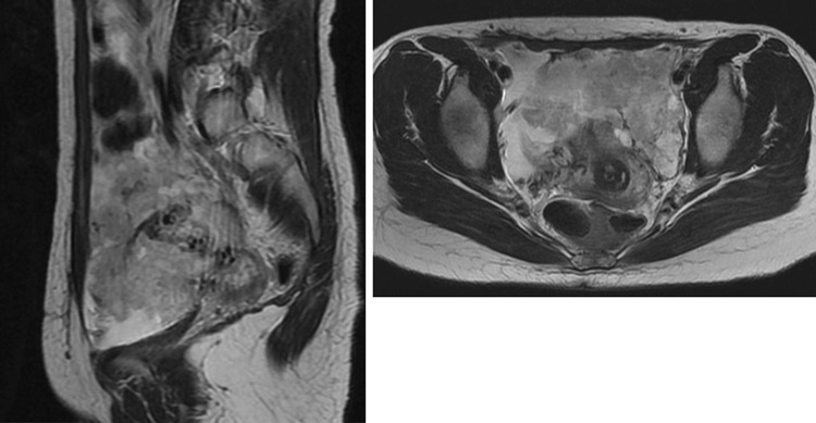
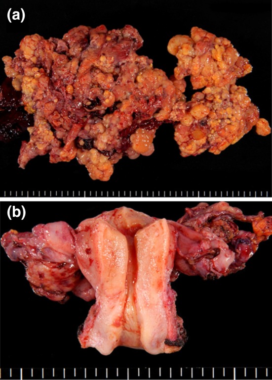
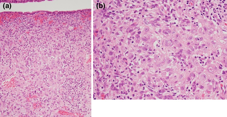
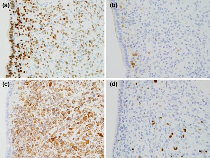
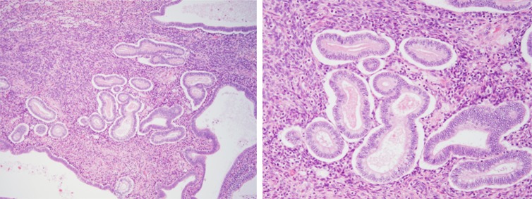
'Polypoid endometriosis' is a rare variant of endometriosis. We describe a case of an extremely large polypoid endometriosis mimicking a malignant tumor. A 37-year-old nulliparous woman was referred due to the rapid growth of an endometriotic cyst of the ovary and a high serum CA125 level. MRI revealed solid components in the pelvic mass. These preoperative clinical data were compatible with an ovarian carcinoma. A frozen section of the tumor biopsy showed as if an adenosarcoma, but finally the diagnosis of polypoid endometriosis with decidual change was made on permanent section. Polypoid endometriosis is a part of the differential diagnosis for malignant tumors in women with endometriosis, and we should consider carefully decision making for treatment.

Keywords: Adenosarcoma; Decidual change; Polypoid endometriosis; Uterine serosa.


Figures

Fig. 1
Fig. 1
Pelvic MRI: T2-weighted imaging shows irregular shaped mass with solid components occupying the whole pelvis
Fig. 2
Fig. 2
Macroscopic findings: a cauliflower-like polypoid mass arising from the serosa of the uterus. b Both ovaries are present separately from the tumor
Fig. 3
Fig. 3
Microscopic findings: the polypoid lesion consisted of endometriosis with decidual change. a The tumor is covered with monolayered columnar epithelia on the surface. A dense cellular layer and relatively loose cellular layer were located under the epithelial layer. b Stromal cells are polygonal having abundant cytoplasm and an enlarged nucleus with prominent nucleoli
Fig. 4
Fig. 4
Immunochemical findings: a estrogen receptor is positive on most epithelial and stromal cells, b a few stromal cells are positive for CD10, c most stromal cells are strongly positive for vimentin, d MIB-1 index is 5 %
Fig. 5
Fig. 5
Some parts of the tumor are composed of endometrium-like tissue showing the secretory phase

References

    1. Mostoufizadeh M, Scully RE. Malignant tumors arising in endometriosis. Clin Obstet Gynecol. 1980;23:951–963. doi: 10.1097/00003081-198023030-00024. - DOI - PubMed
    1. Cantor JO, Fenoglio CM, Richart RM. A case of extensive abdominal endometriosis. Am J Obstet Gynecol. 1979;134:846–847. - PubMed
    1. Chang A, Natarajan S. Polypoid endometriosis. Arch Pathol Lab Med. 2001;125:1257. - PubMed
    1. Kaushal S, Dadhwal V, Mathur SR, et al. Multifocal polypoid endometriosis in a young woman simulating vaginal and pelvic neoplasm. J Clin Pathol. 2010;63:452–454. doi: 10.1136/jcp.2009.073312. - DOI - PubMed
    1. Lambrechts S, Calsteren KV, Capoen A, et al. Polypoid endometriosis of the bladder during pregnancy mimicking urachal carcinoma. Ultrasound Obstet Gynecol. 2011;38:475–478. doi: 10.1002/uog.8985. - DOI - PubMed

Publication types

LinkOut - more resources