. 2019 May 29:2:200.
doi: 10.1038/s42003-019-0455-x. eCollection 2019.

Therapeutic benefit of combining calorie-restricted ketogenic diet and glutamine targeting in late-stage experimental glioblastoma

Affiliations

Therapeutic benefit of combining calorie-restricted ketogenic diet and glutamine targeting in late-stage experimental glioblastoma

Purna Mukherjee et al. Commun Biol. .

Abstract

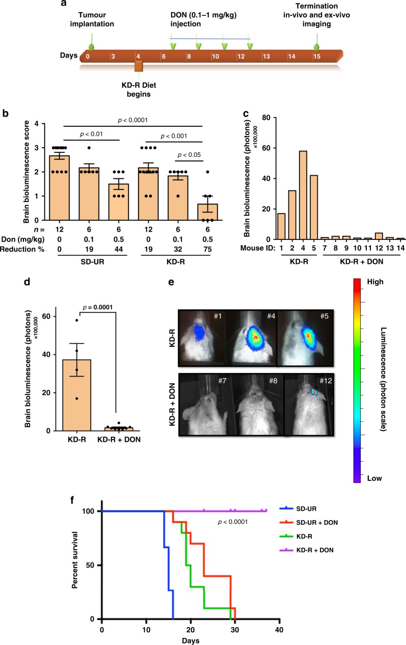
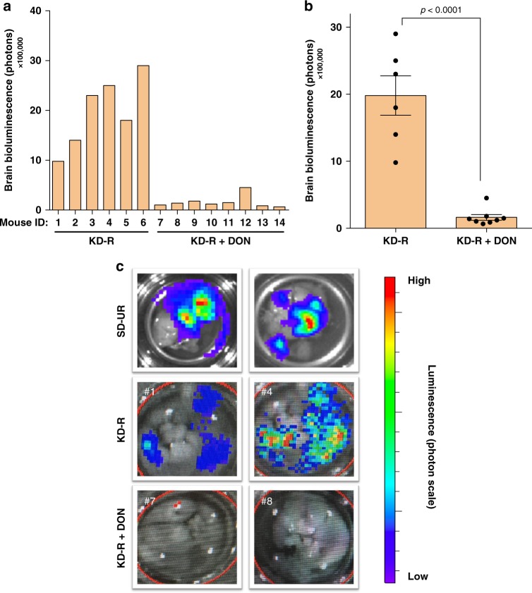
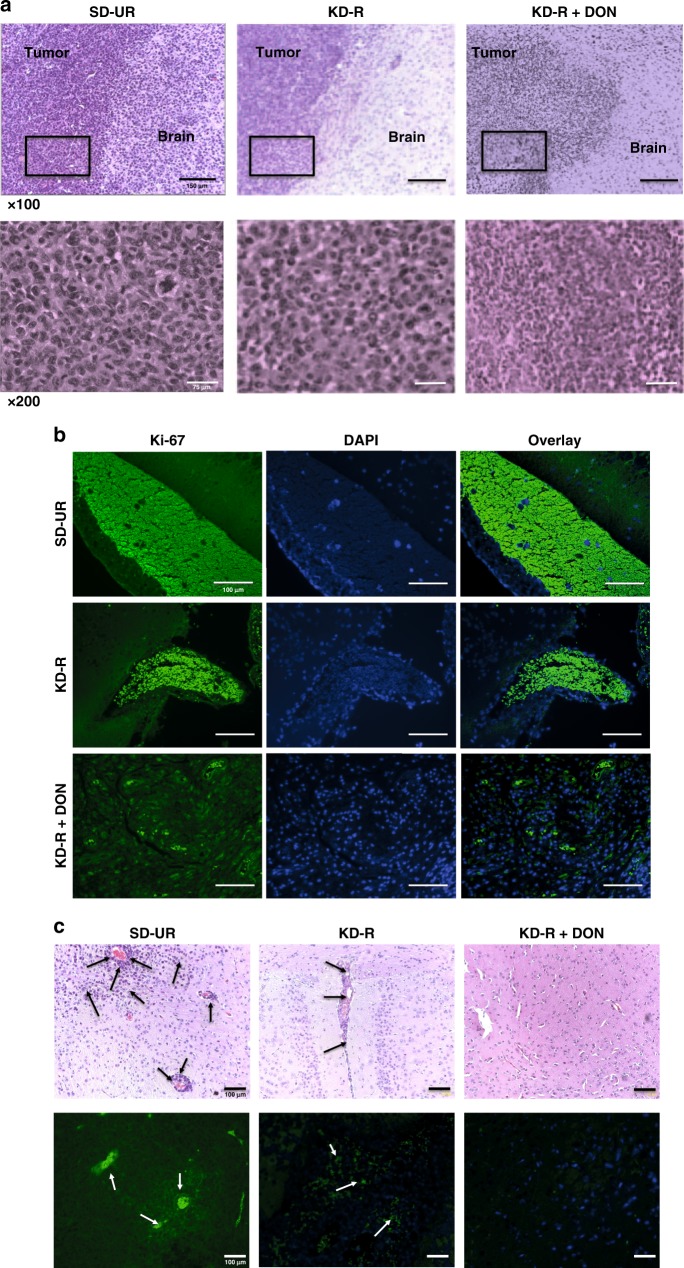
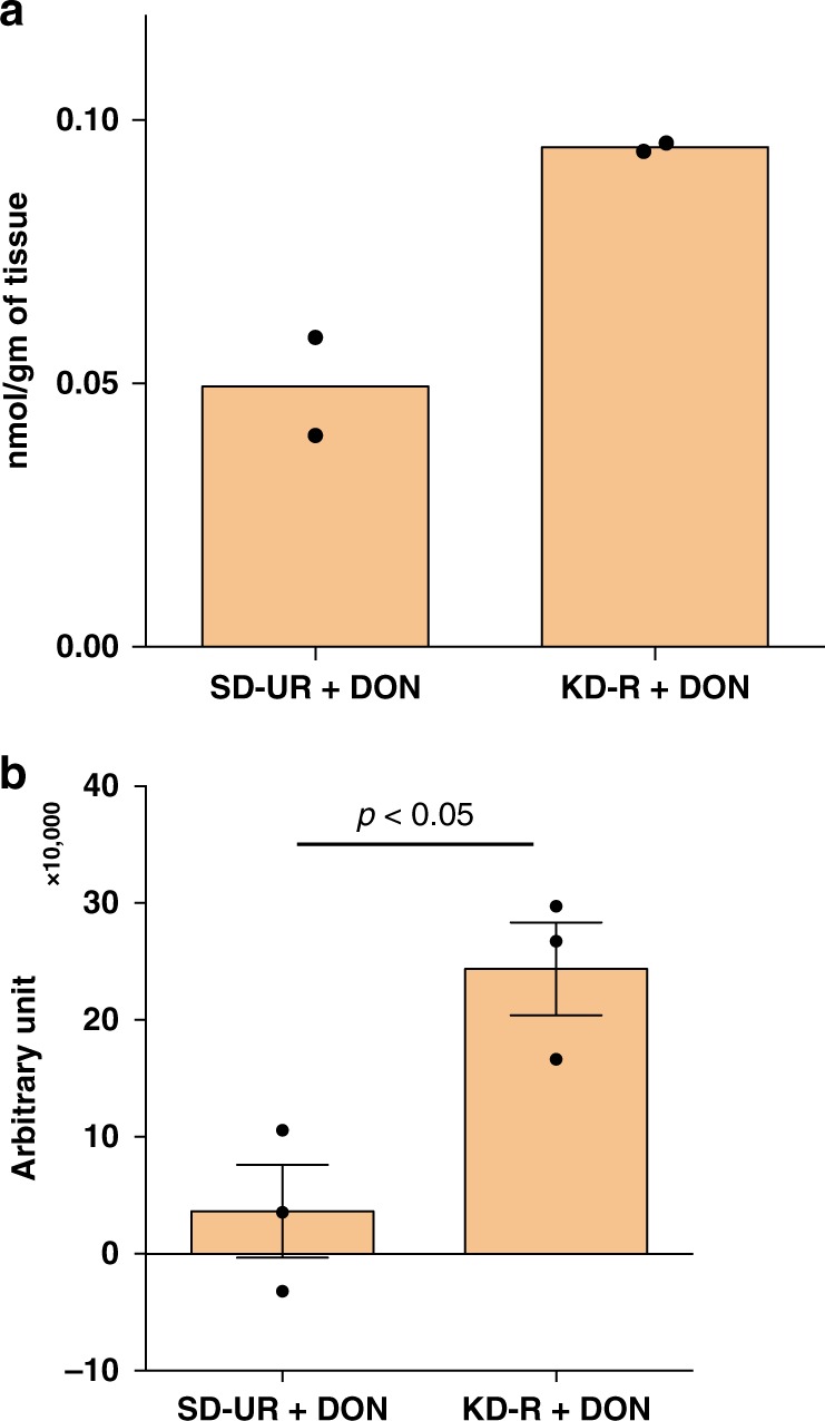
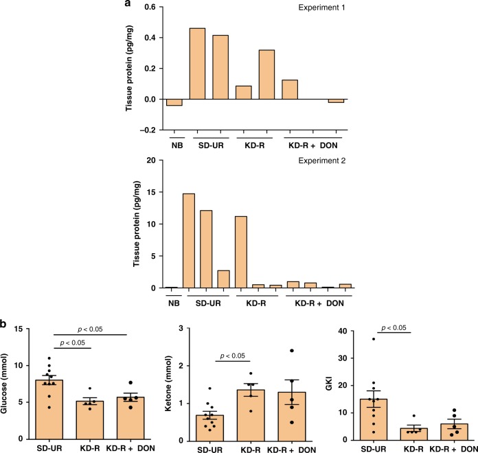
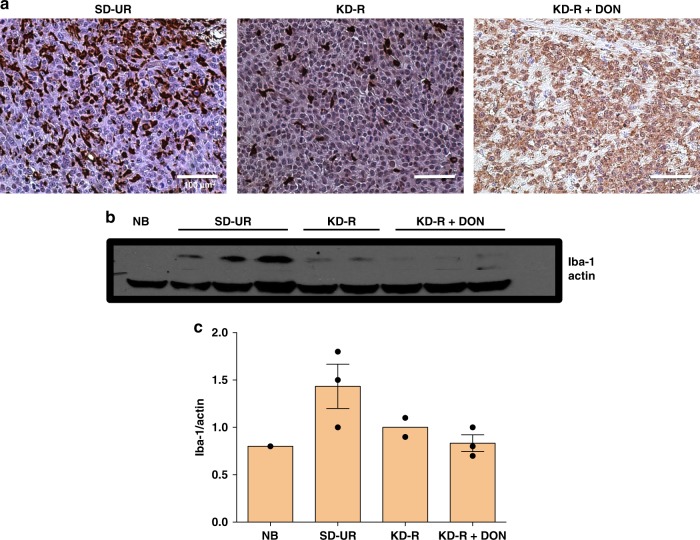
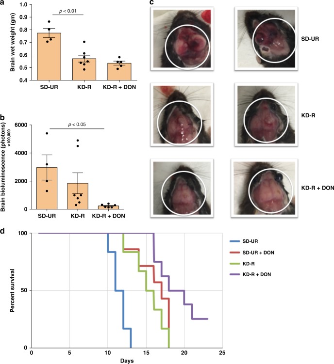
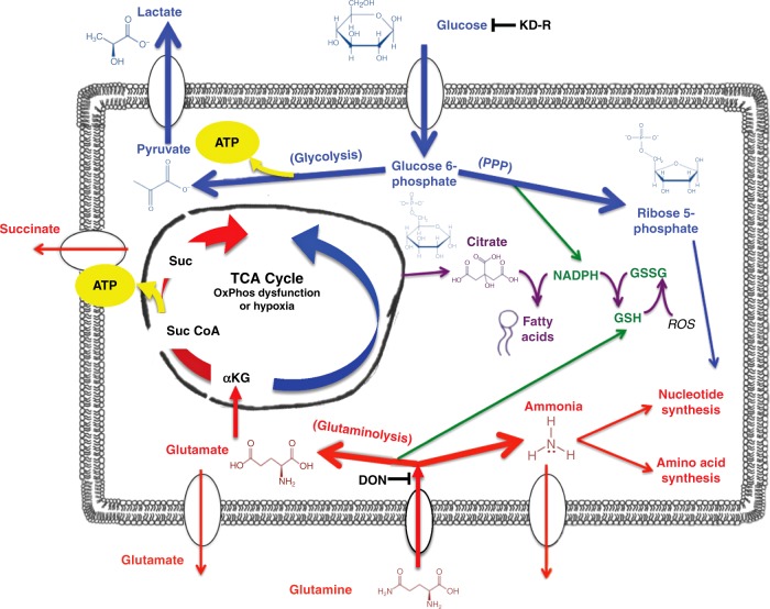
Glioblastoma (GBM) is an aggressive primary human brain tumour that has resisted effective therapy for decades. Although glucose and glutamine are the major fuels that drive GBM growth and invasion, few studies have targeted these fuels for therapeutic management. The glutamine antagonist, 6-diazo-5-oxo-L-norleucine (DON), was administered together with a calorically restricted ketogenic diet (KD-R) to treat late-stage orthotopic growth in two syngeneic GBM mouse models: VM-M3 and CT-2A. DON targets glutaminolysis, while the KD-R reduces glucose and, simultaneously, elevates neuroprotective and non-fermentable ketone bodies. The diet/drug therapeutic strategy killed tumour cells while reversing disease symptoms, and improving overall mouse survival. The therapeutic strategy also reduces edema, hemorrhage, and inflammation. Moreover, the KD-R diet facilitated DON delivery to the brain and allowed a lower dosage to achieve therapeutic effect. The findings support the importance of glucose and glutamine in driving GBM growth and provide a therapeutic strategy for non-toxic metabolic management.

Keywords: CNS cancer; Cancer metabolism.


Conflict of interest statement

Competing interestsThe authors declare no competing interests.

Figures

Fig. 1
Fig. 1
Restricted ketogenic diet with DON reduces progression and mortality of the VM-M3 GBM. VM/Dk inbred mice were implanted orthotopically with a small (1.0 mm × 1.0 mm) tissue fragment from the VM-M3 tumour on day 0. The implanted mice were divided into two groups on day 4 and were fed either a standard chow diet unrestricted or ad libitum (SD-UR), or a ketogenic diet (KD-R) in restricted amounts to reduce body weight by about 15%. DON (0.1–1.0 mg/kg) was injected i.p. 7 days following orthotopic tumour implantation. The diet feeding was continued and DON was injected every day or every alternate day as shown in (a). All mice were imaged in vivo and terminated on day 14 or 15 when all control mice appeared moribund (experiments 1, 2). For experiment 1, bioluminescence was scored from 0–4, following the administration of lower doses of DON (0.1 and 0.5 mg/kg). Values are expressed as the mean ± SEM and a one-way analysis of variance followed by Tukey’s post hoc test was performed to determine the significance between groups (b). In experiment 2, in vivo bioluminescent photon values were obtained following the administration of DON (1.0 mg/kg) in mice under the KD-R (c). The average in vivo bioluminescent photon values were calculated for the KD-R (n = 4 mice) and KD-R + DON (n = 8 mice) in experiment 2. Values are presented as the mean ± SEM and the P value was calculated using a two-tailed student’s t-test (d). In vivo bioluminescent images of three representative mice from both study groups in experiment 2 are shown in (e). For experiment 3, a survival study was performed and a Kaplan–Meier survival plot was configured for SD-UR (n = 15 mice), SD-UR + DON (n = 10 mice), KD-R (n = 10 mice), and KD-R + DON (n = 10 mice) (f). The log-rank statistical analysis test showed a significant difference between groups in this survival study. Source data are provided as  Supplementary Data 1
Fig. 2
Fig. 2
Restricted ketogenic diet with DON reduces VM-M3 GBM bioluminescence. VM/Dk mice were implanted with a VM-M3 tumour, as described in Fig. 1. Mouse brains were imaged ex vivo on day 14 or 15. Mice were imaged ex vivo after in vivo bioluminescent imaging, as described in Fig. 1. Individual ex vivo bioluminescent photon values were measured following the administration of DON (1.0 mg/kg) in KD-R mice (a). From the individual measurements, the average ex vivo bioluminescent photon value was calculated for both the KD-R (n = 6 mice) and KD-R + DON (n = 8 mice) groups. Values are presented as the mean ± SEM and significance of differences was determined following a two-tailed student’s t-test (b). Ex vivo bioluminescent images of brains from representative mice in experiment 2 are shown in comparison to SD-UR (c). Source data are provided as Supplementary Data 1
Fig. 3
Fig. 3
Restricted ketogenic diet with DON kills VM-M3 GBM cells in brain. VM/Dk mice were implanted with VM-M3 cells orthotopically in the brain on day 0 as described in Fig. 1. The brains were fixed in formalin for histology, processed, and stained as described in Methods. Histological analysis (H&E) was used to validate the presence of tumour cells (a). The top panel images show the core of the tumour mass growing in the brain. The black boxes in the top panel’s images are shown in higher magnification in the bottom panels. All scale bars are 150 μm for x100 and 75 μm for x200. Ki-67 positive nuclear staining (×100), as expressed by green fluorescent labeled cells are shown in (b). DAPI is used for the Ki-67 negative nuclear stain. The invasion of tumour cells in the brain tissue is evident (arrows) in both histological (H&E) analysis (×100) and Ki-67 staining (c). All scale bars for b and c are 100 μm
Fig. 4
Fig. 4
Restricted ketogenic diet increases DON delivery to the VM-M3 GBM. The content of DON in the VM-M3 brain tumour tissue was quantified using two LC/MS/MS procedures, as described in Methods. The brain tissue was analyzed for DON content 60 min after i.p. DON injection. The values in experiment a are presented as mean of two independent samples, while the values in experiment b are presented as the mean ± SEM (n = 3 mice). Following a two tailed student's t-test, the difference between the two groups in experiment b was significant (P< 0.05). Both analytical procedures showed that DON content in tumour tissue was greater under KD-R feeding than under SD-UR feeding. Source data are provided as Supplementary Data 1
Fig. 5
Fig. 5
Restricted ketogenic diet with DON reduces TNF-α in the VM-M3 brain tumour tissue, and the Glucose Ketone Index (GKI) in the blood. ELISA was used to measure TNF-α in brain tumour tissue lysates in two different experiments, and the content was expressed as pg/mg of protein. Normal brain (NB) was used as a negative control tissue. The range of TNF-α concentration was −0.04–0.1 pg/mg for NB (n = 2 normal mouse brain tissue); 0.4–14.7 pg/mg for SD-UR (n = 5 mouse tumour brain tissue); 0.09–11.2 pg/mg for KD-R (n = 5 mouse tumour brain tissue); and 0.02–1.0 pg/mg for KD-R + DON (n = 7 mouse tumour brain tissue) (a). Data showing that blood glucose is lower and blood ketones are higher in mice fed the KD-R or KD-R + DON (n = 5 independent mouse blood samples per group) than in mice fed the SD-UR (n = 10 independent mouse blood samples). This shift in blood glucose and ketones causes a significant reduction in the GKI (b). Values are expressed as the mean ± SEM and a one-way analysis of variance followed by Tukey's post hoc test was performed to determine the significance between groups. Source data are provided as Supplementary Data 1
Fig. 6
Fig. 6
Restricted ketogenic diet with DON reduces the Iba-1 expression in the VM-M3 brain tumour tissue. Immunohistochemistry (IHC) of Iba-1 was performed in formalin fixed brain tissues as described in Methods. In comparison to all other groups, the expression of Iba-1 is highest in the SD-UR tumour as seen by the positively stained brown cells. Iba-1 IHC staining was noticeably less in the VM-M3 tumor than in the KD-R-fed mice and the DON-treated mice (a). All scale bars are 100 μm. Western blot analysis of Iba-1 protein expression in the tumours indicates a decrease in the expression of Iba-1 in the DON-treated tumours (n = 3 mouse tumour brain tissue) in comparison to SD-UR (n = 3 mouse tumour brain tissue). A decrease in expression was also seen for KD-R mice (n = 2 mouse tumour brain tissue). Normal brain (NB) was used as a negative control tissue (b and c). Source data are provided as Supplementary Data 1
Fig. 7
Fig. 7
Restricted ketogenic diet with DON reduces progression and mortality of the CT-2A GBM. C57BL/6J mice were implanted with CT-2A tumour fragments on day 0, as described in Fig. 1. Brain wet weights were measured for SD-UR (n = 4 mouse brains), KD-R (n = 7 mouse brains), and KD-R + DON (n = 5 mouse brains) (a). Ex vivo bioluminescent photon values were calculated for the same brain samples and presented as the mean ± SEM (b). All brains were imaged on day 14 or 15. A one-way analysis of variance followed by Tukey's post hoc test was performed to determine the signicance between groups. Representative brain sample images from each study group were portrayed in (c). A Kaplan–Meier survival curve was plotted for the SD-UR (n = 6 mice), SD-UR + DON (n = 8 mice), KD-R (n = 6 mice), and KD-R + DON (n = 8 mice) groups (d). The log-rank test was used to determine the significance. Source data are provided as Supplementary Data 1
Fig. 8
Fig. 8
Targeting glucose and glutamine using KD-R with DON for the Metabolic Management of the VM-M3 and CT-2A Experimental GBM. GBM tumour cells are largely dependent on glucose and glutamine for survival and growth. Energy through substrate level phosphorylation (SLP) in the cytoplasm (glycolysis) and in the TCA cycle (glutaminolysis) will compensate for reduced energy through oxidative phosphorylation (OxPhos) or hypoxia that occurs in these GBM cells. The KD-R will reduce glucose carbons for both the glycolytic and pentose phosphate (PPP) pathways that supply ATP and precursors for lipid and nucleotide synthesis, as well as for glutathione production. DON will inhibit glutaminases thus depleting glutamate and the glutamine-derived amide nitrogen for ammonia and nucleotide synthesis. Depletion of glutamine-derived glutamate will reduce anapleurotic carbons to the TCA cycle through α-KG for protein synthesis, while also reducing ATP synthesis at the succinyl CoA synthase step in the TCA cycle. The glutamine-derived glutamate is also used for glutathione production that protects tumor cells from oxidative stress. The KD-R + DON will thus make the VM-M3 and the CT-2A cells vulnerable to oxidative stress. The simultaneous targeting of glucose and glutamine using the KD-R + DON will starve tumour cells of energy production while blocking their ability to synthesize proteins, lipids, and nucleotides. This metabolic starvation could also reduce extracellular acidification through reduction of lactate and succinate. The elevation of non-fermentable ketone bodies will provide normal cells with an alternative energy source to glucose while also protecting them from oxidative stress. This diagram and legend have been modified from that presented previously under the terms of the Creative Commons Attribution 4.0 International License in Nutrition and Metabolism by Thomas Seyfried et al. (http://creativecommons.org/licenses/by/4.0/)

References

    1. Desjardins A, et al. Recurrent glioblastoma treated with recombinant poliovirus. N. Engl. J. Med. 2018;379:150–161. doi: 10.1056/NEJMoa1716435. - DOI - PMC - PubMed
    1. Shelton LM, et al. A novel pre-clinical in vivo mouse model for malignant brain tumor growth and invasion. J. Neurooncol. 2010;99:165–176. doi: 10.1007/s11060-010-0115-y. - DOI - PubMed
    1. Karsy M, et al. Established and emerging variants of glioblastoma multiforme: review of morphological and molecular features. Folia Neuropathol. 2012;50:301–321. doi: 10.5114/fn.2012.32361. - DOI - PubMed
    1. Huysentruyt, L. C., Akgoc, Z. & Seyfried, T. N. Hypothesis: are neoplastic macrophages/microglia present in glioblastoma multiforme? ASN Neuro.310.1042/AN20110011 (2011). - PMC - PubMed
    1. Yuan X, et al. Isolation of cancer stem cells from adult glioblastoma multiforme. Oncogene. 2004;23:9392–9400. doi: 10.1038/sj.onc.1208311. - DOI - PubMed

Publication types

MeSH terms