. 2019 Aug 1;317(2):H279-H289.
doi: 10.1152/ajpheart.00510.2018. Epub 2019 May 31.

Elevated bone marrow sympathetic drive precedes systemic inflammation in angiotensin II hypertension

Affiliations

Elevated bone marrow sympathetic drive precedes systemic inflammation in angiotensin II hypertension

Niousha Ahmari et al. Am J Physiol Heart Circ Physiol. .

Abstract

Increased sympathetic nervous system activity is a hallmark of hypertension (HTN), and it is implicated in altered immune system responses in its pathophysiology. However, the precise mechanisms of neural-immune interaction in HTN remain elusive. We have previously shown an association between elevated sympathetic drive to the bone marrow (BM) and activated BM immune cells in rodent models of HTN. Moreover, microglial-dependent neuroinflammation is also seen in rodent models of HTN. However, the cause-effect relationship between central and systemic inflammatory responses and the sympathetic drive remains unknown. These observations led us to hypothesize that increase in the femoral BM sympathetic nerve activity (fSNA) initiates a cascade of events leading to increase in blood pressure (BP). Here, we investigated the temporal relationship between the BM sympathetic drive, activation of the central and peripheral immune system, and increase in BP in the events leading to established HTN. The present study demonstrates that central infusion of angiotensin II (ANG II) induces early microglial activation in the paraventricular nucleus of hypothalamus, which preceded increase in the fSNA. In turn, activation of fSNA correlated with the timing of increased production and release of CD4+.IL17+ T cells and other proinflammatory cells into circulation and elevation in BP, whereas infiltration of CD4+ cells to the paraventricular nucleus marked establishment of ANG II HTN. This study identifies cellular and molecular mechanisms involved in neural-immune interactions in early and established stages of rodent ANG II HTN. NEW & NOTEWORTHY Early microglia activation in paraventricular nucleus precedes sympathetic activation of the bone marrow. This leads to increased bone marrow immune cells and their release into circulation and an increase in blood pressure. Infiltration of CD4+ T cells into paraventricular nucleus paraventricular nucleus marks late hypertension.

Keywords: SNS; angiotensin II; hypertension; inflammation; neuroinflammation.


Conflict of interest statement

No conflicts of interest, financial or otherwise, are declared by the authors.

Figures

Fig. 1.
Fig. 1.
Centrally administered ANG II is associated with elevated femoral sympathetic nerve activity (fSNA) and blood pressure (BP) in rats. A: in situ electrophysiology revealed a shift in the fSNA peak activity from the typical inspiratory/postinspiratory (I/P-I) phase to the expiratory (E) phase, suggesting sympathetic respiratory uncoupling typical of that seen in rodent models of hypertension. Left: a schematic of decerebrated artificially perfused rat preparation used to record the activity of fSNA and phrenic nerve activity (PNA) in situ. Middle: shift in the peak of fSNA activity to E phase of the respiratory cycle (arrow) that begins as early as day 3 of intracerebroventricular (ICV) ANG II infusion and elevates at day 7. Right: a marked shift in the peak fSNA from I/P-I to E at day 7 (top graph) and protein levels of bone marrow (BM) norepinephrine (NE) in sympathetic nerve denervated femur (BM SNAX) compared with SHAM femur in rats at day 7 of ICV ANG II infusion (bottom graph). B: significantly increased systolic blood pressure (SBP) at day 14 (left) and mean blood pressure (MBP) at day 7 (right) of ICV ANG II infusion. PP, perfusion pressure. Data are presented as means ± SE, n = 6 rats; *P < 0.05 vs. control, ANOVA or t-test where applicable.
Fig. 2.
Fig. 2.
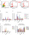
Central administration of ANG II is associated with elevated bone marrow (BM) and circulating inflammatory markers. Intracerebroventricular (ICV) ANG II increased levels of CD4+.IL17+ T cells in the blood (A and B) and BM (C) at day 7, as measured by flow cytometry. In addition, levels of several other T cell subtypes (CD4+.CD25+, CD3+, CD3+.CD45+, and CD68+ macrophage/monocytes were increased in the BM at day 7 of ANG II infusion (C), as measured by flow cytometry. Dennervation of the femoral BM sympathetic nerve (BM SNX) at day 7 of ICV ANG II infusion significantly reduced the ANG II-dependent increase in the inflammatory cells in the BM (C), suggesting BM sympathetic nerve activity-dependent activation of BM immune system by ANG II, as measured by flow cytometry. Real-time PCR showed elevated levels of several inflammatory markers, including TLR4, HIF1a, and ITGAM, in the BM at day 7 of ANG II infusion (D), which was reduced by BM SNX (left); significant elevation in several adrenergic receptors in the BM at day 21 of ANG II infusion, as measured by real-time PCR (right). Data are presented as means ± SE, n = 6; *P < 0.05 vs. control; #P < 0.05 vs. day 7; ANOVA.
Fig. 3.
Fig. 3.
Intracerebroventricular (ICV) administration of ANG II for 3, 7, and 21 days produced graded, time-dependent neuroinflammatory responses in the paraventricular nucleus (PVN) of rats, as evident by microglial morphology and Iba1-positive cell count. A (left): binary masks were used to quantify circular morphology of Iba1+ cells in the PVN of all rats (as represented in B, bottom). Mathematical quantification of cell circularity indicates a change in microglial activation at day 3 of ICV ANG II infusion. Circularity was calculated based on the area and perimeter of each Iba1+ cell and represents shortening of cell processes and thickening of cell bodies (as represented in B, bottom), both properties indicative of microglial activation. A (right): quantification of total number of nucleated Iba1+ cells in the PVN shows a significant increase at days 7 and 21 of ANG II compared with control. B: representative confocal images of rat PVN slices immunolabeled with Iba1 (white) reveal a progressive increase in Iba1+ cells with ANG II infusion. Data are presented as means ± SE, n = 6 rats. *P < 0.05 vs. control, ANOVA.
Fig. 4.
Fig. 4.
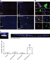
Increased presence of CD4-positive cells in the paraventricular nucleus (PVN) at day 21 of intracerebroventricular (ICV) ANG II infusion. A: representative confocal images of immunolabeling for CD4 (red) counterstained with DAPI (blue) in the PVN at days 3, 7, and 21 following continuous infusion of ANG II (100 ng/h icv) or artificial cerebrospinal fluid (control). White arrows point to the cells counted as CD4-positive cells, which were identified as round nucleated cells present in both the max intensity and three-dimensional projection of each image with their whole length ranging from 6–8 µm (as depicted in C below). Yellow cross marks the apparent positive label that was excluded from the total CD4-positive cell count as per our stringent exclusion methods. The position of the third ventricle (3V) is marked. B: high-magnification confocal microscopy illustrates the proximity of CD4-positive cells to neurons (top at ×90: NeuN in green, CD4 in red, DAPI in blue) and microglia (bottom at ×60: Iba1 in green, CD4 in red, DAPI in blue) in the PVN at day 21 of ANG II infusion. D: quantification of CD4-positive cells shows a statistically significant increase in CD4-positive cells in the PVN at day 21 of ANG II infusion only. Data are presented as means ± SE, n = 5 rats; *P < 0.05 vs. control, #P < 0.05 vs. day 3, &P < 0.05 vs. day 7, ANOVA.

References

    1. Ahmari N, Santisteban M, Baekey D, Raizada M, Zubcevic J. Angiotensin II–dependent increase in the b=bone marrow sympathetic drive initiates the inflammatory and endothelial progenitor cell imbalance and precedes blood pressure increase (Abstract). FASEB J 29, Suppl 1: 1059.1, 2015.
    1. Ahmari N, Schmidt JT, Krane GA, Malphurs W, Cunningham BE, Owen JL, Martyniuk CJ, Zubcevic J. Loss of bone marrow adrenergic beta 1 and 2 receptors modifies transcriptional networks, reduces circulating inflammatory factors, and regulates blood pressure. Physiol Genomics 48: 526–536, 2016. doi:10.1152/physiolgenomics.00039.2016. - DOI - PMC - PubMed
    1. Case AJ, Roessner CT, Tian J, Zimmerman MC. Mitochondrial superoxide signaling contributes to norepinephrine-mediated t-lymphocyte cytokine profiles. PLoS One 11: e0164609, 2016. doi:10.1371/journal.pone.0164609. - DOI - PMC - PubMed
    1. Centenera MM, Selth LA, Ebrahimie E, Butler LM, Tilley WD. New opportunities for targeting the androgen receptor in prostate cancer. Cold Spring Harb Perspect Med 8: a030478, 2018. doi:10.1101/cshperspect.a030478. - DOI - PMC - PubMed
    1. Chi DS, Harris NS. A simple method for the isolation of murine peripheral blood lymphocytes. J Immunol Methods 19: 169–172, 1978. doi:10.1016/0022-1759(78)90176-X. - DOI - PubMed

Publication types

MeSH terms