. 2019 May 30;177(6):1448-1462.e14.
doi: 10.1016/j.cell.2019.04.025.

Defining the Independence of the Liver Circadian Clock

Affiliations

Defining the Independence of the Liver Circadian Clock

Kevin B Koronowski et al. Cell. .

Abstract

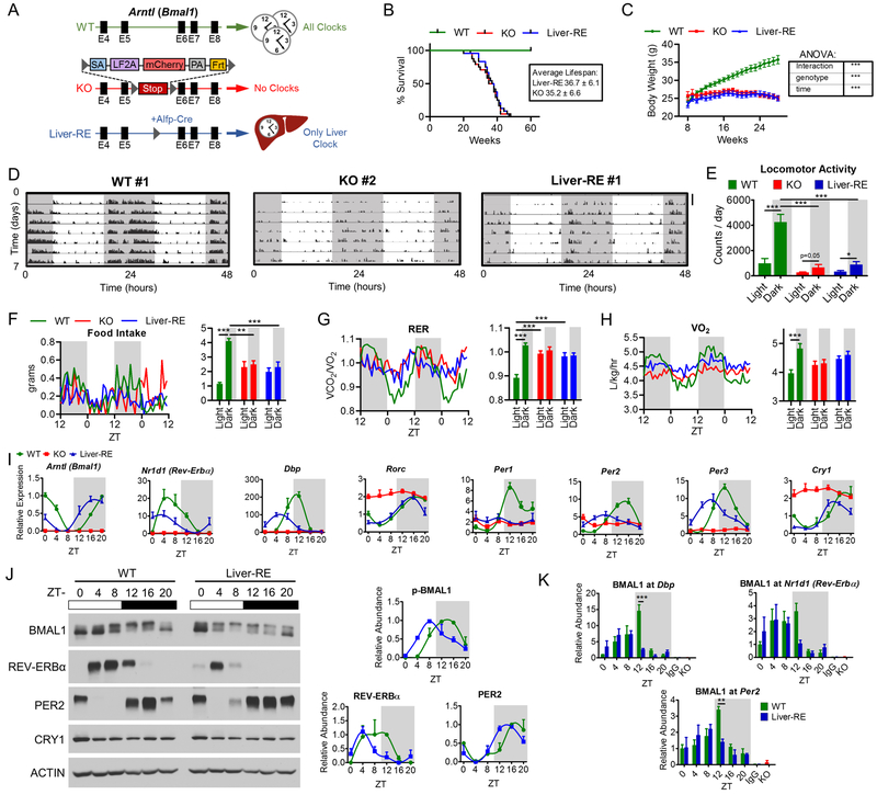
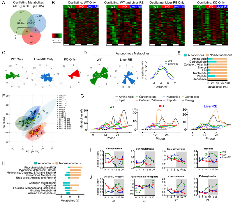
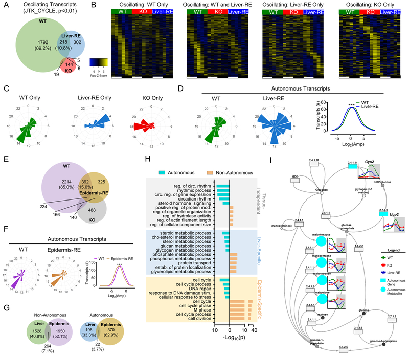
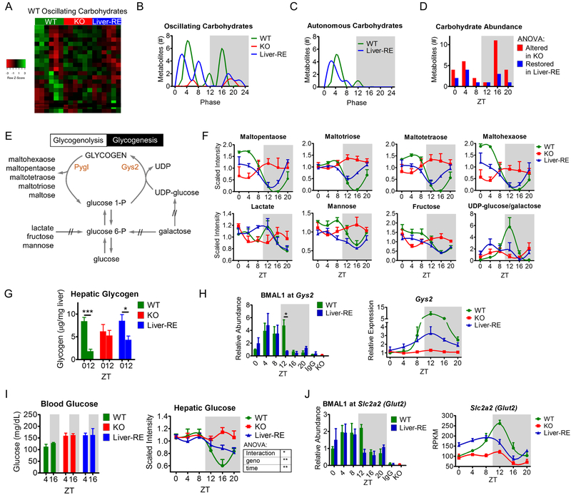
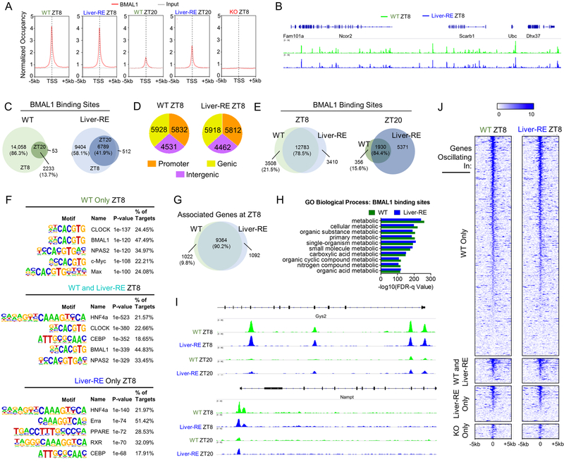
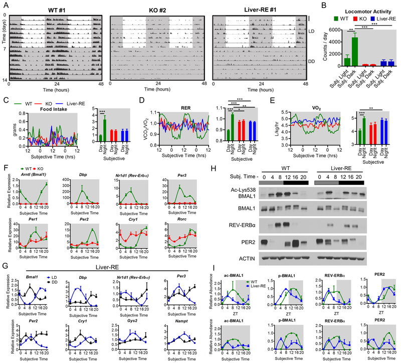
Mammals rely on a network of circadian clocks to control daily systemic metabolism and physiology. The central pacemaker in the suprachiasmatic nucleus (SCN) is considered hierarchically dominant over peripheral clocks, whose degree of independence, or tissue-level autonomy, has never been ascertained in vivo. Using arrhythmic Bmal1-null mice, we generated animals with reconstituted circadian expression of BMAL1 exclusively in the liver (Liver-RE). High-throughput transcriptomics and metabolomics show that the liver has independent circadian functions specific for metabolic processes such as the NAD+ salvage pathway and glycogen turnover. However, although BMAL1 occupies chromatin at most genomic targets in Liver-RE mice, circadian expression is restricted to ∼10% of normally rhythmic transcripts. Finally, rhythmic clock gene expression is lost in Liver-RE mice under constant darkness. Hence, full circadian function in the liver depends on signals emanating from other clocks, and light contributes to tissue-autonomous clock function.

Keywords: autonomous; bmal1; chromatin; circadian; clock; diurnal physiology; epigenetics; light; metabolism; systemic signaling.


Conflict of interest statement

DECLARATION OF INTERESTS

J.M.K is an employee of Metabolon, Inc. (no role in the study design)

Figures

Fig. 1.
Fig. 1.. Reconstitution of the liver clock using Bmal1-Stop-FL mice
A) Scheme of genetic reconstitution of the liver clock. See also Fig. S1A–B. B) Survival curves, WT n=25; KO n=24; Liver-RE n=24. C) Progression of body weight, WT n=27; KO n=39; Liver-RE n=27. Two-way ANOVA, ***=p<0.001. D-E) Locomotor activity in LD cycle. D) Representative trace, duplicated over 2 days for visualization; scale bar = 30 counts. E) Group quantification, n=5. Two-way ANOVA, *=p<0.05; ***=p<0.001. See also Fig. S1C. F-H) Metabolic cage assessment of mice in LD. Traces of group averages (left) and light phase (ZT0–12)/dark phase (ZT12–24) averages (right). RER – respiratory exchange ratio. WT n=8; KO n=5; Liver-RE n=7. Two-way ANOVA, **=p<0.01; ***=p<0.001. See also Fig. S1D–E. I) Gene expression in liver; n=4. See also Fig. S1F. J) Protein levels in liver whole-cell extracts. Right, quantification of n=3. See also Fig. S1G. J) ChIP-qPCR for BMAL1 recruitment to promoters, n=4. IgG and KO negative controls are from ZT20. Two-way ANOVA, **=p<0.01; ***=p<0.001.
Fig. 2.
Fig. 2.. Diurnal metabolome reveals autonomous metabolic output
A) Overlap of oscillating metabolites from liver obtained at 6 time-points over 12 hr LD cycle. Percentages are of the total number of Bmal1-dependent metabolites oscillating in WT, n=4. See also Fig. S2A. B) Phase sorted heatmaps of distinct groups of oscillating metabolites. C) Polar histogram plots of peak phase for oscillating metabolites. D) Polar histogram plots of peak phase for autonomously oscillating metabolites (left) and amplitude histogram (right). See also S2B. E) Chemical classification of oscillating metabolites, presented as % of total oscillating for that class. See also S2C–D. F) Principal component analysis of metabolites at each time-point. G) Histograms showing the peak phase of oscillating metabolites in each class. H) Metabolic process or pathway for oscillating metabolites (top 5), see also Table S1. I-J) Examples of autonomously (I) and non-autonomously (J) oscillating metabolites. See also S2E.
Fig. 3.
Fig. 3.. Autonomous transcriptional output of peripheral clocks is tissue-specific
(A) Overlap of oscillating liver transcripts in a 12 hr LD cycle. Percentages are of the total number of Bmal1-dependent transcripts oscillating in WT; n=3. See also Fig. S3A. B) Phase sorted heatmaps of distinct groups of transcripts. C) Polar histogram plots of peak phase for oscillating transcripts. D) Polar histogram plots of peak phase for autonomously oscillating transcripts (left) and amplitude histogram (right). See also S3D. E) Overlap of oscillating transcripts in epidermis of WT, KO and epidermis reconstituted (Epidermis-RE) mice (Welz et al.). Percentages are of the total number of oscillating transcripts in WT, JTK – p<0.01; n=3–4. F) Polar histogram plots of peak phase and amplitude histogram for autonomously oscillating transcripts in epidermis. See also Fig. S3F. G) Overlap of oscillating transcripts between Liver-RE and Epidermis-RE. Percentages are of the total number of oscillating transcripts. H) GO Biological Process enrichment analysis of oscillating transcripts, grouped by tissue-specificity and autonomy (top 5), see also Table S4. I) Example of cohesively autonomous pathway – glycogen metabolism (KEGG). Metabolites = circles; genes = rectangles, EC number is given for specific enzymatic activity. See also Fig. S3G.
Fig. 4.
Fig. 4.. Liver clock drives diurnal glycogen metabolism
A) Phase-sorted heatmap showing all oscillating carbohydrates in WT livers. B-C) Peak phases of all oscillating and autonomously oscillating carbohydrates. D) Differential abundance at each ZT (as percent of total metabolites altered in KO). ANOVA with Fisher’s LSD, altered in KO = WT vs KO – FDR<0.05; restored in Liver-RE = WT vs KO – FDR<0.05, WT vs Liver-RE – FDR>0.05, Liver-RE vs KO – FDR<0.05. E) Simplified schematic of hepatic glycogen metabolism. Rate-limiting enzymes are orange, Pygl – glycogen phosphorylase, liver form; Gys2 – glycogen synthase 2 (liver). // - indicates multiple steps. F) Examples of glycogen-related metabolites. G) Hepatic glycogen content at indicated ZTs, as μg/mg tissue, n=4. Student’s t-test was performed to determine a diurnal difference within each genotype, *=p<0.05; ***=p<0.001. H) Left - BMAL1 recruitment to Gys2 promoter region; two-way ANOVA, *=p<0.05. Right – Gys2 expression validated by qPCR. I) Left – blood glucose measurements at indicated ZTs, n=3–6. Right – hepatic glucose levels, Two-Way ANOVA – *=p<0.05; **=p<0.01. J) Left - BMAL1 recruitment to Slc2a2 (Glut2) promoter. Two-way ANOVA, p>0.05. Right – Slc2a2 (Glut2) expression (RNA-sequencing).
Fig. 5.
Fig. 5.. Clock regulation of hepatic NAD+ metabolism
A) Schematic of NAD+ metabolism showing the effect of organism-wide clock deficiency and reconstitution of liver clock. One-way ANOVA, *=p<0.01. Metabolite names are black: NR, nicotinamide riboside; NMN, nicotinamide mononucleotide; NA, nicotinate; NAD+, nicotinamide adenine dinucleotide; NAM, nicotinamide. Enzymes are gray: Nmrk1 – nicotinamide riboside kinase 1; Nampt – nicotinamide phosphoribosyltransferase; Nmnat – nicotinamide mononucleotide adenylyltransferase; Tdo2 – tryptophan 2,3-dioxygenase; Naprt – nicotinate phosphoribosyltransferase; Nadsyn1 – glutamine-dependent NAD+ synthetase 1; Nnmt – NAM N-methyltransferase; Nadk - NAD+ kinase; Aox1 – aldehyde oxidase 1. // - multiple enzymatic steps. B) Key enzymes of NAD+ metabolism validated by qPCR. See also Fig. S4A. C) BMAL1 recruitment to NAD+-related genes, IgG and KO negative controls at ZT20. Two-way ANOVA, *=p<0.05. D) Amplitude of circadian SIRT1 target genes. One-way ANOVA, n.s.=not significant; **=p<0.01; ***=p<0.001. E) Representative western blot from liver whole-cell extracts, n=3. BMAL1 and ACTIN blots are the same as Fig. 1 and S1.
Fig. 6.
Fig. 6.. BMAL1 recruitment to chromatin in the liver
A) Normalized occupancy of BMAL1 genome-wide. TSS – transcription start site. See also Fig. S5A–B. B) Example section of genome. C) Overlap of binding sites between ZT8 and ZT20. Percentages are of the total number of sites at ZT8. D) Genomic distribution of binding sites. See also Fig. S5C. E) Overlap of binding sites in WT and Liver-RE. Percentages of total number of sites in WT at that ZT. F) Motif enrichment analysis at ZT8. See also Fig. S5G and Table S6. G) Overlap of genes targeted in WT and Liver-RE at ZT8. H) GO enrichment analysis of all binding sites in WT or in Liver-RE. “Process” was removed from terms for clarity. See also Table S6. I) Example of binding peaks for WT and Liver-RE at autonomously oscillating genes. See also Fig. S5D–F, J) Heatmap of BMAL1 occupancy at the distinct groups of oscillating genes. 0 = transcription start site. See also Fig. S5H.
Fig. 7.
Fig. 7.. Characterization of Liver-RE mice under constant conditions
(A-B) Locomotor activity in 12:12 hr light-dark (LD) and dark-dark (DD) cycles. A) Representative actogram (double plotted for visualization, LD same as in Fig. 1 for comparison), scale bar = 30 counts. B) Group quantification, n=5. Two-way ANOVA, **=p<0.01; ***=p<0.001. See also Fig. S1B; C-E) Metabolic cage assessment of mice at 3–4 days in DD. Left – traces of group averages; Right – subjective light phase (CT0–12)/subjective dark phase (CT12–24) values. RER = respiratory exchange ratio. For food intake, n=4–6. Otherwise, WT n=8; KO n=5; Liver-RE n=7. Two-way ANOVA, *=p<0.05; **=p<0.01; ***=p<0.001. See Fig. S6A–B. F) Clock genes expression after 7 days of DD. See also Fig. S6D, G) Direct comparison of LD and DD conditions in Liver-RE; n=3–4. See Fig. S6E–F. H) Liver whole-cell extracts from DD. I) Quantification of core clock proteins in LD (top, as in Fig. 1) and DD (bottom) for n=3.

Comment in

References

    1. Adamovich Y, Rousso-Noori L, Zwighaft Z, Neufeld-Cohen A, Golik M, Kraut-Cohen J, Wang M, Han X, and Asher G (2014). Circadian clocks and feeding time regulate the oscillations and levels of hepatic triglycerides. Cell Metab 19, 319–330. - PMC - PubMed
    1. Agostinelli F, Ceglia N, Shahbaba B, Sassone-Corsi P, and Baldi P (2016). What time is it? Deep learning approaches for circadian rhythms. Bioinformatics 32, i8–i17. - PMC - PubMed
    1. Akhtar RA, Reddy AB, Maywood ES, Clayton JD, King VM, Smith AG, Gant TW, Hastings MH, and Kyriacou CP (2002). Circadian cycling of the mouse liver transcriptome, as revealed by cDNA microarray, is driven by the suprachiasmatic nucleus. Curr Biol 12, 540–550. - PubMed
    1. Asher G, Gatfield D, Stratmann M, Reinke H, Dibner C, Kreppel F, Mostoslavsky R, Alt FW, and Schibler U (2008). SIRT1 regulates circadian clock gene expression through PER2 deacetylation. Cell 134, 317–328. - PubMed
    1. Asher G, and Sassone-Corsi P (2015). Time for food: the intimate interplay between nutrition, metabolism, and the circadian clock. Cell 161, 84–92. - PubMed

Publication types