. 2019 Jul 9;11(4):045005.
doi: 10.1088/1758-5090/ab25f9.

Thermal inkjet bioprinting triggers the activation of the VEGF pathway in human microvascular endothelial cells in vitro

Affiliations

Thermal inkjet bioprinting triggers the activation of the VEGF pathway in human microvascular endothelial cells in vitro

Luis H Solis et al. Biofabrication. .

Abstract

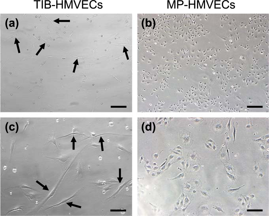
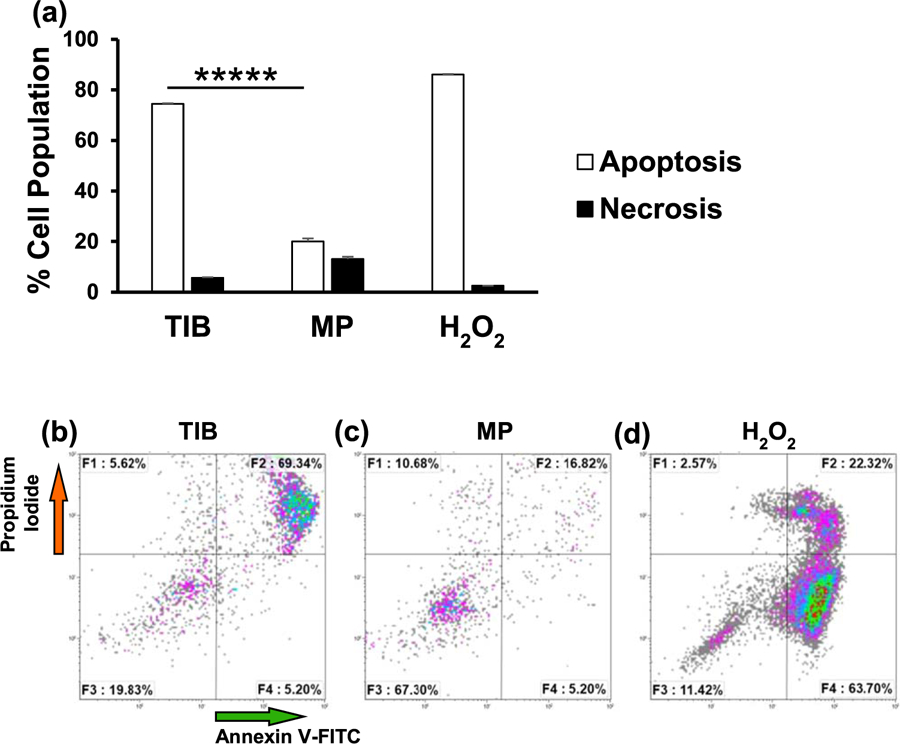
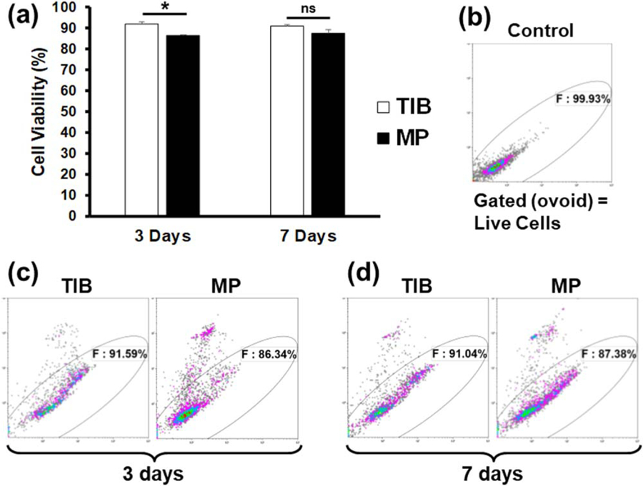
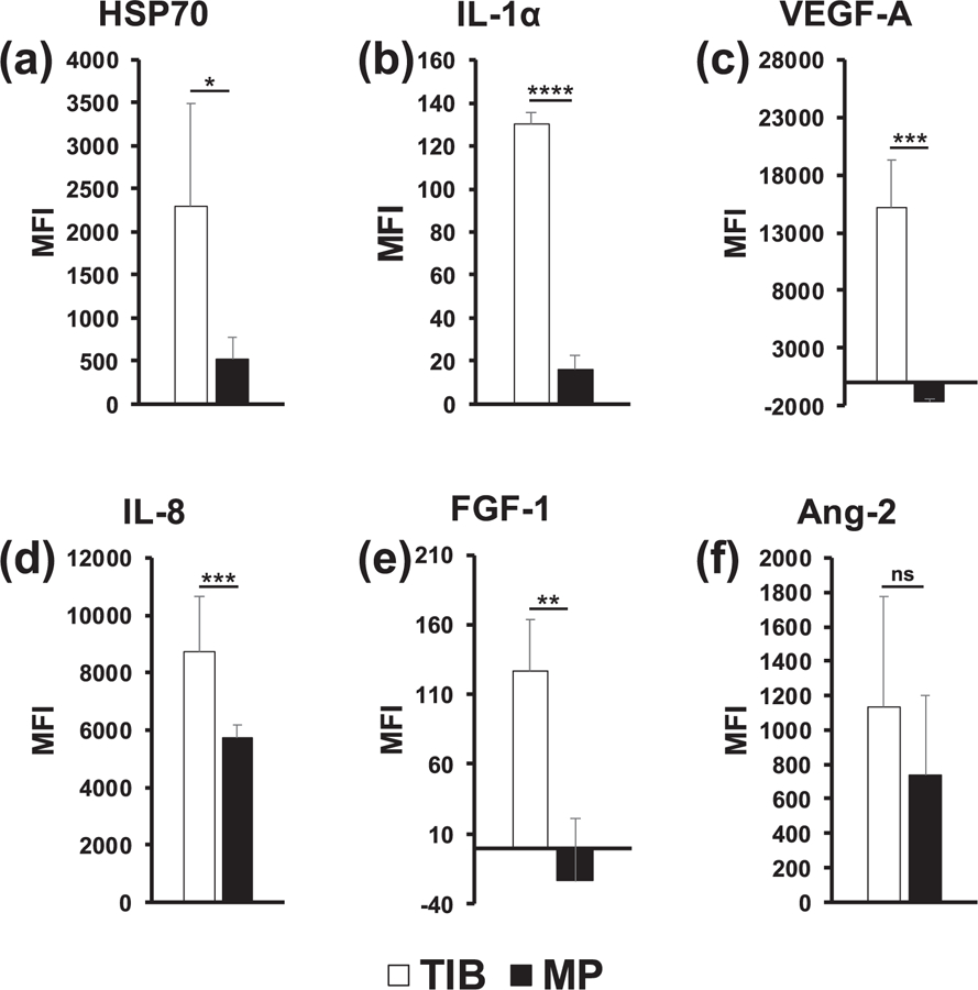
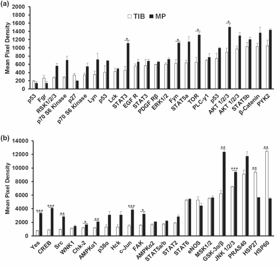
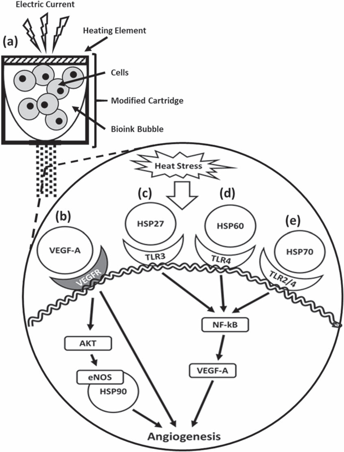
One biofabrication process that has gained tremendous momentum in the field of tissue engineering and regenerative medicine is cell-printing or most commonly bioprinting. We have shown that thermal inkjet bioprinted human microvascular endothelial cells were recruited or otherwise involved in the formation of microvasculature to form graft-host anastomoses upon implantation. The present study aims to quantify and characterize the expression and activation of specific cytokines and kinases in vitro. Morphological characteristics demonstrate elongated protrusions of TIB-HMVECs at 5-6 times the size of manually pipetted cells. Moreover, annexin V-FITC and propidium iodide apoptosis assay via flow cytometry demonstrated a 75% apoptosis among printed cells as compared to among control cells. Cell viability at a 3 d incubation period was significantly higher for printed cells as compared to control. Milliplex magnetic bead panels confirmed significant overexpression of HSP70, IL-1α, VEGF-A, IL-8, and FGF-1 of printed cells compared to control. In addition, a Human phospho-kinase array displayed a significant over activation of the heat-shock proteins HSP27 and HSP60 of printed cells compared to the manually seeded cells. Collectively, it is suggested that the massive appearance of capillary blood vessels upon implantation that has been reported elsewhere may be due to the activation of the HSP-NF-κB pathway to produce VEGF. This cell activation may be used as a new strategy for vascularization of tissue engineered constructs which are in high demand in regenerative medicine applications.


Figures

Figure 1.
Figure 1.
Cell morphology between TIB and MP HMVECs after a 24 h incubation period. (a) and (c) demonstrate TIB-HMVECs at 10× and 20×, respectively. Arrows indicate elongated cells. Images (b) and (d) show MP-HMVECs at 10× and 20×, respectively. Their appearance is almost diamond-shaped with some cells demonstrating minor elongation. TIB = thermal inkjet bioprinted. MP = manually pipetted, HMVECs = human microvascular endothelial cells, Scale bar 10× = 100 μm and 20× = 200 μm.
Figure 2.
Figure 2.
Flow cytometry analysis demonstrating phosphatidylserine (PtdSer) externalization for TIB and MP HMVECs after a 24 h incubation period. F1 quadrant represents necrotic cells (PI positive and annexin negative). F2 quadrant represents cells that are in late apoptosis (both PI and annexin positive). F3 quadrant represents viable cells (both annexin and PI negative). F4 represents cells in early apoptosis (annexin positive and PI negative). (a) Bar graph demonstrating the differences in percentage rates of apoptosis and necrotic cells among TIB and MP HMVECs. The total percent of apoptotic cells is expressed as the sum of the early and late stages of apoptosis (white bars), as determined by the percentage of annexin V-FITC positive cells. Each bar represents the average of three independent measurements and the error bars represent their corresponding standard deviations. Dot plots show ratios of apoptosis among TIB-HMVECs. (b) Dot plots show ratios of apoptosis in TIB-HMVECs. (c) Dot plots show ratios of apoptosis in MP-HMVECs. (d) Dot plots show apoptosis of positive control (HMVECs in 1 mM H2O2). TIB = thermal inkjet bioprinted, MP = manually pipetted, HMVECs = human microvascular endothelial cells. *****P < 0.000 01.
Figure 3.
Figure 3.
Flow cytometry analysis of a PI exclusion assay demonstrating cell viability percentages between TIB and MP-HMVECs at 3 and 7 d incubation periods. (a) Bar graph depicting the differences in cell viability by percent among TIB and MP-HMVECs. Each bar represents the average of three independent measurements and the error bars represent their corresponding standard deviations. (b) Flow cytometric dot plot of untreated HMVECs without PI used to demark the gate of viable cells. (c) Dot plots demonstrating the differences in cell viability by percent between TIB and MP-HMVECs at a 3 d incubation period. (d) Dot plot showing the differences in cell viability by percent between TIB and MP-HMVECs at a 7 d incubation period. PI = propidium iodide, TIB = thermal inkjet bioprinted, MP = manually pipetted, HMVECs = human microvascular endothelial cells, ns = no significant difference. *P < 0.05.
Figure 4.
Figure 4.
In vitro Milliplex magnetic bead panel analysis of 6 specifically selected cytokines after a 12 h incubation period. (a) HSP70 was significantly overexpressed in TIB-HMVECs as compared to MP-HMVECs. (b) IL-1α was significantly overexpressed among TIB-HMVECs in comparison to MP-HMVECs. (c) VEGF-A was significantly overexpressed in TIB-HMVECs as compared to MP-HMVECs. (d) IL-8 was significantly overexpressed in TIB-HMVECs as compared to MP-HMVECs. (e) FGF-1 was significantly overexpressed among TIB-HMVECs in comparison to MP-HMVECs. (f) Ang-2 demonstrated no significant differences in expression between TIB and MP-HMVECs. HSP70 = heat-shock protein 70, IL-1α = interleukin 1α, VEGF-A = vascular endothelial growth factor A, IL-8 = interleukin 8, FGF-1 = fibroblast growth factor 1, Ang-2 = Angiopoietin 2, TIB = thermal inkjet bioprinted, MP = manually pipetted, HMVECs = human microvascular endothelial cells, MFI = median fluorescent intensity, ns = no significant difference. *P < 0.05; **P < 0.01; ***P < 0.001; ****P < 0.0001.
Figure 5.
Figure 5.
In vitro human phospho-kinase array analysis of the activation of 45 kinases in HMVECs after a 12 h incubation period. (a) STAT3, Fyn, TOR, AKT 1/2/3 were significantly overactivated among MP-HMVECs as compared to TIB-HMVECs.(b) Yes, CREB, Src, Chk-2, AMPKα1, c-JUN, FAK, GSK-3α/β, and JNK 1/2/3 were significantly overactivated among MP-HMVECs as compared to TIB-HIMVECs while HSP27 and HSP60 were overactivated significantly in TIB-HMVECs in comparison MP-HMVECs. STAT = signal transducer and activator of transcription proteins, EGF R = epidermal growth factor receptor, PDGF R = platelet-derived growth factor receptor, AKT = protein kinase B, PYK-2 = proline-rich tyrosine kinase 2, CREB = cAMP response element-binding protein, WNK1 = lysine deficient protein kinase 1, AMPK = adenosine monophosphate-activated protein kinase, eNOS = endothelial nitric oxide synthase, MSK = mitogen- and stress-activated protein kinase, GSK = glycogen synthase kinase, JNK = Jun N-terminal kinase, PRAS = proline-rich AKT substrate, HSP = heat-shock protein, TIB = thermal inkjet bioprinted, MP = manually pipetted, HMVECs = human microvascular endothelial cells. *P < 0.05; **P < 0.01; ***P < 0.001.
Figure 6.
Figure 6.
Schematic of how thermal inkjet bioprinting triggers the activation of the VEGF pathway. Heat from the printing process causes cellular heat stress leading to various pathways in which extracellular heat-shock proteins play an angiogenic role. (a) The electric current heats a heating element producing a bubble that collapses causing the ejection of droplets containing cells. (b) VEGF-A binds VEGFR and either activates the AKT-eNOS-HSP90 pathway or promotes angiogenesis directly [72]. (c) HSP27 binds the TLR-3 activating the NF-κB pathway to produce VEGF and induce angiogenesis [64]. (d) Mitochondrial HSP60 binds the TLR-4 [68] leading to the activation of angiogenesis [63, 73]. (e) HSP70 binds TLR 2 and TLR-4 [71] leading to the activation of the NF-κB pathway to produce VEGF and induce angiogenesis [64, 74]. HSP = heat-shock protein, VEGFR = vascular endothelial growth factor receptor, eNOS = endothelial nitric oxide synthase, TLR = toll-like receptor, NF-κB = nuclear factor kappa-light-chain-enhancer of activated B cells.

References

    1. Chang R, Nam J and Sun W 2008. Effects of dispensing pressure and nozzle diameter on cell survival from solid freeform fabrication–based direct cell writing Tissue Eng. A 14 41–8 - PubMed
    1. Murphy SV and Atala A 2014. 3D bioprinting of tissues and organs Nat. Biotechnol 32 773–85 - PubMed
    1. Li J, Chen M, Fan X and Zhou H 2016. Recent advances in bioprinting techniques: approaches, applications and future prospects J. Transl. Med 14 271–86 - PMC - PubMed
    1. Zhang YS et al. 2017. 3D bioprinting for tissue and organ fabrication Ann. Biomed. Eng 45 148–63 - PMC - PubMed
    1. Ringeisen BR, Pirlo RK, Wu PK, Boland T, Sun W, Hamid Q, Huang Y and Chrisey DB 2013. Cell and organ printing turns 15: diverse research to commercial transitions MRS Bull 38 834–43

Publication types