Review
. 2019 May 30;11(6):497.
doi: 10.3390/v11060497.

Reverse Engineering Provides Insights on the Evolution of Subgroups A to E Avian Sarcoma and Leukosis Virus Receptor Specificity

Affiliations
Review

Reverse Engineering Provides Insights on the Evolution of Subgroups A to E Avian Sarcoma and Leukosis Virus Receptor Specificity

Mark J Federspiel. Viruses. .

Abstract

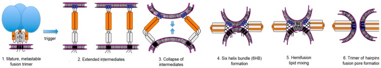
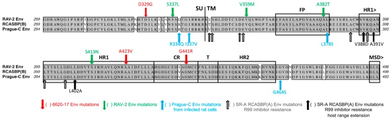
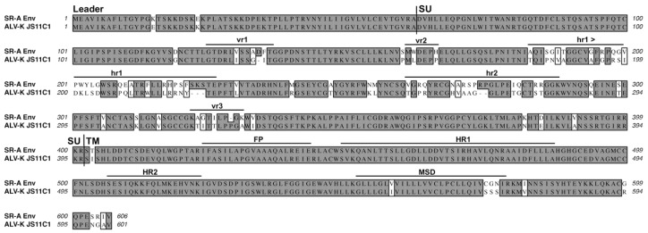
The initial step of retrovirus entry-the interaction between the virus envelope glycoprotein trimer and a cellular receptor-is complex, involving multiple, noncontiguous determinants in both proteins that specify receptor choice, binding affinity and the ability to trigger conformational changes in the viral glycoproteins. Despite the complexity of this interaction, retroviruses have the ability to evolve the structure of their envelope glycoproteins to use a different cellular protein as receptors. The highly homologous subgroup A to E Avian Sarcoma and Leukosis Virus (ASLV) glycoproteins belong to the group of class 1 viral fusion proteins with a two-step triggering mechanism that allows experimental access to intermediate structures during the fusion process. We and others have taken advantage of replication-competent ASLVs and exploited genetic selection strategies to force the ASLVs to naturally evolve and acquire envelope glycoprotein mutations to escape the pressure on virus entry and still yield a functional replicating virus. This approach allows for the simultaneous selection of multiple mutations in multiple functional domains of the envelope glycoprotein that may be required to yield a functional virus. Here, we review the ASLV family and experimental system and the reverse engineering approaches used to understand the evolution of ASLV receptor usage.

Keywords: Avian Sarcoma and Leukosis Viruses; envelope glycoprotein evolution; receptor usage.


Conflict of interest statement

The author declares no conflicts of interest.

Figures

Figure 1
Figure 1
Current model of Class 1 viral fusion protein mediated membrane fusion pathway.
Figure 2
Figure 2
Schematic representations of the major functional domains and comparison of representative Avian Sarcoma and Leukosis Virus (ASLV) subgroups A to E envelope glycoprotein sequences. The envelope glycoprotein leader sequence (Leader), surface glycoprotein sequence (SU), transmembrane glycoprotein sequence (TM) are indicated. The variable region (vr1, vr2, and vr3) and the host range region (hr1 and hr2) sequences in the surface glycoprotein, and the fusion peptide (FP), heptad repeat (HR1 and HR2), and the membrane spanning domain (MSD) sequences in the transmembrane glycoprotein are indicated. The cysteine residues are highlighted in red boxes; the one unpaired cysteine residue at position 100 is highlighted with a blue box. The sequence alignments were done using the ClustalW program in MacVector 14.5.3: identical residues are shaded; conserved residue differences are in boxes, and nonconserved residue differences are unmarked. SR-A: Schmidt–Ruppin A subgroup A ASLV strain UniProt P03397; SR-B: Schmidt–Ruppin B subgroup B ASLV Genbank AAC08989; RAV-2, this study and Genbank AAA87241; Prague-C subgroup C ASLV Genbank AAB59934.1; SR-D: Schmidt–Ruppin D subgroup D ASLV Genbank BAD98245.1; RAV-0* is a partial sequence of a subgroup E ASLV and is the combination of two partial sequences: Genbank AAA87242 and CAA30677.
Figure 3
Figure 3
The proposed secondary structure of the subgroup A to E ASLV envelope glycoproteins. Shown are the disulfide bonds determined using a His-tagged, secreted form of the RCASBP(A) envelope glycoprotein expressed using chicken DF-1 cells and purified. The bonds to C11 and C12 (indicated with a box) could not be assigned further. The free cysteine is labeled C2. The N-linked glycosylation sites actually containing carbohydrate are marked in blue. The unglycosylated sites are underlined, N8 and N13. For reference, the hypervariable regions are marked: hr1 in green boxes; hr2 in an orange box; and vr3 in a pink box.
Figure 4
Figure 4
Schematic representations and protein sequences of critical domains of the three families of cellular surface proteins used as subgroup A to E ASLV receptors.
Figure 5
Figure 5
Schematic representations and binding affinities of ASLV receptor and glycoprotein immunoadhesins. (A) Western immunoblot analysis of the soluble forms of the chicken Tva receptor, sTva-mIgG (sTva), the TvbS1 receptor, sTvbS1-mIgG (sTvb) and the Tvc receptor, sTvc-mIgG (sTvc), immunoprecipitated with anti-mouse IgG-agarose beads, and the secreted forms of the SU glycoproteins SU(C)-rIgG (SUC), SU(A)-rIgG (SUA), and SU(B)-rIgG (SUB) immunoprecipitated with anti-rabbit IgG-agarose beads. The precipitated proteins were denatured, separated by SDS-12% PAGE, and transferred to nitrocellulose. The filters were probed with either peroxidase-conjugated goat anti-mouse IgG or goat anti-rabbit IgG, and the bound protein–antibody complexes visualized by chemiluminescence using Kodak X-Omat film. Molecular sizes (in kilodaltons) are given on the left. (BE) Uninfected DF-1 cells (B) and DF-1 cells chronically infected with either ASLV(A) (C), ASLV(B) (D), or ASLV(C) (E) and uninfected DF-1 cells (B) were fixed with paraformaldehyde and incubated with different amounts of each secreted SU-rIgG (B) or each soluble receptor–mIgG (C–E). The receptor–viral glycoprotein complexes were bound to either goat anti-mouse IgG or goat anti-rabbit IgG linked to phycoerythrin. The amount of phycoerythrin bound to the cells was measured by FACS, and the maximum fluorescence was estimated. The data were plotted as percent maximum fluorescence bound versus concentration of the soluble receptor–mIgG or secreted SU-rIgG. The values shown are averages and standard deviations (error bars) of three experiments. (B) A, SU(A)-rIgG; B, SU(B)-rIgG; C, SU(C)-rIgG. (CE): A, sTva-mIgG; B, sTvbS1-mIgG; C, sTvc-mIgG. (F) Estimated binding affinities of the soluble forms of the ASLV receptors for ASLV envelope glycoproteins expressed on the surface of infected DF-1 cells, and soluble forms of the ASLV surface glycoproteins for endogenous levels of the ASLV receptors expressed on DF-1 cells. a Apparent Kd values were estimated by fitting the data via nonlinear least squares to a log logistic growth curve function as described in Materials and Methods. Each result is the average and standard deviation from three experiments. b n.d.b.—no detectable binding. Gray fields indicate binding reactions not performed.
Figure 6
Figure 6
Examples of analyses of receptor usage of the wild type and mutant ASLVs. Infectious titers were determined using 10-fold serial dilutions of wild type RCASBP(A), RCASBP(B), and RCASBP(C) viruses, the parental Δ155–160 virus, and the Δ155–160 mutant virus supernatants produced using DF-1 cells. The infectious titer was determined by the AP assay. No infectious units detected are denoted with (∗). The results shown are an average of three different experiments; error bars show standard deviations. (A) ASLV receptor interference patterns of the ASLVs infecting parental DF-1 cells, and DF-1 cells chronically infected with ASLV(A), ASLV(B), ASLV(C), or subgroup J ASLV, HPRS103. The replicative abilities and receptor usage of RAV-2/Del136–142 mutants. (B,C) The abilities of the ASLV parental viruses and mutants to alter receptor usage in avian cells using a receptor interference assay (B) using virus supernatants produced from transfected DF-1 cells (see above) titered on parental DF-1 cells (DF-1) and DF-1 cells previously infected by ASLV(A), ASLV(B), ASLV(C) or ASLV(J). The same viral supernatants were also assayed for their abilities to infect mammalian cells (C) that do not express ASLV receptors.
Figure 7
Figure 7
Current model of ASLV(A) Env mediated membrane fusion highlighting some of the stable intermediate stages that can be achieved experimentally with examples of biochemical assays that support the two-step fusion/entry mechanism of ASLV. For clarity, not all of the SU glycoproteins in the trimer are shown in steps 1–3; the SU glycoproteins are not shown in steps 4 and 5 but published reports indicate the disulfide bonds between SU-TM remain stable throughout the fusion process.
Figure 8
Figure 8
A summary and comparison of the ASLV SU glycoprotein wild type and escape mutations identified using various genetic-based selections strategies on limiting the entry and subsequent production of replication-competent ASLVs. Comparison of the SR-A, Prague C, and RAV-2 hr1, hr2, and vr3 hypervariable regions of the SU glycoproteins. The sequence alignments were done using the ClustalW program in MacVector 14.5.3: gaps are noted with black dashes (−). The identified mutations are shown in red text; deletions are shown with red dashes (−). The sequence numbering is provided relative to each ASLV Env protein.
Figure 9
Figure 9
A summary and comparison of the ASLV c-terminal SU and N-terminal TM glycoprotein wild type and escape mutations identified using various genetic-based selections strategies limiting the entry and subsequent production of replication-competent ASLVs. Comparison of the C-terminal end of SU and N-terminal end of TM glycoproteins of RAV-2, RCASBP(B)SR-A, and Prague C. The sequence alignments were done using the ClustalW program in MacVector 14.5.3: identical residues are shaded; conserved residue differences are in boxes, and nonconserved residue differences are unmarked.
Figure 10
Figure 10
A comparison of the SR-A subgroup A and the JS11C1 subgroup K envelope glycoproteins. See Figure 2 legend for analysis details. JS11C1, Genbank #KF46200.1.

Similar articles

Cited by

References

    1. Cardone G., Brecher M., Fontana J., Winkler D.C., Butan C., White J.M., Steven A.C. Visualization of the two-step fusion process of the retrovirus avian sarcoma/leukosis virus by cryo-electron tomography. J. Virol. 2012;86:12129–12137. doi: 10.1128/JVI.01880-12. - DOI - PMC - PubMed
    1. Cosset F.L., Lavillette D. Cell entry of enveloped viruses. Adv. Genet. 2011;73:121–183. - PMC - PubMed
    1. Harrison S.C. Viral membrane fusion. Nat. Struct. Mol. Biol. 2008;15:690–698. doi: 10.1038/nsmb.1456. - DOI - PMC - PubMed
    1. Plemper R.K. Cell entry of enveloped viruses. Curr. Opin. Virol. 2011;1:92–100. doi: 10.1016/j.coviro.2011.06.002. - DOI - PMC - PubMed
    1. White J.M., Delos S.E., Brecher M., Schornberg K. Structures and mechanisms of viral membrane fusion proteins: Multiple variations on a common theme. Crit. Rev. Biochem. Mol. Biol. 2008;43:189–219. doi: 10.1080/10409230802058320. - DOI - PMC - PubMed

Publication types

LinkOut - more resources