Review
. 2019 May 30;24(11):2071.
doi: 10.3390/molecules24112071.

Development of the Inhibitors that Target the PD-1/PD-L1 Interaction-A Brief Look at Progress on Small Molecules, Peptides and Macrocycles

Affiliations
Review

Development of the Inhibitors that Target the PD-1/PD-L1 Interaction-A Brief Look at Progress on Small Molecules, Peptides and Macrocycles

Katarzyna Guzik et al. Molecules. .

Abstract

Cancer immunotherapy based on antibodies targeting the immune checkpoint PD-1/PD-L1 pathway has seen unprecedented clinical responses and constitutes the new paradigm in cancer therapy. The antibody-based immunotherapies have several limitations such as high production cost of the antibodies or their long half-life. Small-molecule inhibitors of the PD-1/PD-L1 interaction have been highly anticipated as a promising alternative or complementary therapeutic to the monoclonal antibodies (mAbs). Currently, the field of developing anti-PD-1/PD-L1 small-molecule inhibitors is intensively explored. In this paper, we review anti-PD-1/PD-L1 small-molecule and peptide-based inhibitors and discuss recent structural and preclinical/clinical aspects of their development. Discovery of the therapeutics based on small-molecule inhibitors of the PD-1/PD-L1 interaction represents a promising but challenging perspective in cancer treatment.

Keywords: PD-1/PD-L1 pathway; cancer immunotherapy; cocrystal structures; lead optimization; peptide-based and small synthetic molecule inhibitors; rational drug design; scaffold hopping; structure-activity relationship.


Conflict of interest statement

The authors declare no conflict of interest.

Figures

Figure 1
Figure 1
Structure of AUNP-12 (1)—A 29-residue peptide sequence.
Figure 2
Figure 2
An example of modified heptapeptide (SNTSEFS-NH2) derived from 1 (modified moiety is coloured with blue).
Figure 3
Figure 3
The structures of BMS peptides 3 (BMS-57) and 4 (BMS-71) with their IC50 values reported in the patent.
Figure 4
Figure 4
Geldanamycin (5) and Rifabutin (6) as the PD-L1 inhibitors.
Figure 5
Figure 5
Representative structures of the Aurigene peptidomimetics 7 and 8. The SAR-affected parts are marked in blue.
Figure 6
Figure 6
Linear 9 and cyclic peptide 10 showing the highest MSPA rates.
Figure 7
Figure 7
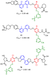
Examples of the macrocyclic peptidomimetic compounds 11 and 12 with glycol-derived linker.
Figure 8
Figure 8
Examples of Aurigene’s small-molecule inhibitors.
Figure 9
Figure 9
Modifications of structures 13 and 14 [55,56].
Figure 10
Figure 10
Examples of the small-molecules patented by Aurigene in 2018/2019.
Figure 11
Figure 11
SAR of structures from Aurigene patents [56,57].
Figure 12
Figure 12
General structure and examples of the PD-1/PD-L1 sulfamonomethoxine 24 and sulfamethizole 25 inhibitors [59].
Figure 13
Figure 13
General structures of the compounds disclosed by Bristol-Myers Squibb company and their representative examples [60,61,62,63,64,65].
Figure 14
Figure 14
(A) Crystal structure of the 26/PD-L1 complex. The asymmetric unit contains four molecules of PD-L1 (ribbon representation), which are organized into two dimers (APD-L1—green, BPD-L1—purple-blue, cPD-L1—salmon, DPD-L1—bright orange). Each dimer binds a single molecule of 26 (yellow) at the dimer interface. (B) Detailed interactions of 26 at the binding cleft of PD-L1 dimer. 26 binds at a hydrophobic cavity formed upon PD-L1 dimerization (PDB: 5J89). Compound 30 cocrystal structure is consistent with structural data for 26.
Figure 15
Figure 15
(A) Detailed interactions of 31 (light yellow) at the binding cleft of PD-L1. Compound 31 binds at a hydrophobic cavity formed upon PD-L1 binding. (B) Detailed interactions of 32 (yellow) at the binding cleft of PD-L1. 32 binds at a hydrophobic tunnel formed upon the PD-L1 dimerization. The movement of the aromatic ring of ATyr56 is induced by the 2,3-dihydro-1,4-benzdioxine moiety. (C) The movement of ATyr56 (green) that is induced by the 2,3-dihydro-1,4-benzdioxine group of 32 (yellow) compared with the ATyr56 (salmon) arrangement in the complex of 31/PD-L1.
Figure 16
Figure 16
Examples of the BMS class compounds, for which structural data were provided. (A, B) 2,3-dihydro-1,4-benzodioxine group of 33 and 34 induced transformation of the binding pocket into the binding tunnel across the transverse vertical axis of the dimer. Both compounds trigger formation of a subpocket binding the benzonitrile moiety [41,43,44]. (C) The binding cleft of 35 is closed from one side by the AY56 residue. The arrangement of this inhibitor follows patterns for compounds 26, 30, and 31.
Figure 17
Figure 17
(A) Fragment 36 bound to dimeric PD-L1. Detailed interactions of 36 (yellow) at the binding cleft of PD-L1. 36 binds at a hydrophobic tunnel formed upon the PD-L1 dimerization. PDB: 6NM7 (B) Overlay of fragment 36 (yellow), BMS small molecule inhibitor 33 (light pink) and BMS macrocyclic inhibitor 4 (plum) (PDB codes: Fragment 1: 6NM7, BMS small molecule 5NIU: 6NM8, BMS peptide-71: 6NNV).
Figure 18
Figure 18
Examples of hits 3644 found in the fragment-based screening published by Perry et al. [44]. Fragments were able to displace PD-1 from PD-L1 in the NMR-based AIDA assay [66]. The PD-1 rescue score was estimated on the percent of the G90 signal rescued of the 15N labeled PD-1 at 800 µM fragment concentration. Score 1 indicates 1–15% signal rescue and score 2 indicates >15% signal rescue (some of the Kd values were not determined due to solubility limits and/or resonance peak broadening).
Figure 19
Figure 19
The general structure and examples of the PD-1/PD-L1 inhibitors (45 and 46) reported by Incyte Corporation. The biphenyl fragment is shown in blue and the fused heteroaromatic system is shown in red. The next figure follows the same color pattern unless stated otherwise.
Figure 20
Figure 20
General structure and examples of PD-1/PD-L1 inhibitors (4749) reported by Incyte Corporation in the second patent. Aryl ether fragment is shown in green.
Figure 21
Figure 21
General structure and examples of the PD-1/PD-L1 inhibitors (50 and 51) reported by Incyte Corporation [71].
Figure 22
Figure 22
General structure and examples of the PD-1/PD-L1 inhibitors (50 and 51) reported by Incyte Corporation [71].
Figure 23
Figure 23
General structure and examples of the PD-1/PD-L1 inhibitors (54 and 55) reported by Incyte Corporation, based on the fused six-membered heteroaromatic rings linked with the biphenyl scaffold.
Figure 24
Figure 24
General structure and examples of the PD-1/PD-L1 inhibitors (56 and 57) reported by Incyte Corporation, based on the picolinamide-biphenyl scaffold.
Figure 25
Figure 25
General structure and examples of the PD-1/PD-L1 inhibitors (58 and 59) reported by Incyte Corporation and based on the N-methyl-2-pyridone-6-carboxamide moiety (depicted with red).
Figure 26
Figure 26
General structures and examples of the PD-1/PD-L1 inhibitors patented by Incyte Corporation, based on heterocyclic five-membered aromatic rings (60 and 61) and on heterocyclic five-membered aromatic rings fused with piperidine (62 and 63).
Figure 27
Figure 27
General structure and examples of the PD-1/PD-L1 inhibitors patented by Incyte Corporation (64 and 65). The 3′ biphenyl’s substituent is depicted in gold.
Figure 28
Figure 28
The structures of inhibitors 66, 67 disclosed by ChemoCentryx.
Figure 29
Figure 29
Examples of the benzyl phenyl ether derivatives 68 reported by Feng et al., [82]; the resorcinol (69) and isonicotinic acid (70) derivatives reported, respectively, by Li et al. [83] and Sun et al. [84].
Figure 30
Figure 30
Examples of the structures patented by Arising International LLC.
Figure 31
Figure 31
Examples of inhibitors 7475 designed by Polaris Pharmaceuticals company [86].
Figure 32
Figure 32
Compounds reported by Aktoudianakis et al. [87] (Gilead Sciences).
Figure 33
Figure 33
Examples of the compounds published by Qin et al. [88].
Figure 34
Figure 34
Structures of PD-1 binders reported by Patil and co-workers [89].
Figure 35
Figure 35
General structures 21–24 and examples of compounds 8588 described by Dömling.
Figure 36
Figure 36
General structure and examples of the PD-1/PD-L1 inhibitors patented by the Maxinovel Corporation (8991). Biphenyl fragments are shown in blue, phenyl fragments in red, the ethenyl and acetylenyl linkers in green.
Figure 37
Figure 37
The general pattern in the development of PD-L1 inhibitors.

Similar articles

Cited by

References

    1. Agata Y., Kawasaki A., Nishimura H., Ishida Y., Tsubat T., Yagita H., Honjo T. Expression of the PD-1 antigen on the surface of stimulated mouse T and B lymphocytes. Int. Immunol. 1996;8:765–772. doi: 10.1093/intimm/8.5.765. - DOI - PubMed
    1. Nishimura H., Agata Y., Kawasaki A., Sato M., Imamura S., Minato N., Yagita H., Nakano T., Honjo T. Developmentally regulated expression of the PD-1 protein on the surface of double-negative(CD4(−)CD8(−)) thymocytes. Int. Immunol. 1996;8:773–780. doi: 10.1093/intimm/8.5.773. - DOI - PubMed
    1. Simon S., Labarriere N. PD-1 expression on tumor-specific T cells: Friend or foe for immunotherapy? Oncoimmunology. 2018;7:e1364828. doi: 10.1080/2162402X.2017.1364828. - DOI - PMC - PubMed
    1. Zak K.M., Grudnik P., Magiera K., Dömling A., Dubin G., Holak T.A. Structural Biology of the Immune Checkpoint Receptor PD-1 and Its Ligands PD-L1/PD-L2. Structure. 2017;25:1163–1174. doi: 10.1016/j.str.2017.06.011. - DOI - PubMed
    1. Lee H.T., Lee S.H., Heo Y.-S. Molecular Interactions of Antibody Drugs Targeting PD-1, PD-L1, and CTLA-4 in Immuno-Oncology. Molecules. 2019;24:1190. doi: 10.3390/molecules24061190. - DOI - PMC - PubMed

MeSH terms