

Magnetic resonance image findings in pug dogs with thoracolumbar myelopathy and concurrent caudal articular process dysplasia
- PMID: 31151444
- PMCID: PMC6544997
- DOI: 10.1186/s12917-019-1866-0
Magnetic resonance image findings in pug dogs with thoracolumbar myelopathy and concurrent caudal articular process dysplasia
Abstract
Background: A retrospective case series study was undertaken to describe the magnetic resonance imaging (MRI) findings in Pug dogs with thoracolumbar myelopathy and concurrent caudal articular process (CAP) dysplasia. Electronic clinical records were searched for Pug dogs who underwent MRI for the investigation of a T3-L3 spinal cord segment disease with subsequent confirmation of CAP dysplasia with computed tomography between January 2013 and June 2017. Clinical parameters age, gender, neuter status, body weight, urinary or faecal incontinence, severity and duration of clinical signs were recorded. MRI abnormalities were described. Univariable non-parametric tests investigated the association between the clinical parameters and evidence of extra- or intra-dural spinal cord compression on MRI.
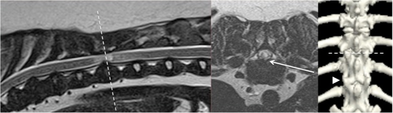
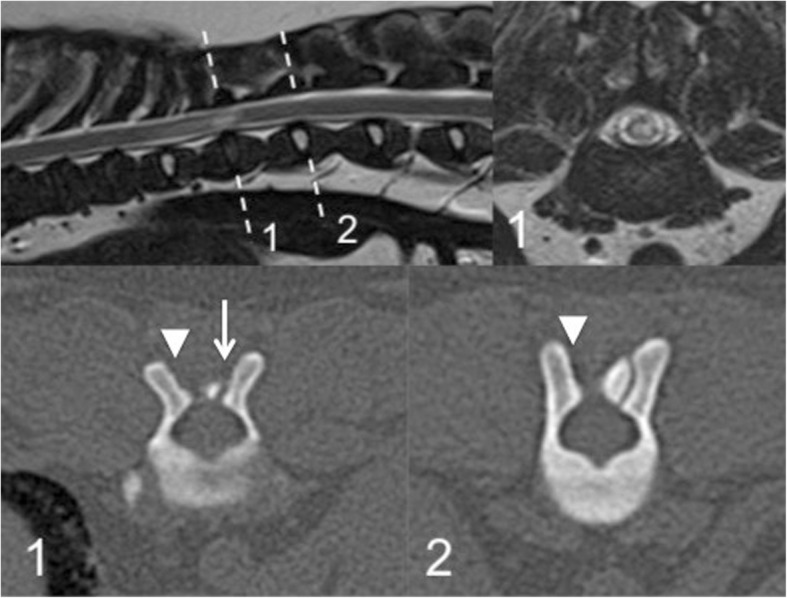
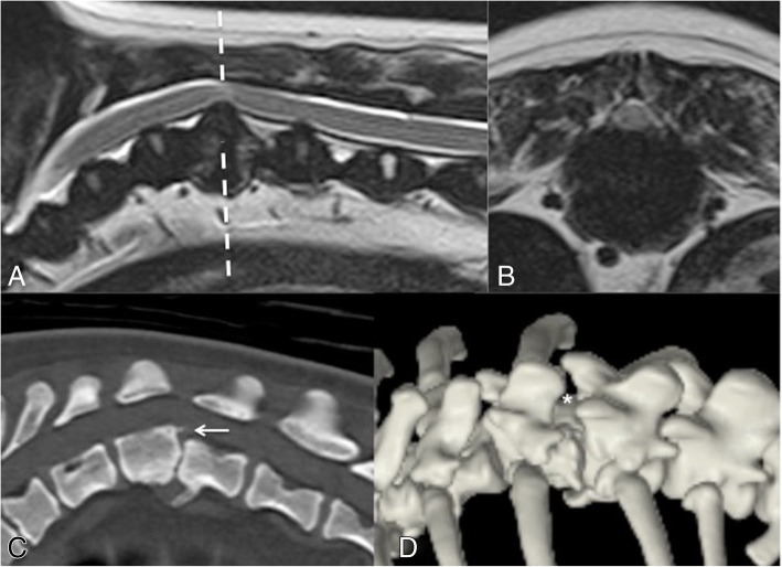
Results: 18 Pug dogs were included. The median age was 106 months with median duration of clinical signs 5 months. All presented with variable severity of spastic paraparesis and ataxia; 50% suffered urinary/faecal incontinence. In all cases, MRI revealed a focal increase in T2-weighted signal intensity within the spinal cord at an intervertebral level where bilateral CAP dysplasia was present; this was bilateral aplasia in all but one case, which had one aplastic and one severely hypoplastic CAP. MRI lesions were associated with spinal cord compression in all but one case; intervertebral disc protrusion resulted in extra-dural compression in 10 (56%) cases; intra-dural compression was associated with a suspected arachnoid diverticulum in 4 (22%) cases and suspected pia-arachnoid fibrosis in 3 cases (17%). There was no association between clinical parameters and a diagnosis of intra-dural vs extra-dural compression. CAP dysplasia occurred at multiple levels in the T10-13 region with bilateral aplasia at T11-12 most often associated with corresponding spinal cord lesions on MRI.
Conclusions: All Pugs dogs in this study were presented for chronic progressive ambulatory paraparesis; incontinence was commonly reported. Although intervertebral disc disease was the most common radiologic diagnosis, intra-dural compression associated with arachnoid diverticulae/fibrosis was also common. Bilateral CAP aplasia was present in all but one Pug dog at the level of MRI detectable spinal cord lesions. A causal relationship between CAP dysplasia and causes of thoracolumbar myelopathy is speculated but is not confirmed by this study.
Keywords: Facet dysplasia; MRI; Pug dogs; Vertebral malformation.
Conflict of interest statement
The University of Surrey and Fitzpatrick Referrals did not play a role in the study design, data collection and analysis, decision to publish, or preparation of the manuscript and only provided financial support in the form of authors’ salaries and/or research materials. None of the authors have personal or financial relationships with other people or organizations that might inappropriately influence or bias the content of the paper. There are no patents, products in development, or marketed products to declare.
Figures

Similar articles
-
Clinical features and MRI characteristics of presumptive constrictive myelopathy in 27 pugs.Vet Radiol Ultrasound. 2020 Sep;61(5):545-554. doi: 10.1111/vru.12890. Epub 2020 Jun 25. Vet Radiol Ultrasound. 2020. PMID: 32583954
-
Thoracolumbar myelopathies in pug dogs.J Vet Intern Med. 2023 Mar;37(2):618-625. doi: 10.1111/jvim.16639. Epub 2023 Feb 6. J Vet Intern Med. 2023. PMID: 36744714 Free PMC article.
-
Clinical and magnetic resonance imaging characterization of cervical spondylomyelopathy in juvenile dogs.J Vet Intern Med. 2019 Sep;33(5):2160-2166. doi: 10.1111/jvim.15602. Epub 2019 Aug 30. J Vet Intern Med. 2019. PMID: 31469206 Free PMC article.
-
The Role of Fenestration in Management of Type I Thoracolumbar Disk Degeneration.Vet Clin North Am Small Anim Pract. 2018 Jan;48(1):187-200. doi: 10.1016/j.cvsm.2017.08.012. Epub 2017 Oct 23. Vet Clin North Am Small Anim Pract. 2018. PMID: 29074336 Review.
-
Vertebral and spinal malformations in small brachycephalic dog breeds: Current knowledge and remaining questions.Vet J. 2024 Apr;304:106095. doi: 10.1016/j.tvjl.2024.106095. Epub 2024 Mar 6. Vet J. 2024. PMID: 38458418 Review.
Cited by
-
Multiple Genetic Loci Associated with Pug Dog Thoracolumbar Myelopathy.Genes (Basel). 2023 Feb 1;14(2):385. doi: 10.3390/genes14020385. Genes (Basel). 2023. PMID: 36833311 Free PMC article.
-
Comparison of standard T2-weighted turbo spin echo and volumetric interpolated breath-hold examination magnetic resonance imaging sequences in the assessment of articular process dysplasia in Pug dogs with thoracolumbar myelopathy.Front Vet Sci. 2023 Sep 27;10:1265665. doi: 10.3389/fvets.2023.1265665. eCollection 2023. Front Vet Sci. 2023. PMID: 37829356 Free PMC article.
-
Surgical techniques used in the management of intra-arachnoid diverticula in dogs across four referral centres and their immediate outcome.J Small Anim Pract. 2022 Jul;63(7):520-525. doi: 10.1111/jsap.13486. Epub 2022 Feb 9. J Small Anim Pract. 2022. PMID: 35137433 Free PMC article.
-
Thoracolumbar meningeal fibrosis in pugs.J Vet Intern Med. 2020 Mar;34(2):797-807. doi: 10.1111/jvim.15716. Epub 2020 Jan 31. J Vet Intern Med. 2020. PMID: 32003496 Free PMC article.
-
Surgical Outcomes of Laminectomy, Durotomy and a Non-Synthetic Dura Substitute Application in Ten Dogs with a Spinal Subarachnoid Diverticulum.Vet Sci. 2024 Mar 14;11(3):128. doi: 10.3390/vetsci11030128. Vet Sci. 2024. PMID: 38535862 Free PMC article.
References
MeSH terms
Supplementary concepts
LinkOut - more resources
Full Text Sources
Medical
Research Materials
Miscellaneous
