

Pathophysiology of Lacunar Stroke: History's Mysteries and Modern Interpretations
- PMID: 31151839
- PMCID: PMC7416422
- DOI: 10.1016/j.jstrokecerebrovasdis.2019.05.006
Pathophysiology of Lacunar Stroke: History's Mysteries and Modern Interpretations
Abstract
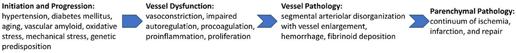
Since the term "lacune" was adopted in the 1800s to describe infarctions from cerebral small vessels, their underlying pathophysiological basis remained obscure until the 1960s when Charles Miller Fisher performed several autopsy studies of stroke patients. He observed that the vessels displayed segmental arteriolar disorganization that was associated with vessel enlargement, hemorrhage, and fibrinoid deposition. He coined the term "lipohyalinosis" to describe the microvascular mechanism that engenders small subcortical infarcts in the absence of a compelling embolic source. Since Fisher's early descriptions of lipohyalinosis and lacunar stroke (LS), there have been many advancements in the understanding of this disease process. Herein, we review lipohyalinosis as it relates to modern concepts of cerebral small vessel disease (cSVD). We discuss clinical classifications of LS as well as radiographic definitions based on modern neuroimaging techniques. We provide a broad and comprehensive overview of LS pathophysiology both at the vessel and parenchymal levels. We also comment on the role of biomarkers, the possibility of systemic disease processes, and advancements in the genetics of cSVD. Lastly, we assess preclinical models that can aid in studying LS disease pathogenesis. Enhanced understanding of this highly prevalent disease will allow for the identification of novel therapeutic targets capable of mitigating disease sequelae.
Keywords: Stroke; cerebral small vessel disease; genetics; lacunar stroke.
Copyright © 2019 Elsevier Inc. All rights reserved.
Conflict of interest statement
Conflicts of Interest/Disclosures
The authors report no conflicts of interest or disclosures.
Figures
References
-
- Pearce J Dechambre’s description of lacunes, 1838. J Neurol Neurosurg Psychiatry. 1990;53(2):134.
-
- Dechambre A Memoire sur la curabilite du ramollissement cerebral. Gaz Medicale, Paris: 1838;6:305–14.
-
- Roman GC. The original description of lacunes. Neurology. 1986;36:85.
-
- Roman GC. On the history of lacunes, etat crible, and the white matter lesions of vascular dementia. Cerebrovasc Dis. 2002;13 Suppl 2:1–6. - PubMed
-
- Durand-Fardel M Traite du ramollissement du cerveau. JB Baillière, Paris.
Publication types
MeSH terms
Substances
Grants and funding
LinkOut - more resources
Full Text Sources
