. 2019 Jul 11;75(1):13-25.e5.
doi: 10.1016/j.molcel.2019.05.004. Epub 2019 May 28.

Arc Oligomerization Is Regulated by CaMKII Phosphorylation of the GAG Domain: An Essential Mechanism for Plasticity and Memory Formation

Affiliations

Arc Oligomerization Is Regulated by CaMKII Phosphorylation of the GAG Domain: An Essential Mechanism for Plasticity and Memory Formation

Wenchi Zhang et al. Mol Cell. .

Abstract

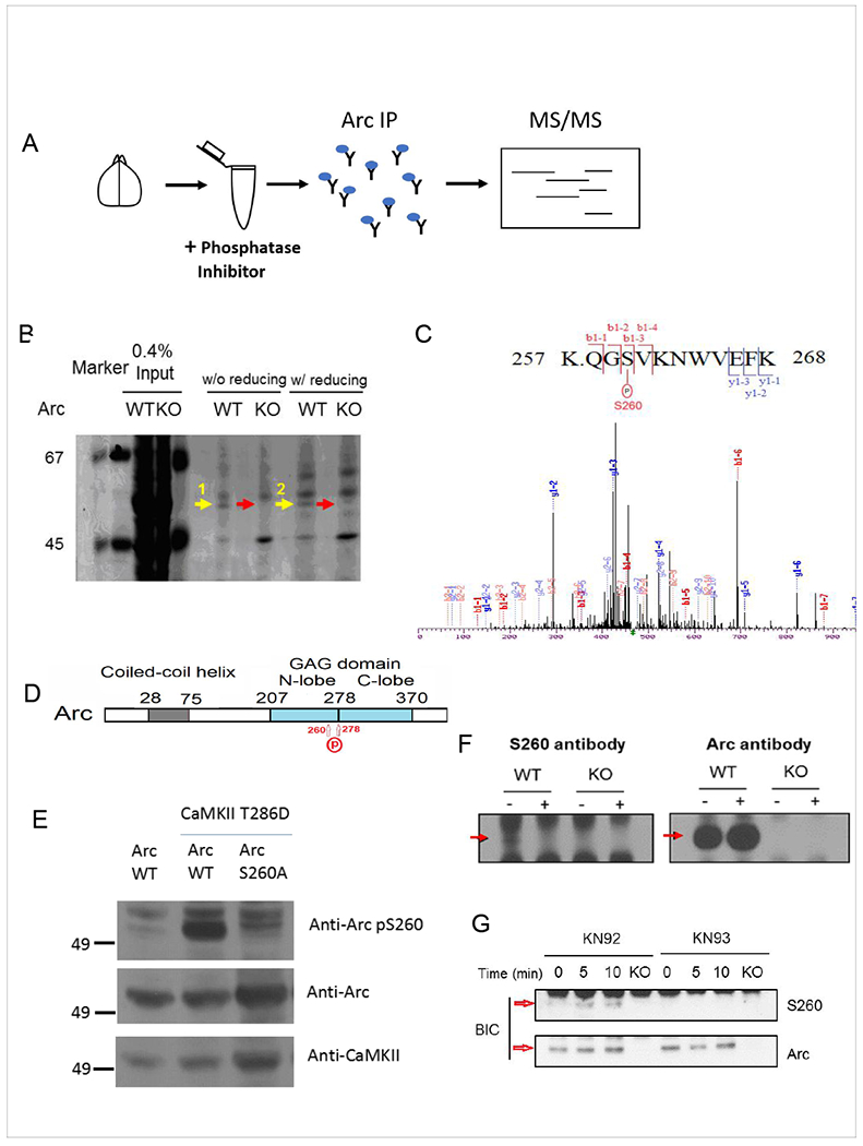
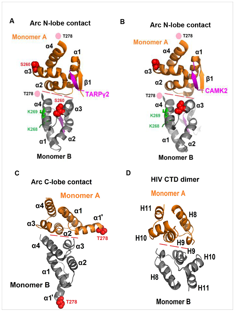
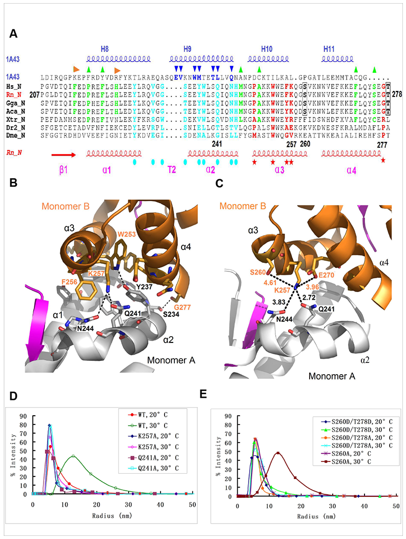
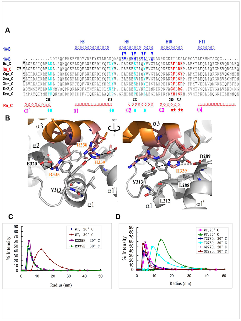
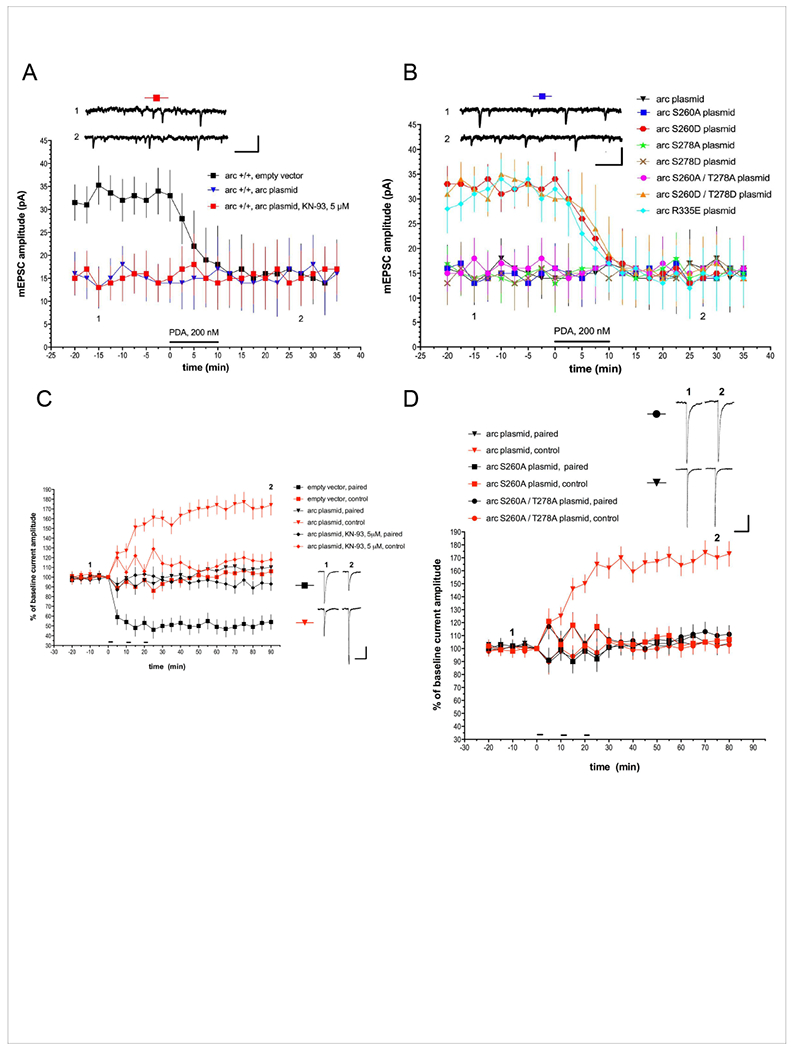
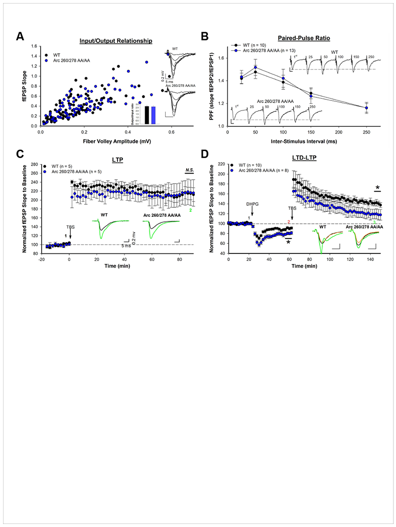
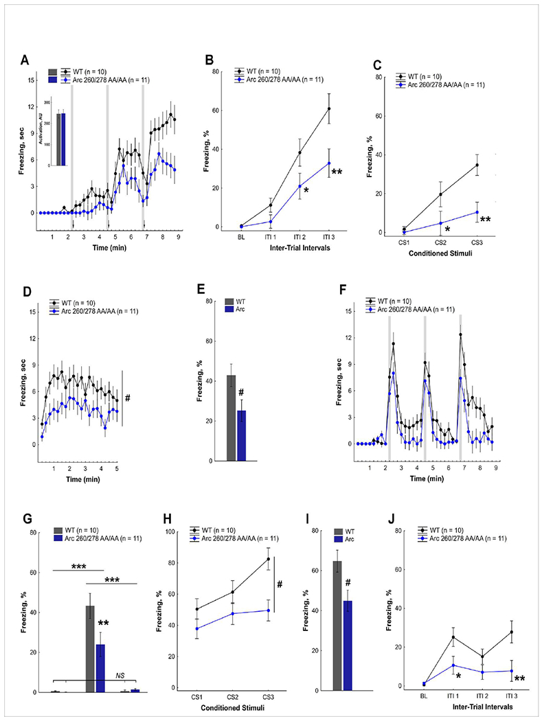
Arc is a synaptic protein essential for memory consolidation. Recent studies indicate that Arc originates in evolution from a Ty3-Gypsy retrotransposon GAG domain. The N-lobe of Arc GAG domain acquired a hydrophobic binding pocket in higher vertebrates that is essential for Arc's canonical function to weaken excitatory synapses. Here, we report that Arc GAG also acquired phosphorylation sites that can acutely regulate its synaptic function. CaMKII phosphorylates the N-lobe of the Arc GAG domain and disrupts an interaction surface essential for high-order oligomerization. In Purkinje neurons, CaMKII phosphorylation acutely reverses Arc's synaptic action. Mutant Arc that cannot be phosphorylated by CaMKII enhances metabotropic receptor-dependent depression in the hippocampus but does not alter baseline synaptic transmission or long-term potentiation. Behavioral studies indicate that hippocampus- and amygdala-dependent learning requires Arc GAG domain phosphorylation. These studies provide an atomic model for dynamic and local control of Arc function underlying synaptic plasticity and memory.

Keywords: Arc; CAMKII; capsid; evolution; learning and memory; phosphorylation; polymerization; synapse plasticity.


Conflict of interest statement

Declaration of Interests

The authors declare no competing interests.

Figures

Figure 1
Figure 1. Identification of Arc phosphorylation sites
(A) Arc immunoprecipitation from naïve adult mouse brain and MS analysis. (B) Immunoprecipitates from detergent lysates of WT and Arc KO brain were sequentially eluted with 2% SDS and 2% SDS with 10 mM BME and analyzed by Coomassie. Arc band was excised and submitted for MS. Yellow arrows: Arc, Red arrows: Arc knockout control. (C) Mass spectrometry profile of the sequence of Arc from residues 257-268 is shown. Analysis by MS/MS revealed the presence of phosphorylated modification. The fragment ions whose m / z value corresponds to y or b ions (peptide fragments from C-terminus and from N-terminus, respectively) are indicated. The observed fragment ions are labeled on the spectrum and peptide sequence. (D) schematic of Arc indicating position of phosphorylation sites. (E) CaMKII phosphorylates ArcS260. Arc or ArcS260A were co-transfected with constitutively active CamKIIα (CamKII T286D) to HEK293 cells and cell lysates were blotted with ArcS260 phospho-specific antibody. (F) ArcS260 is phosphorylated in mouse brain. A parallel blot with Arc monoclonal antibody confirmed Arc immunoprecipitation and insensitivity to lambda phosphatase. (G) ArcS260 phosphorylation is induced in cultured neurons. Phospho-specific antibody revealed bicuculline-induced phosphorylation of ArcS260 that was blocked by 30 min pretreatment with CamKII inhibitor (KN93) but not control (KN92).
Figure 2
Figure 2. Comparison of dimers of Arc N-lobe, C-lobe and HIV C-lobe deduced from crystal structures.
Dimers are generated by application of crystal symmetry to the protomer present in the asymmetric unit. Red dashes indicate dimer interface of N-lobe or C-lobe. (A, B) Structures of complex crystals of Arc N-lobe with TARPγ2 peptide (PDB ID, 4X3H) or CamKII fragment (PDB ID, 4X3I). TARPγ2 peptide and CamKII fragment are colored purple. 260S is indicated in red ball. Unstructured 278T is displayed as a pink ball. Ubiquitination sites K268 and K269 are show in green. (C) Structure of Arc C-Lobe crystal (PDB ID, 4X3X). 278T is indicated in red ball. (D) HIV capsid CTD dimer (PDB ID, 1A43). In each case, the monomers that comprise the dimer are colored differently. Helix labels in parentheses follow references for Arc (Zhang et al., 2015) and HIV capsid domain (Pornillos et al., 2009).
Figure 3
Figure 3. Arc N-lobe and oligomerization.
(A) The sequence alignment of Arc N-lobe (PDB ID, 4X3H) from typical species based on their 3D structural superposition with HIV capsid CTD (PDB ID, 1A43). Hs, Homo sapiens, NP_056008.1; Rn, Rattus norvegicus, NP_062234.1; Gga, Gallus gallus, NP_989763.1; Aca, Anolis carolinensis, XP_003223977.1; Xtr, Xenopus (Silurana) tropicalis, XP_002934511.1; Dme, Drosophila melanogaster, Arc1, Q7K1U0; Dr2, Danio rerio, XP_001919627; _N refer to N-lobe. Secondary structures of Arc N-lobe and HIV capsid CTD (PDB 1A43) are shown on the bottom in red and on the top in blue, respectively. The cyan solid circles (monomer A) and red stars (monomer B) below the sequence alignment mark amino acids that are predicted to mediate dimerization of Arc N-lobe. The green up triangles indicate hydrophobic pocket of Arc N-lobe, and the pair of aromatic residues supporting beta strand formation of N-lobe are highlighted with orange right deltoids. Blue down triangles indicate residues in the HIV C-lobe dimer interface. The phosphorylation sites S260 and T278 are shown in bold black with box. (B-C) magnified view of the interaction between two subunits of Arc N-lobe in grey (monomer A) and brown (monomer B), separately (PDB ID, 4X3H). The residues important for interaction are shown as a stick representation. (D-E) Polymerization status of various full-length Arc proteins in solution (10uM) examined by dynamic light scattering (DLS) with increasing temperature (1°C/min) from 20°C to 30°C and assayed after an additional 20 min at 30°C. (D) Mutation of predicted dimerization sites prevents oligomerization. (E) Phosphomimic mutations of S260 prevent oligomerization.
Figure 4
Figure 4. Arc C-Lobe and oligomerization.
(A) The sequence alignment of Arc C-lobe (PDB ID, 4X3X) for typical species based on their 3D structural superposition with HIV capsid CTD (PDB ID, 1A43). Hs, Homo sapiens, NP_056008.1; Rn, Rattus norvegicus, NP_062234.1; Gga, Gallus gallus, NP_989763.1; Aca, Anolis carolinensis, XP_003223977.1; Xtr, Xenopus (Silurana) tropicalis, XP_002934511.1; Dme, Drosophila melanogaster, Arcl, Q7K1U0; Dr2, Danio rerio, XP_001919627; _C refer to C-lobe. Secondary structures of Arc C-lobe and HIV capsid CTD (PDB 1A43) are shown on the bottom in red and on the top in blue, respectively. The cyan solid circles (monomer A) and red stars (monomer B) below the sequence alignment mark amino acids that mediate inter-subunit interactions of Arc C-lobe. The blue down triangles indicate residues in the HIV C-lobe dimer interface. The phosphorylation site T278 is shown in bold black with box. (B) Detailed view of the interaction between two subunits of Arc C-lobe in grey (monomer A) and brown (monomer B), separately (PDB ID, 4X3X). The residues important for interaction are shown as a stick representation. (C, D) Polymerization status of various full-length Arc proteins examined in solution (10uM) with increasing temperature (1°C/min) from 20°C to 30°C. (C) Mutations in the putative hinge region. After an additional 6 minutes at 30°C the mutant ArcT278D peaks ~1276 KDa (hexamer of tetramers) calculated based on gyration radius, while WT Arc peaks ~2458 KDa (dodecamer of tetramers). (D) Mutation of the C-lobe interface blocks oligomerization of full-length Arc.
Figure 5
Figure 5. Arc oligomerization, synaptic function and regulation by CaMKII.
Whole-cell voltage-clamp recordings from mouse Purkinje cells expressing Arc transgenes in culture. (A) Basal mEPSC amplitude was driven low and chemical LTD evoked by PDA was occluded in Arc-transfected Purkinje cells. This effect was not blocked by pretreatment with the CaMKII inhibitor KN-93. N= 10 cells/group. (B) Phosphomimic mutant at the site S260 and C-lobe mutant R335E did not reduce the basal amplitude of mEPSCs and did not occlude subsequent PDA-evoked chemical LTD. ArcS260A mimics WT Arc by reducing mEPSC and occluding chemical LTD. N= 10 cells/group. The scale bars for the insets on (A) and (B) are both 500 msec, 20 pA. (C) Purkinje cells in primary cerebellar culture were transfected with Arc transgene or an empty vector control plasmid and examined using a standard protocol that induces dendrite-specific LTD in Purkinje cells treated with the control plasmid (n=8; or untransfected cells, not shown). In Arc expressing cells (n=7) LTD was occluded in the paired pathway that received glutamate/depolarization conjunction as indicated by the horizontal bars (paired pathway), while LTP was induced in the control pathway. LTP was abolished by pretreatment with the CamKII inhibitor KN-93 (n=8). Scale bars = 2 sec, 50 pA. (D) LTD induced by glutamate/depolarizing pairing (indicated by horizontal bars) was occluded by both WT, ArcS260A and ArcS260,T278A. LTP of control pathway was abolished in neurons expressing de-phosphorylation mimic mutants at ArcS260A. Arc plasmid (n=7); Arc S260A plasmid (n=8); Arc S260A,T278A plasmid (n=6). Scale bars = 2 sec, 60 pA.
Figure 6
Figure 6. Enhanced DHPG-induced LTD in Arc 260/278 AA/AA mutant mice.
(A) The input-output ratio is not altered in Arc 260/278 AA/AA mutant mice. For input-output ratio: WT n = 86, I/O =2.70 ± 0.15 ms−1; Mutant n=81, I/O = 2.64 ± 0.14 ms−1. P>0.05 with two-tailed student’s t-test. (B) Paired pulse facilitation is not altered in Arc 260/278 AA/AA mutant mice. WT n=13; Mutant n = 10. P>0.05 with two-way repeated measures ANOVA with Bonferoni post-hoc test. (C) Theta-burst stimulation (TBS) induced LTP is not altered in Arc 260/278 AA/AA mutant mice. n=5 for each group. P>0.05 with two-tailed student’s t-test. (D) DHPG-induced LTD is significantly enhanced in Arc 260/278 AA/AA mutant mice. The TBS-induced LTP following DHPG-LTD is significantly reduced in Arc 260/278 AA/AA mutant mice when normalized to original baseline, but not to last 6 min of LTD (see supplement figure S5). WT n=10; mutant n=8. * P<0.05 with two-tailed student’s t-test.
Figure 7
Figure 7. Arc 260/278 AA/AA mutant mice are deficient in acquisition of fear memory.
(A) Time course of freezing to context and conditioned stimulus, CS (shaded areas), observed in Arc 260/278 AA/AA mutant mice and littermate control WT mice during training in a delayed fear conditioning (Session 1). Arrows indicate presentation of unconditioned stimulus, US (a foot-shock) at the end of each CS. Insert shows equal sensitivity to a foot-shock in both genotypes (ANOVA, F(1, 19)=0.1, P>0.9). (B) Acquisition of fear to context is delayed in Arc 260/278 AA/AA mice (ANOVA, effect of genotype F(1,19)=5.63, P<0.028; genotype × intertrial interval (ITI) interaction F(3,57)=4.23, P<0.009). Pre-training basal levels (BL) of freezing were low in both genotypes. (C) Acquisition of fear to a conditioned stimulus (CS) is impaired in Arc 260/278 AA/AA mice (ANOVA, effect of genotype F(1,19)=7.60, P<0.013; genotype × inter-trial interval (ITI) interaction F(2,38)=3.98, P<0.027). (D) Time course of freezing to the training context after a 24-hr delay (Session 2). (E) Long-term contextual fear memory was impaired in Arc 260/278 AA/AA mice (ANOVA, effect of genotype F(1,19)=5.05, P<0.037; genotype × time block interaction, P>0.7). Average levels of freezing in Session 2 are shown. (F) Time course of freezing after a 28-hr delay tested in a new context before and after presentation of the CS (shown as shaded areas) (Session 3). (G) Freezing (time %) in the 1st 2 min of each session (S1, S2, S3) indicated no generalization of fear to a new context in either genotype. Initial levels of freezing in Session1 and Session 3 were comparable (P>0.8, NS) but dramatically lower than in Session 2 (ANOVA, effect of genotype F(1,19)=4.29, P<0.052, genotype × session interaction F(2,38)=5.22), P<0.009). (H) Long-term fear memory to the CS was impaired in Arc 260/278 AA/AA mice (ANOVA, effect of genotype F(1,19)=6.77, P<0.018; genotype × inter-trial interval (ITI) interaction, P>0.11). (I) Average levels of freezing to the CS were lower in Arc 260/278 AA/AA mice than in their WT littermates. (J) Acquisition of secondary fear to the new context was impaired in Arc 260/278 AA/AA mice (ANOVA, effect of genotype F(1,19)=5.34, P<0.032, genotype × session interaction F(3,57)=4.49), P<0.007). ITI - intertrial intervals in Session 3. Single and double asterisks in panels B-C, G, and J indicate significant deficits in Arc 260/278 AA/AA mice at p levels 0.05 (*) or 0.001 (**) (LSD post-hoc tests applied to a significant interaction in ANOVA). Pound signs in panels D-E and H-J indicate significant differences between genotypes (ANOVA, main effect of genotype, P<0.05). Numbers of cases are shown next to the labels.

Similar articles

Cited by

References

    1. Ashley J, Cordy B, Lucia D, Fradkin LG, Budnik V, and Thomson T (2018). Retrovirus-like Gag Protein Arc1 Binds RNA and Traffics across Synaptic Boutons. Cell 172, 262–274 e211. - PMC - PubMed
    1. Barklis E, Alfadhli A, McQuaw C, Yalamuri S, Still A, Barklis RL, Kukull B, and Lopez CS (2009). Characterization of the in vitro HIV-1 capsid assembly pathway. Journal of molecular biology 387, 376–389. - PMC - PubMed
    1. Barylko B, Wilkerson JR, Cavalier SH, Binns DD, James NG, Jameson DM, Huber KM, and Albanesi JP (2018). Palmitoylation and Membrane Binding of Arc/Arg3.1: A Potential Role in Synaptic Depression. Biochemistry 57, 520–524. - PMC - PubMed
    1. Byers CE, Barylko B, Ross JA, Southworth DR, James NG, Taylor C.A.t., Wang L, Collins KA, Estrada A, Waung M, et al. (2015). Enhancement of dynamin polymerization and GTPase activity by Arc/Arg3.1. Biochim Biophys Acta 1850, 1310–1318. - PMC - PubMed
    1. Campillos M, Doerks T, Shah PK, and Bork P (2006). Computational characterization of multiple Gag-like human proteins. Trends Genet 22, 585–589. - PubMed

Publication types

MeSH terms

Substances