. 2019 Jul 18;134(3):277-290.
doi: 10.1182/blood.2018893404. Epub 2019 May 31.

EFL1 mutations impair eIF6 release to cause Shwachman-Diamond syndrome

Affiliations

EFL1 mutations impair eIF6 release to cause Shwachman-Diamond syndrome

Shengjiang Tan et al. Blood. .

Abstract

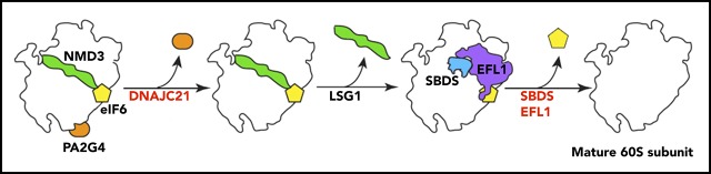
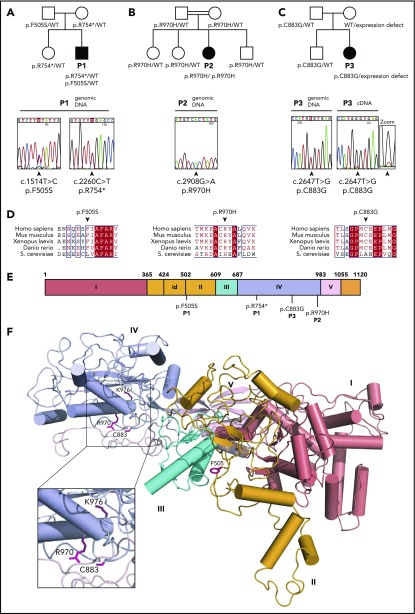
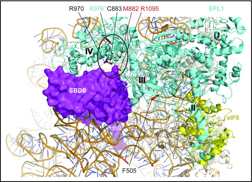
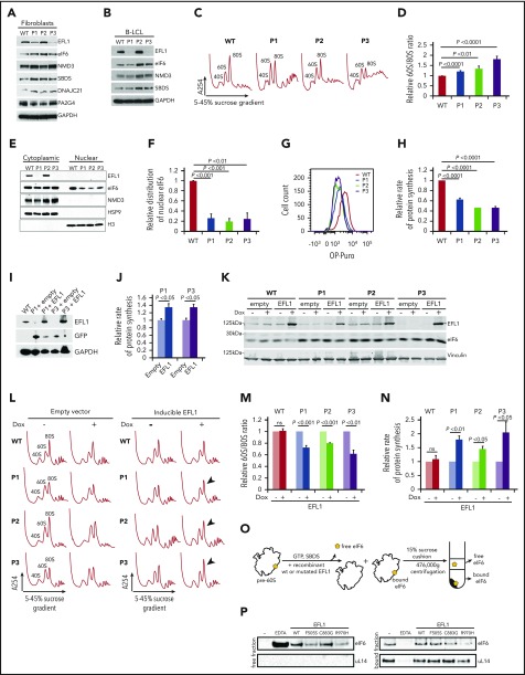
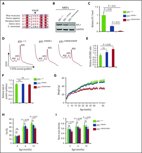
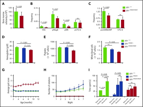
Shwachman-Diamond syndrome (SDS) is a recessive disorder typified by bone marrow failure and predisposition to hematological malignancies. SDS is predominantly caused by deficiency of the allosteric regulator Shwachman-Bodian-Diamond syndrome that cooperates with elongation factor-like GTPase 1 (EFL1) to catalyze release of the ribosome antiassociation factor eIF6 and activate translation. Here, we report biallelic mutations in EFL1 in 3 unrelated individuals with clinical features of SDS. Cellular defects in these individuals include impaired ribosomal subunit joining and attenuated global protein translation as a consequence of defective eIF6 eviction. In mice, Efl1 deficiency recapitulates key aspects of the SDS phenotype. By identifying biallelic EFL1 mutations in SDS, we define this leukemia predisposition disorder as a ribosomopathy that is caused by corruption of a fundamental, conserved mechanism, which licenses entry of the large ribosomal subunit into translation.


Conflict of interest statement

Conflict-of-interest disclosure: The authors declare no competing financial interests.

Figures

None
Graphical abstract
Figure 1.
Figure 1.
Identification of EFL1 mutations in 3 individuals with SDS. Family pedigrees and direct Sanger sequencing of the EFL1 gene in individuals 1 (P1) (A), 2 (P2) (B), and 3 (P3) (C). Arrows indicate the position of the mutations. (D) Multiple sequence alignment of EFL1 proteins from representative species. Identical amino acids are shown in red with white characters, similar amino acids are in red character, and a blue frame represents similarity across groups. (E) Schematic of the domain architecture of human EFL1 showing the position of disease-associated mutations. Domains I to V are indicated; id, insertion domain (residues 424-502) that distinguishes EFL1 from other translational GTPases. (F) Mapping of SDS-associated mutations onto human EFL1 (Protein Data Bank accession number 5anc), shown in ribbon representation. Domains are colored deep salmon (I), light orange (id, II), cyan (III), and light blue (IV). Residue K976 corresponds to K983 mutated in the N-ethyl-N-nitrosourea mutant mouse model (see below). WT, wild-type.
Figure 2.
Figure 2.
SDS-associated EFL1 mutations map to key functional interfaces. SBDS C-terminal domain, magenta, space-filling representation; EFL1, cyan; eIF6, yellow (Protein Data Bank accession number 5anb), ribbon representation. EFL1 domains II, III, and IV are indicated. EFL1 residues targeted by mutations that map to the interface with SBDS are encircled. Residues mutated in SDS are indicated in black (this study) or red (Stepensky et al) text; residue K976 (cyan) is targeted in the mouse model (this study). H69, rRNA helix 69.
Figure 3.
Figure 3.
SDS-associated EFL1 mutations impair eIF6 release. (A) Total cell lysates from fibroblasts and (B) B-LCL from wild-type and 3 individuals (P1, P2, and P3) with SDS were immunoblotted to visualize the indicated proteins. (C) Defective ribosome assembly in EFL1 mutant fibroblasts. Polysome profiles from fibroblast extracts from 3 unrelated individuals with SDS compared with wild-type control. Quantification of the 60S:80S ribosomal subunit ratios is indicated as a bar chart (n ≥ 5) (D). EFL1 genotypes are provided in supplemental Table 1. (E) EFL1 is required for eIF6 recycling in human cells. Indicated proteins were visualized by immunoblotting in cytoplasmic or nuclear fractions from wild-type and EFL1 mutant fibroblasts. Histone H3, nuclear marker; HSP9, cytoplasmic marker. (F) Relative amount of eIF6 in the nucleus of EFL1 mutant cells compared with wild-type (n = 5). (G-H) EFL1 deficiency attenuates protein synthesis. OP-Puro incorporation in fibroblast cells lines from individuals P1, P2, and P3 relative to wild-type control cells quantified 1 hour after OP-Puro administration (n = 6). (I) Complementation of EFL1-deficient fibroblasts with wild-type EFL1. Lysates from EFL1 mutant fibroblasts transduced with a vector expressing GFP alone (+empty) or GFP + EFL1 (+EFL1) and from wild-type fibroblasts as a control. The indicated proteins were visualized by immunoblotting. (J) Wild-type EFL1 rescues global translation in patient fibroblast cell lines from P1 and P3 (n = 4). (K) Complementation of EFL1-deficient fibroblasts with inducible vector allows wild-type EFL1 expression after doxycycline (Dox) treatment. Lysates from EFL1 mutant fibroblasts transduced with an empty vector or EFL1-expressing vector with (+) or without (−) doxycycline. Vinculin is used as a loading control. (L) Comparison of polysome profiles from wild-type and EFL1-mutant fibroblasts transduced with empty vector or inducible EFL1-expression vector treated (+) or not (−) with doxycycline. Arrows indicate increased 80S formation in complemented cells from individuals P1, P2, and P3. (M) Quantification of the 60S:80S ribosomal subunit ratios in cells transduced with inducible EFL1-expression vector treated (+) or not (−) with doxycycline (n = 3). (N) Inducible expression of wild-type EFL1 rescues global protein translation rates in EFL1-deficient fibroblast cell lines (n = 6). (O) Schematic of eIF6 release assay. Pre-60S subunits extracted from P3-derived fibroblasts were incubated with the indicated release factors and pelleted through a 15% sucrose cushion. Immunoblotting reveals the eIF6 distribution in the supernatant (“free”) and pellet (“bound”). (P) Release of eIF6 by SDS-associated EFL1 variants. P3-derived pre-60S subunits were incubated with guanosine triphosphate (GTP), SBDS, and the indicated EFL1 variants. EDTA was added as a positive control for eIF6 release. eIF6 and uL14 were visualized by immunoblotting. (−) indicates the negative control lacking EFL1. All data represent mean ± standard error. Statistical significance between samples was assessed by a 2-tailed Student t test. ns, not significant.
Figure 4.
Figure 4.
Efl1 K983R mutation leads to pleiotropic effects in mice. (A) Multiple sequence alignment of EFL1 proteins from representative species indicates that the murine EFL1 K983R mutation targets a highly conserved residue. Identical amino acids are indicated by a red box with white characters, similar amino acids are in red characters, and a blue frame represents similarity across groups. (B) Representative total cell lysates of mouse embryonic fibroblasts (MEFs) from wild-type (Efl1+/+), heterozygous Efl1+/K983R, and homozygous Efl1K983R /K983R mice were immunoblotted to visualize EFL1 protein. Glyceraldehyde-3-phosphate dehydrogenase (GAPDH) is used as a loading control. (C) Quantifications show the EFL1/glyceraldehyde-3-phosphate dehydrogenase ratio. (D) Representative polysome profiles of MEF extracts from Efl1K983R /K983R (n = 4) and heterozygous Efl1+/K983R (n = 9) mice compared with wild-type control (Efl1+/+) (n = 9). (E) Quantification of the 60S:80S ribosomal subunit ratios is indicated as a bar chart (n ≥ 4). (F) Global protein translation rates in MEFs from Efl1 K983R /K983R and heterozygous Efl1+/K983R mice compared with wild-type control (Efl1+/+) (n = 6). EFL1 K983R mutation reduces body weight from 3 weeks of age (G), affecting the percentage of fat mass (H) and bone mass density (I) from 3 months of age. For all tests, P values are indicated.
Figure 5.
Figure 5.
Impaired hematopoiesis, motor abnormalities, and cognitive deficits in Efl1K983R/K983Rmice. (A) Bone marrow cellularity in wild-type and homozygous Efl1K983R/K983R mice is indicated. (B-C) Frequencies of hematopoietic stem cells (HSCs); megakaryocyte, granulocyte, erythroid progenitors (pMegE); megakaryocyte progenitors (MkP); myeloid progenitors (pre–granulocyte-monocyte/granulocyte-monocyte progenitors); and erythroid progenitors (pCFU-E and CFU-E) in the bone marrow in the indicated genotypes are shown (n = 8 per genotype). EFL1 K983R mutation affects hematopoiesis. Hemoglobin levels (D), platelets (E), and total white blood cell counts (F). (G) Gait abnormalities in K983R mutant mice. Gait was analyzed using a qualitative scoring system at selected time points. Scores are explained in Materials and methods. (H) Motor deficits in EFL1 K983R mutant mice. Free wheel-running activity (over 7 nights, 3 months of age) for the indicated genotypes. (I) Cognitive deficits in K983R mutant mice. Y maze habituation testing of the indicated genotypes at 3 months of age. For all tests, P values are indicated.

References

    1. Warren AJ. Molecular basis of the human ribosomopathy Shwachman-Diamond syndrome. Adv Biol Regul. 2017;S2212-4926(17):30153-30157. - PMC - PubMed
    1. Ruggero D, Shimamura A. Marrow failure: a window into ribosome biology. Blood. 2014;124(18):2784-2792. - PMC - PubMed
    1. Yelick PC, Trainor PA. Ribosomopathies: global process, tissue specific defects. Rare Dis. 2015;3(1):e1025185. - PMC - PubMed
    1. Weis F, Giudice E, Churcher M, et al. . Mechanism of eIF6 release from the nascent 60S ribosomal subunit. Nat Struct Mol Biol. 2015;22(11):914-919. - PMC - PubMed
    1. Donadieu J, Fenneteau O, Beaupain B, et al. ; Associated investigators of the French Severe Chronic Neutropenia Registry* . Classification of and risk factors for hematologic complications in a French national cohort of 102 patients with Shwachman-Diamond syndrome. Haematologica. 2012;97(9):1312-1319. - PMC - PubMed

Publication types

MeSH terms