. 2019 Jun 21;146(13):dev170522.
doi: 10.1242/dev.170522.

SOX2 is required for inner ear growth and cochlear nonsensory formation before sensory development

Affiliations

SOX2 is required for inner ear growth and cochlear nonsensory formation before sensory development

Aleta R Steevens et al. Development. .

Abstract

The transcription factor sex determining region Y-box 2 (SOX2) is required for the formation of hair cells and supporting cells in the inner ear and is a widely used sensory marker. Paradoxically, we demonstrate via fate mapping that, initially, SOX2 primarily marks nonsensory progenitors in the mouse cochlea, and is not specific to all sensory regions until late otic vesicle stages. SOX2 fate mapping reveals an apical-to-basal gradient of SOX2 expression in the sensory region of the cochlea, reflecting the pattern of cell cycle exit. To understand SOX2 function, we undertook a timed-deletion approach, revealing that early loss of SOX2 severely impaired morphological development of the ear, whereas later deletions resulted in sensory disruptions. During otocyst stages, SOX2 shifted dramatically from a lateral to medial domain over 24-48 h, reflecting the nonsensory-to-sensory switch observed by fate mapping. Early loss or gain of SOX2 function led to changes in otic epithelial volume and progenitor proliferation, impacting growth and morphological development of the ear. Our study demonstrates a novel role for SOX2 in early otic morphological development, and provides insights into the temporal and spatial patterns of sensory specification in the inner ear.

Keywords: Cochlea; Inner ear; Mouse; Otocyst; SOX2; Sensory; Specification.


Figures

Fig. 1.
Fig. 1.
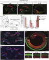
SOX2-expressing cells initially contribute to nonsensory regions in the cochlea but later contribute exclusively to the cochlear floor regions, including organ of Corti. (A-C) Cross-section through the E18.5 cochlea showing tdT/SOX2 expression and hair cell labeling (MYO6). (A) E8.5 tdT/SOX2-expressing cells primarily contributed to the roof of the cochlear duct with the exception of a few interdental (id) cells (arrows). oC, organ of Corti (bracket). (B) E10.5 tdT/SOX2 was downregulated in some cells in the roof of the cochlear duct (arrowheads), and expanded into the floor of the nonsensory cochlea, including the id cell region and some cells in Kölliker's organ (Ko, bracket). (C) By E12.5, expression of tdT/SOX2 was exclusively in the floor of the cochlea, including the oC. (D) Illustration of a cochlea cross-section with respect to the entire inner ear anatomy. (E) The area of nonsensory and sensory regions labeled by tdT/SOX2 was quantified. A trend is apparent, in which SOX2 initially contributes to the nonsensory regions, but switches over time to contribute exclusively to the floor of the cochlea. Comparison of roof nonsensory E8.5 tdT/SOX2 with E8.5 floor nonsensory tdT/SOX2: ****P<0.00001. Comparison of nonsensory E8.5 tdT/SOX2 with E8.5 sensory tdT/SOX2: ****P<0.00001. Comparison of roof nonsensory E12.5 tdT/SOX2 with E12.5 floor nonsensory tdT/SOX2: **P=0.004. Comparison roof nonsensory E12.5 tdT/SOX2 with E12.5 sensory tdT/SOX2: ****P<0.00001. Significance determined using a two-tailed Student's t-test. ns, not significant. Data are mean±s.e.m. (F-G) Midmodiolar regions in Sox2CreERT2/+ E18.5 control cochleae showed that the contribution of tdT/SOX2-expressing cells to the cochlea at E8.5 (F) and E10.5 (G) decreased along the apical–basal axis. At E8.5, tdT/SOX2 was largely excluded from the oC (bracket), with the exception of the apical domain, in which a much smaller negative floor region is observed (arrow) (F). At E10.5, tdT/SOX2 expression expanded along the cochlear floor but remained excluded from the oC in the middle and basal turns (I-bars) (G). (H) Whole-mount cochlea showed E10.5 tdT/SOX2 expression with respect to the sensory region. Boxed areas from different apical and/or basal regions are shown at a higher magnification below. In the apex, tdT/SOX2 was extensively expressed throughout the sensory region, but was gradually excluded more basally (I-bar). IHC, inner hair cell; OHC, outer hair cell. Scale bars: 100 µm.
Fig. 2.
Fig. 2.
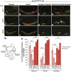
SOX2-expressing cells contribute initially to both sensory and nonsensory regions in the vestibule, but later are specific to the sensory regions. (A-L) Cross-sections through the E18.5 vestibular regions showing tdT/SOX2 expression and hair cell labeling (MYO6). (A-D) E8.5 tdT/SOX2-expressing cells (red) contributed widely to both nonsensory tissue and sensory regions (marked by MYO6, green). (E-H) The extent of E10.5 tdT/SOX2-expressing cells marking nonsensory tissue was reduced, and more cells contributed to the sensory regions. (I-L) E12.5 tdT/SOX2 fate-mapped cells almost exclusively contributed to the sensory regions in the vestibule. (M) Illustration of the vestibular regions shown in cross-section. (N) Quantification of the area of tdT/SOX2 expression in nonsensory and sensory regions over time showed a substantial contribution of SOX2 to vestibular nonsensory regions that decreased over time; by contrast, labeling in the sensory regions increased over time and became specific to these regions. No significant difference was detected in sensory tdT/SOX2 in the vestibule over time. Comparison of nonsensory tdT/SOX2 in the saccule: E8.5-E10.5, *P<0.05; E8.5-E12.5, **P<0.001; E10.5-E12.5, ****P<0.0001. Comparison of nonsensory tdT/SOX2 in the utricle: E8.5-E10.5, ns; E8.5-E12.5, *P<0.05; E10.5-E12.5, *P<0.05. Comparison of nonsensory tdT/SOX2 in the ampullae: E8.5-E10.5, **P<0.001; E8.5-E12.5, ****P<0.0001; E10.5-E12.5, ***P<0.001. Significance determined using a two-tailed Student's t-test. Data are mean±s.e.m. ns, not significant. Scale bar: 100 µm.
Fig. 3.
Fig. 3.
Paint-filling reveals a severe inner ear malformation resulting from the early deletion of SOX2, whereas later deletion has little effect on the overall morphology. (A-F) Paint-filled E15.5 inner ears with SOX2 deletion at indicated time points during development. Control (A) and two examples of E8.5 Sox2-deleted mutants (B,C) demonstrate severe loss of otic tissue caused by early loss of SOX2. (D,E) Medial and lateral views of an E10.5-deleted mutant show loss of the lateral and posterior ampullae, smaller maculae and an undercoiled cochleae. (F) Deletion of SOX2 at E12.5 did not show overt morphological defects. The smaller saccule (sac) is marked by an arrow (B-E), the utricle (ut) is marked by an arrowhead (D,E), missing ampullae or truncated canals are indicated by an asterisk (B-E). aa, anterior ampulla; asc, anterior semicircular canal; cd, cochlear duct; ed, endolymphatic duct; la, lateral ampulla; lsc, lateral semicircular canal; pa, posterior ampulla; psc, posterior semicircular canal. Scale bar: 500 μm.
Fig. 4.
Fig. 4.
Despite severe morphological defects in early Sox2-deleted cochlea, sensory development still occurs. (A) Time points for deletion (E8.5 or E10.5) and harvest (E18.5). (B-G″) Sections through midmodiolar regions of E18.5 ears. (B) Midmodiolar section in Sox2+/+ controls. The number of openings seen in cross-section reflects the length of the cochlear spiral. Loss of SOX2 at E8.5 (C) or E10.5 (D) resulted in shortened cochleae that still developed sensory regions in the more basal regions, although the patterning was abnormal. Sensory formation was abolished in more apical cochlear turns (arrows) in which SOX2 was deleted. Hair cells only formed where SOX2 was present in the otic epithelium (E-G″, magnification of boxed areas in C and D). Scale bars: 100 µm (in D, representing B-D); 50 µm (in F″, representing E-G″).
Fig. 5.
Fig. 5.
Later deletion of SOX2 severely impairs sensory development in the cochlea but has little impact on cochlear morphology. (A) Time point for deletion (E12.5) and harvest (E18.5). (B) Low power section through the midmodiolar region of a Sox2CreER/+ control at E18.5 shows that E12.5 tdT/SOX2 contributed exclusively to the floor of the cochlear duct in all turns, overlapping with the normal SOX2 protein expression in the supporting cells (white). (C) Section through an E12.5 Sox2-deleted mutant shows relatively normal cochlear morphology but an absence of sensory formation. The missing ganglia in the mutant (tdT labeled in the modiolus in B, marking SOX2-expressing glia at E18.5) probably occurred because of the missing sensory regions, given that it was present earlier at E14.5 (not shown). Magnification of the cochlear turns from the boxed areas in control (D-G) and E12.5-deleted Sox2 mutant (H-K) show the absence of the organ of Corti in all turns, with the exception of an occasional abnormally shaped MYO6-positive cell in the basal turn (H). GER, greater epithelial ridge; id, interdental cell area; LER, lesser epithelial ridge; oC, organ of Corti. Scale bars: 100 µm (in C, representing B,C); 50 µm (K, representing D-K).
Fig. 6.
Fig. 6.
Marker analysis in the cochlea indicates that SOX2 is required for both prosensory specification and sensory cell differentiation. (A) Time points for deletion (E10.5 or E12.5) and harvest (E14.5). (B-B″) Control E14.5 midmodiolar region shows typical expression of JAG1 and CDKN1B (marking the future organ of Corti). Unlike JAG1, CDKN1B expression was not always present in control cochlea in the apex. (C-C″) Representative sections of a midmodiolar region of E14.5 cochlea after deletion at E10.5, with indicated prosensory markers and tdT, reflecting SOX2 expression at E10.5. JAG1 expression was mildly expanded in the basal turn of the cochlea (compare brackets in C′ and B′ control) and was significantly downregulated from the middle turn (asterisk) (C′). The shifted JAG1 expression in the apical domain was frequently observed in controls, probably because of a difference in the plane of sectioning rather than a shifted domain. CDKN1B expression was also downregulated in the middle turn (arrowhead) (C″). (D-D″) Representative section of an E14.5 midmodiolar cochlea deleted for SOX2 at E12.5. After deletion of SOX2 at E12.5, JAG1 expression was present in all turns, although, again, mildly expanded in the basal turn of the cochlea (compare bracket in D′ with B′ control). In the absence of SOX2 at E12.5, CDKN1B expression was significantly downregulated in both middle and basal turns (arrowheads) (D″). Arrows and arrowheads indicate either the presence or absence of CDKN1B, respectively. Scale bar: 100 µm.
Fig. 7.
Fig. 7.
Early SOX2 deletion results in both nonsensory and sensory loss in the vestibule, whereas later deletion has few effects on the vestibular regions. (A-D) Sections through the vestibular organs of an E18.5 control. Sensory regions are marked by MYO6 (hair cells) and SOX2 (supporting cells). (E-H) In the absence of SOX2 at E8.5, the vestibule largely failed to form, with the exception of a very small saccule (E), which contained a few MYO6- and SOX2-positive cells. Asterisks indicate missing vestibular sensory organs (F-H). (I-L) The vestibule from E10.5 deletion resulted in a significantly underdeveloped saccule (J), a smaller utricle devoid of sensory formation (K), a posterior crista lacking an ampulla and fused to the common crus (arrow), and a missing anterior crista and ampulla (asterisk) (L). (M-P) Deletion of SOX2 at E12.5 did not produce an overt phenotype in any of the vestibular organs. SCC, semicircular canal. Scale bar: 100 µm (in P, representing A-P).
Fig. 8.
Fig. 8.
Analysis of NEUROG1-deficient ears indicates that the nonsensory defects are not caused secondarily from reductions in the otic ganglion. (A-C) Low-power midmodiolar sections from control (A), Neurog1−/ (B) and E8.5 Sox2-deleted cochleae (C). The nonsensory phenotype was more severe in the absence of E8.5 SOX2 than of NEUROG1, and some neuronal formation still occurred [arrows mark neuronal expression (TUJ1; white) in C,F,I]. (D-K) Higher magnification views of the boxed regions in A-C highlight that sensory formation occurred fairly normally in the Neurog1-deficient mutant. (L-T) Examples of cross-sections through each vestibular organ in control (L-O), Neurog1−/ mutant (P-S) and E8.5 SOX2-deleted inner ears (T). The vestibule forms relatively normally in Neurog1-deficient mutants, with the exception of a smaller saccule devoid of sensory markers (arrow, P). By contrast, the only vestibular structure seen in E8.5 SOX2-deficient inner ears was an underdeveloped saccule (T). Scale bars: 50 µm.
Fig. 9.
Fig. 9.
Simultaneous fate mapping and expression of SOX2 demonstrate a dramatic shift from nonsensory to sensory progenitors in the otocyst between E8.5 and E11.5. (A-C″) Sections at indicated ages demonstrating previous fate-mapped SOX2 expression (tdT; red) versus current SOX2 expression (green). The SOX2 expression domain switched from a lateral, primarily nonsensory associated tdT/SOX2 domain at E8.5/E9.5 (A-B′) to a medial, sensory-associated domain of SOX2 protein expression at E11.5 (A″,B″,C″). (B′) The contribution of E9.5 SOX2-expressing cells labeled by tdT/SOX2 decreased in the lateral otocyst (arrowhead), and increased medially (B″). At E10.5, the contribution of tdT/SOX2 to nonsensory progenitors in the cochlear roof decreased (arrowheads in C′ and E) and began to contribute to cochlear floor nonsensory progenitors (arrows in C′ and E). (D-F) Demonstration of how the pattern of early SOX2 expression in the otocyst contributes to the mature cochlea. The region in the posterior medial otocyst negative for tdT/SOX2 tdT early on (dashed lines in A′,B′,C′) probably contributes to the future organ of Corti (bracket in D), which was positive by E11.5 (solid lines in B″,C″,F). M, medial; OV, otic vesicle; P, posterior. Scale bars: 100 µm.
Fig. 10.
Fig. 10.
SOX2 is required for otic growth and progenitor proliferation and/or viability. (A-F) Sections through E10.5 otocysts of a Sox2+/+ control (A-C) and an E8.5 Sox2CreERT2/fl mutant (D-F) showing a decrease in the proliferation marker pHH3 along the dorsoventral axis (D↔V) in Sox2-deficient mutants. Brackets indicate the thickness of the otic epithelium. (G-H′) The increase in cleaved caspase 3 in the E8.5 Sox2-deleted mutant. Insets show magnification of areas within dashed boxes that display caspase 3-expressing cells in areas where SOX2 was deleted. (I) Quantifications of the reductions in otic epithelium volume (Sox2+/+ n=15; Sox2CreERT2/fl n=12; *P<0.05), pHH3-expressing cells (Sox2+/+ n=9; Sox2CreERT2/fl n=6; ****P<0.0001) and a corresponding increase in caspase 3 (Sox2+/+ n=6; Sox2CreERT2/fl n=6; **P<0.01) in Sox2CreERT2/fl mutants after deletion at E8.5 compared with controls. Significance determined using a two-tailed Student's t-test. Data are mean±s.e.m. L, lateral; P, posterior. Scale bars: 100 µm.

Similar articles

Cited by

References

    1. Abelló G., Khatri S., Radosevic M., Scotting P. J., Giráldez F. and Alsina B. (2010). Independent regulation of Sox3 and Lmx1b by FGF and BMP signaling influences the neurogenic and non-neurogenic domains in the chick otic placode. Dev. Biol. 339, 166-178. 10.1016/j.ydbio.2009.12.027 - DOI - PubMed
    1. Adams M. E., Hurd E. A., Beyer L. A., Swiderski D. L., Raphael Y. and Martin D. M. (2007). Defects in vestibular sensory epithelia and innervation in mice with loss of Chd7 function: implications for human CHARGE syndrome. J. Comp. Neurol. 504, 519-532. 10.1002/cne.21460 - DOI - PubMed
    1. Ahmed M., Wong E. Y. M., Sun J., Xu J., Wang F. and Xu P.-X. (2012a). Eya1-Six1 interaction is sufficient to induce hair cell fate in the cochlea by activating Atoh1 expression in cooperation with Sox2. Dev. Cell 22, 377-390. 10.1016/j.devcel.2011.12.006 - DOI - PMC - PubMed
    1. Ahmed M., Xu J. and Xu P.-X. (2012b). EYA1 and SIX1 drive the neuronal developmental program in cooperation with the SWI/SNF chromatin-remodeling complex and SOX2 in the mammalian inner ear. Development 139, 1965-1977. 10.1242/dev.071670 - DOI - PMC - PubMed
    1. Amador-Arjona A., Cimadamore F., Huang C. T., Wright R., Lewis S., Gage F. H. and Terskikh A. V. (2015). SOX2 primes the epigenetic landscape in neural precursors enabling proper gene activation during hippocampal neurogenesis. Proc. Natl. Acad. Sci. USA 112, E1936-E1945. 10.1073/pnas.1421480112 - DOI - PMC - PubMed

Publication types

MeSH terms

Substances