. 2019 Jul 18;39(7):BSR20190763.
doi: 10.1042/BSR20190763. Print 2019 Jul 31.

Down-regulation of microRNA-138 improves immunologic function via negatively targeting p53 by regulating liver macrophage in mice with acute liver failure

Affiliations

Down-regulation of microRNA-138 improves immunologic function via negatively targeting p53 by regulating liver macrophage in mice with acute liver failure

You-Qiang Wang et al. Biosci Rep. .

Retraction in

Abstract

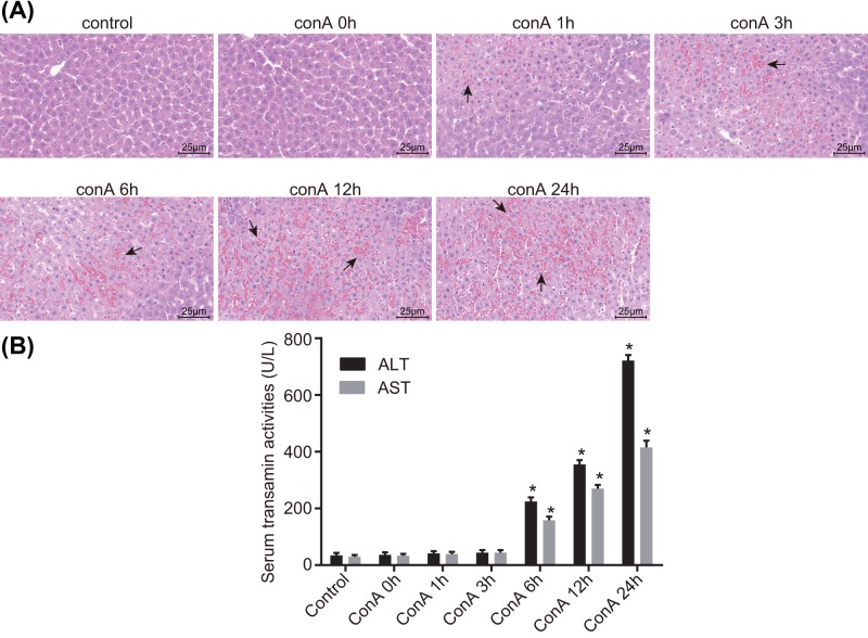
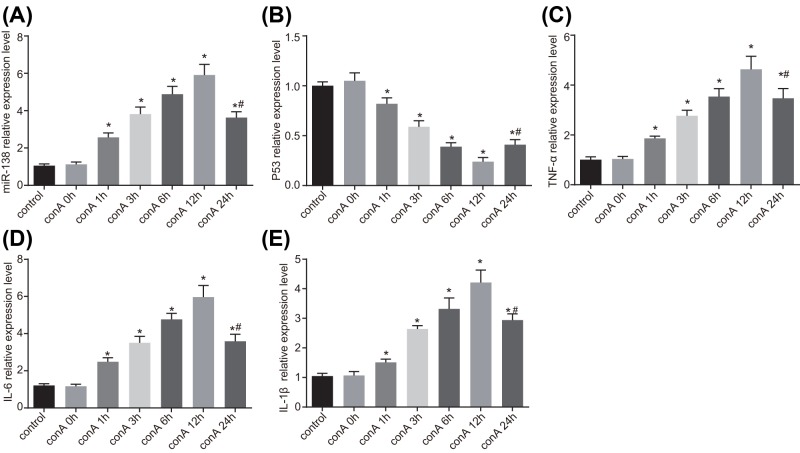
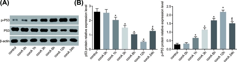
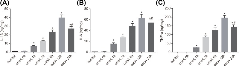
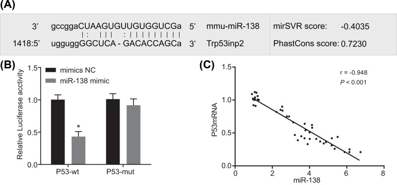
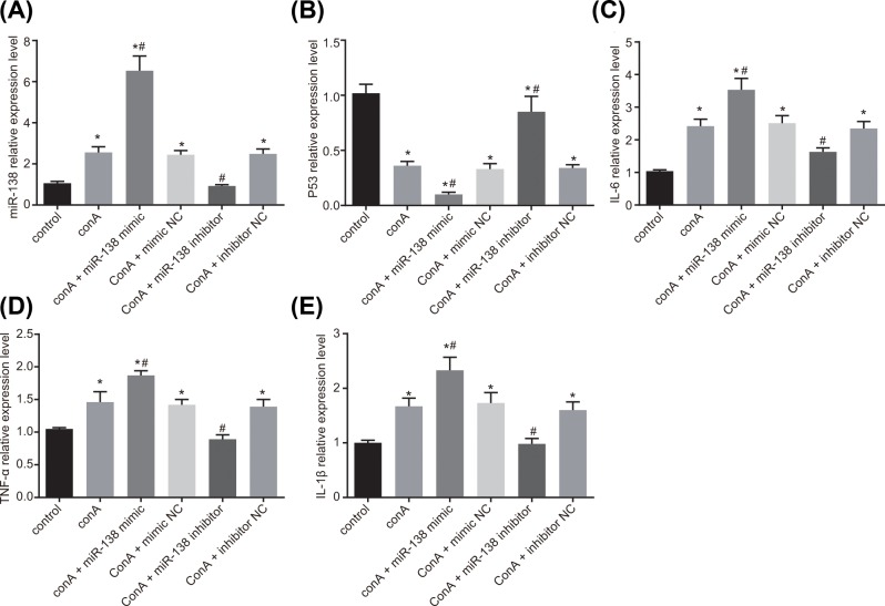
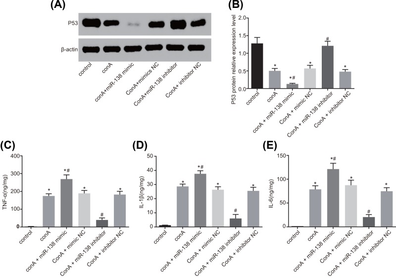
MicroRNAs (miRNAs) have been frequently identified as key mediators in almost all developmental and pathological processes, including those in the liver. The present study was conducted with aims of investigating the role of microRNA-138 (miR-138) in acute liver failure (ALF) via a mechanism involving p53 and liver macrophage in a mouse model. The ALF mouse model was established using C57BL/6 male mice via tail vein injection of Concanamycin A (Con A) solution. The relationship between miR-138 and p53 was tested. The mononuclear macrophages were infected with mimic and inhibitor of miR-138 in order to identify roles of miR-138 in p53 and levels of inflammatory factors. Reverse transcription quantitative polymerase chain reaction (RT-qPCR), Western blot analysis and ELISA were conducted in order to determine the levels of miR-138, inflammatory factors, and p53 during ALF. The results showed an increase in the levels of miR-138 and inflammatory factors in ALF mice induced by the ConA as time progressed and reached the peak at 12 h following treatment with ConA, while it was on the contrary when it came to the level of p53. Dual-luciferase reporter gene assay revealed that p53 was a target gene of miR-138. Furthermore, the results from the in vitro transfection experiments in primary macrophages of ALF mouse showed that miR-138 down-regulated p53 and enhanced levels of inflammatory factors; thus, improving immune function in ALF mice. In conclusion, by negatively targeting p53, the decreased miR-138 improves immunologic function by regulating liver macrophage in mouse models of ALF.

Keywords: Acute liver failure; Immune function; Macrophage; P53; microRNA-138.


Conflict of interest statement

The authors declare that there are no competing interests associated with the manuscript.

Figures

Figure 1
Figure 1. ConA induced ALF in mouse
HE staining images of liver tissues of mice in each group (yellow arrows indicate necrotic inflammatory cells) (400×) (A). The serum ALT and AST levels of mice in each group (B). The control group: liver tissues without treatment. The ConA treatment groups were tissue-stained after their processing time. n=6. *P<0.05 vs the control group. ConA, Concanamycin A
Figure 2
Figure 2. The primary liver macrophages were identified in mice
Positive liver macrophage ratio labeled by IgG2b after mice were treated with ConA for 12 h determined by flow cytometry (A). The positive liver macrophage ratio labeled by F4/80 after mice were treated with ConA for 12 h by flow cytometry (B). The positive liver macrophage ratio labeled by IgG2b in mice of the control group by flow cytometry (C). The positive liver macrophage ratio labeled by F4/80 in mice of the control group by flow cytometry (D). n=3. ConA, Concanamycin A.
Figure 3
Figure 3. miR-138 expression and mRNA levels of TNF-α, IL-6, and IL-1β were increased, while mRNA level of p53 was decreased in liver macrophages
The miR-138 level in response to ConA 0 h, ConA 1 h, ConA 3 h, ConA 6 h, ConA 12 h, ConA 24 h (A). The mRNA level of p53 in response to ConA 0 h, ConA 1 h, ConA 3 h, ConA 6 h, ConA 12 h, ConA 24 h (B). The mRNA level of TNF-α in response to ConA 0 h, ConA 1 h, ConA 3 h, ConA 6 h, ConA 12 h, ConA 24 h (C). The mRNA level of IL-6 in response to ConA 0 h, ConA 1 h, ConA 3 h, ConA 6 h, ConA 12 h, ConA 24 h (D). The mRNA level of IL-1β in response to ConA 0 h, ConA 1 h, ConA 3 h, ConA 6 h, ConA 12 h, ConA 24 h (E). The ConA treatment groups were cell-treated after their processing time. *P<0.05 vs the control group. #P<0.05 vs the ConA 12 h group. n=6. One-way analysis of variance was used for multi-group comparisons followed by Tukey’s post hoc test.ConA, Concanamycin A.
Figure 4
Figure 4. Protein levels of p53 were reduced following treatment of ConA
The protein level of p53 and the extent of p53 phosphorylation in primary macrophages of ALF mice (A). The gray value of p53 protein bands (B). The ConA treatment groups were cells treated after their processing time. *P<0.05 vs the control group. #P<0.05 vs the ConA 12 h group. n=6. One-way analysis of variance was used for multi-group comparisons followed by Tukey’s post hoc test. ConA, Concanamycin A.
Figure 5
Figure 5. Protein level of p53 after ConA treatment in ALF mice
Increased levels of TNF-α (C), IL-6 (B), and IL-1β (A) after treatment of ConA. The ConA treatment groups were cells treated after their processing time. *P<0.05 vs the control group. #P<0.05 vs the ConA 12 h group. n=6. One-way analysis of variance was used for multi-group comparisons followed by Tukey’s post hoc test. ConA, Concanamycin A.
Figure 6
Figure 6. miR-138 targets p53
The binding sites of miR-138 and p53-3′UTR predicted by public databases (A). The dual-luciferase reporter assay for confirmation of the targeting relationship between miR-138 and p53 (B). Pearson’s analysis of the relationship between miR-138 and p53 (the data from panel 3A and B) (C). *P<0.05 vs the NC group. The experiment was repeated 3 times. The cells in each group were detected 24 h after transfection. t test was used in comparisons between two groups. ConA, Concanamycin A.
Figure 7
Figure 7. Elevated mRNA levels of TNF-α, IL-6, and IL-1β and reduced level of p53 after infection of miR-138 inhibitor in RAW264.7 cells
The miR-138 level of cells treated with ConA alone and with miR-138 mimic or inhibitor (A). The mRNA level of p53 of cells treated with ConA alone and with miR-138 mimic or inhibitor (B). The mRNA level of IL-6 of cells treated with ConA alone and with miR-138 mimic or inhibitor (C). The mRNA level of TNF-α of cells treated with ConA alone and with miR-138 mimic or inhibitor (D). The mRNA level of IL-1β of cells treated with ConA alone and with miR-138 mimic or inhibitor (E). *P<0.05 vs the control group; #P<0.05 vs the ConA group. The experiment was repeated 3 times. The cells in each group were detected 24 h after transfection. One-way analysis of variance was used for multi-group comparisons followed by Tukey’s post hoc test. ConA, Concanamycin A.
Figure 8
Figure 8. Protein level of p53 was decreased by miR-138
The p53 protein level in cells among the six groups (A). The gray value of p53 protein bands using the Image J software (B). The TNF-α protein level in cells among the six groups (C). The IL-6 protein level in cells among the six groups (D). The IL-1β protein level in cells among the six groups (E). *P<0.05 vs the control group. #P<0.05 vs the ConA group. The experiment was repeated 3 times. The cells in each group were detected 24 h after transfection. One-way analysis of variance was used for multi-group comparisons followed by Tukey’s post hoc test. ConA, Concanamycin A.

Similar articles

Cited by

References

    1. Sugawara K., Nakayama N. and Mochida S. (2012) Acute liver failure in Japan: definition, classification, and prediction of the outcome. J. Gastroenterol. 47, 849–86110.1007/s00535-012-0624-x - DOI - PMC - PubMed
    1. Wei G., Bergquist A., Broome U., Lindgren S., Wallerstedt S., Almer S.. et al. (2007) Acute liver failure in Sweden: etiology and outcome. J. Intern. Med. 262, 393–40110.1111/j.1365-2796.2007.01818.x - DOI - PubMed
    1. Chen J.G. and Zhang S.W. (2011) Liver cancer epidemic in China: past, present and future. Semin. Cancer Biol. 21, 59–6910.1016/j.semcancer.2010.11.002 - DOI - PubMed
    1. Siveen K.S., Nguyen A.H., Lee J.H., Li F., Singh S.S., Kumar A.P.. et al. (2014) Negative regulation of signal transducer and activator of transcription-3 signalling cascade by lupeol inhibits growth and induces apoptosis in hepatocellular carcinoma cells. Br. J. Cancer 111, 1327–133710.1038/bjc.2014.422 - DOI - PMC - PubMed
    1. Bosoi C.R. and Rose C.F. (2013) Brain edema in acute liver failure and chronic liver disease: similarities and differences. Neurochem. Int. 62, 446–45710.1016/j.neuint.2013.01.015 - DOI - PubMed

Publication types