. 2019 Aug;86(2):279-292.
doi: 10.1002/ana.25512. Epub 2019 Jun 14.

Different microvascular alterations underlie microbleeds and microinfarcts

Affiliations

Different microvascular alterations underlie microbleeds and microinfarcts

Susanne J van Veluw et al. Ann Neurol. 2019 Aug.

Abstract

Objective: Cerebral amyloid angiopathy (CAA) is characterized by the accumulation of amyloid β (Aβ) in the walls of cortical vessels and the accrual of microbleeds and microinfarcts over time. The relationship between CAA severity and microbleeds and microinfarcts as well as the sequence of events that lead to lesion formation remain poorly understood.

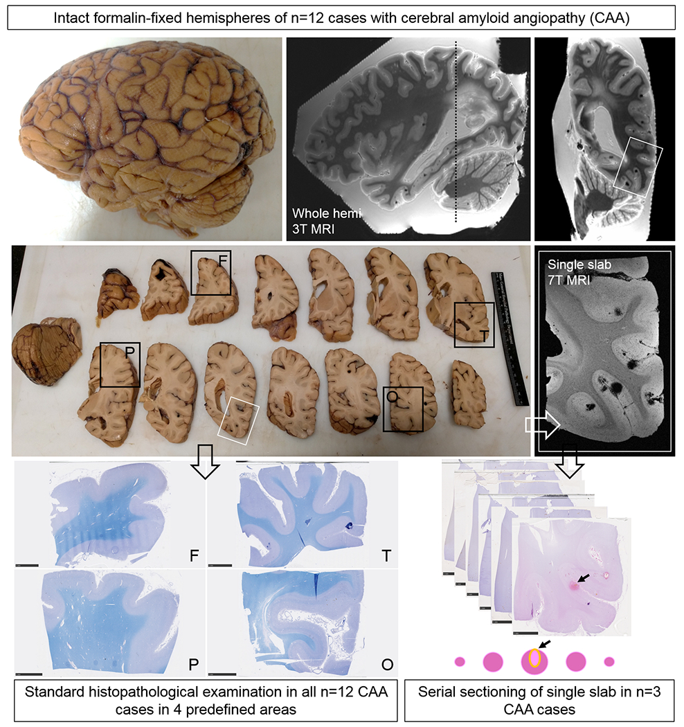
Methods: We scanned intact formalin-fixed hemispheres of 12 CAA cases with magnetic resonance imaging (MRI), followed by histopathological examination in predefined areas and serial sectioning in targeted areas with multiple lesions.

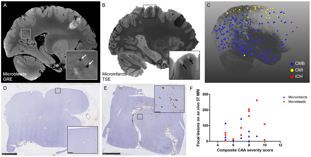
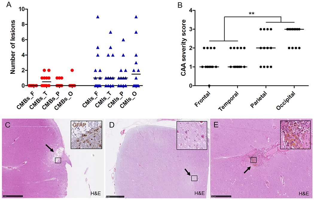
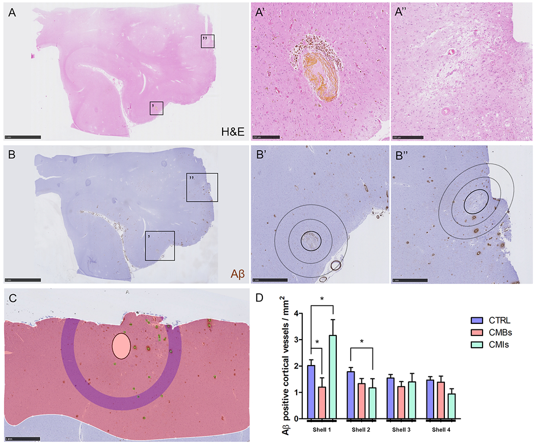
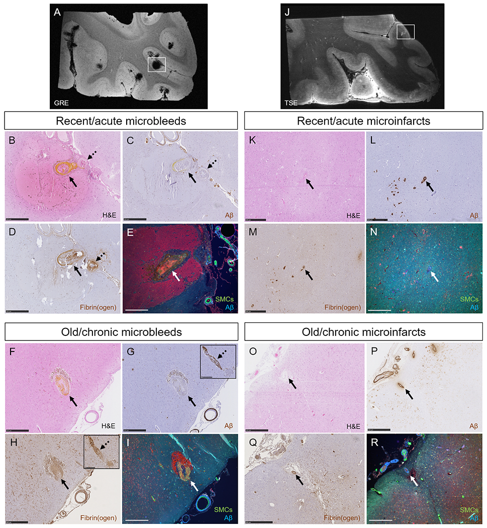
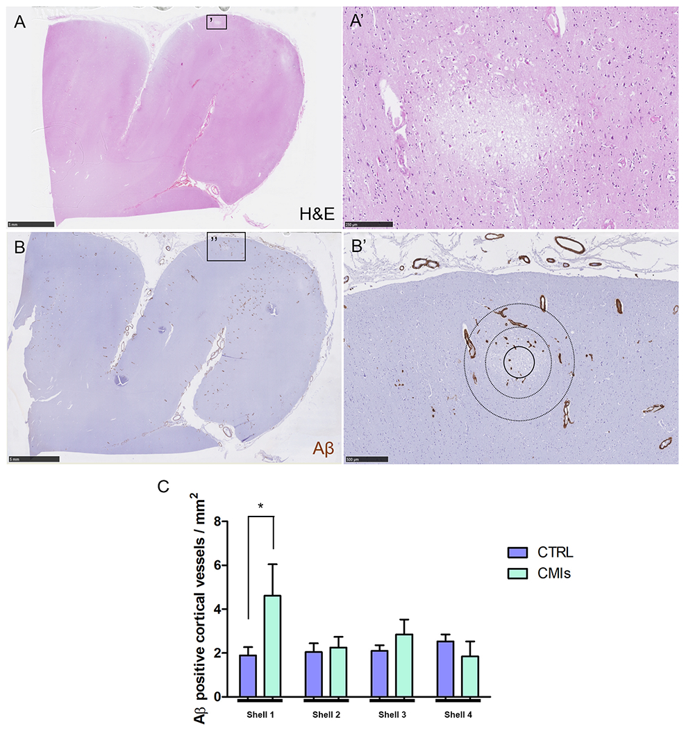
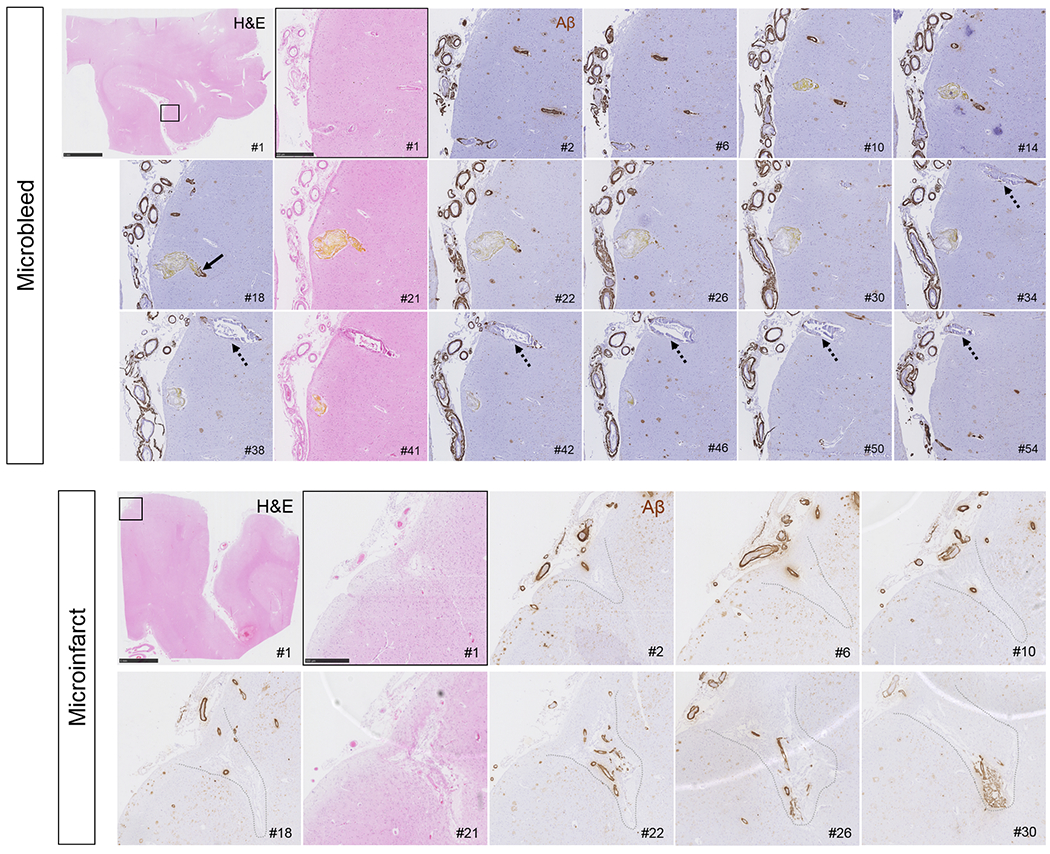
Results: In total, 1,168 cortical microbleeds and 472 cortical microinfarcts were observed on ex vivo MRI. Increasing CAA severity at the whole-brain or regional level was not associated with the number of microbleeds or microinfarcts. However, locally, the density of Aβ-positive cortical vessels was lower surrounding a microbleed compared to a simulated control lesion, and higher surrounding microinfarcts. Serial sectioning revealed that for (n = 28) microbleeds, both Aβ (4%) and smooth muscle cells (4%) were almost never present in the vessel wall at the site of bleeding, but Aβ was frequently observed upstream or downstream (71%), as was extensive fibrin(ogen) buildup (87%). In contrast, for (n = 22) microinfarcts, vascular Aβ was almost always observed at the core of the lesion (91%, p < 0.001) as well as upstream or downstream (82%), but few vessels associated with microinfarcts had intact smooth muscle cells (9%).

Interpretation: These observations provide a model for how a single neuropathologic process such as CAA may result in hemorrhagic or ischemic brain lesions potentially through 2 different mechanistic pathways. ANN NEUROL 2019;86:279-292.


Conflict of interest statement

Potential conflicts of interest

Nothing to report.

Figures

Figure 1.
Figure 1.
Study design. Intact formalin-fixed hemispheres of 12 CAA cases were subjected to high-resolution ex vivo 3T MRI to detect microbleeds and microinfarcts. Next, the hemispheres were cut in 10 mm-thick coronal slabs and samples were taken from pre-defined standard areas of frontal (F), temporal (T), parietal (P), and occipital (O) cortex, to fit a standard tissue cassette (black squares). Four adjacent 6 μm-thick sections were cut from these samples and stained with hematoxylin & eosin (H&E) or Luxol fast blue H&E (depicted here) and underwent immunohistochemistry to detect Aβ and GFAP. Finally, three additional samples were taken from three cases in an area with multiple microbleeds (n=2, from temporal cortex) or microinfarcts (n=1, from parieto-occipital cortex) guided by the ex vivo 3T MR images (white square). These samples underwent ultra-high-resolution ex vivo 7T MRI scanning to confirm the high number of lesions in those areas. After 7T MRI, the samples were cut in half to fit two standard tissue cassettes and underwent complete serial sectioning. Sections 1, 21, 41, 61 etc. were stained with H&E to identify microbleeds and microinfarcts, and sections 2, 6, 10, 14 etc. underwent immunohistochemistry against Aβ. If present, the vessel that could be traced through the core of the lesion was identified as the presumed culprit vessel. In addition, for each identified lesion on H&E, sections adjacent to a microbleed or microinfarct underwent immunohistochemistry to detect fibrin(ogen) or SMCs.
Figure 2.
Figure 2.
Whole-brain associations of microbleeds and microinfarcts with CAA severity. Cortical microbleeds were assessed on the ex vivo 3T MRI gradient-echo (GRE) scans (A) and cortical microinfarcts on the ex vivo 3T MRI T2-weighted turbo-spin echo (TSE) scans (B). The projection of annotated lesions on a 3D reconstruction of a representative hemisphere (case #2) demonstrates that microbleeds (CMB, blue dots) were more often observed in posterior parts of the brain, whereas microinfarcts (CMI, yellow dots) frequently involved the areas of the brain perfused by end arteries (C). Note: red dots are areas affected by intracerebral hemorrhages (ICH). CAA severity was assessed on standard sections from frontal (D, example of score 1), temporal, parietal, and occipital (E, example of score 3) cortex to create a composite CAA severity score. Neither the number of microbleeds (red circles, Spearman’s ρ 0.426, p=0.17) nor the number of microinfarcts (blue triangles, Spearman’s ρ −0.278, p=0.38) were associated with composite CAA severity score (F). Scale bar in D and E = 5 mm.
Figure 3.
Figure 3.
Regional associations on standard histopathologic examination of microbleeds and microinfarcts with CAA severity. On average, CAA severity across all 12 cases followed an anterior-to-posterior distribution (B), whereas number of histopathologically-observed microbleeds and microinfarcts did not (A), explaining weak regional associations. From the total number of 94 microinfarcts observed on standard histopathology, 61 (65%) were considered old/chronic based on GFAP positivity (inset) (C, this example follows the perfusion area of a penetrating cortical vessel) and 33 (35%) recent/acute based on the presence of ‘red’ neurons (inset) (D, this example is located deeper (within cortical layer III-VI)). From the total number of 13 microbleeds observed on standard histopathology, 12 (92%) were considered old/chronic based on the presence of hemosiderin-containing macrophages (inset) (E), and 1 (8%) recent/acute (not shown). Median is indicated in A and B. Scale bar in C and D = 1 mm, scale bar in E = 500 μm.
Figure 4.
Figure 4.
Local associations of microbleeds and microinfarcts with Aβ positive cortical vessels. Based on the H&E-stained serial sections (A) from the additional samples taken from the temporal cortex in two CAA cases, 28 microbleeds (A’) and 18 microinfarcts (A’’) were included. Lesions were localized on the adjacent Aβ-stained sections (B) to perform Sholl analysis (B’B’’, inner circle with solid outline indicates masked area, circles with dotted outlines indicate first two shells). After masking of the lesion, Aβ positive cortical vessels were manually annotated (green markers), the cortical ribbon was outlined (red shaded area), and the density of Aβ positive cortical vessels was generated by the software for four concentric circles extending from the outer border of the masked area (C, purple concentric circle in this example is shell 4). Significantly fewer Aβ positive cortical vessels were observed in the first shell immediately adjacent to a microbleed compared to a simulated control lesion (p=0.023), whereas more Aβ positive cortical vessels were observed in the first shell immediately adjacent to a microinfarct compared to a simulated control lesion (p=0.054) (D). For the second shell significantly fewer Aβ positive cortical vessels were observed for microinfarcts compared to simulated control lesions (p=0.039). Scale bar in A and B = 5 mm, scale bar in B’, B’’, and C = 1 mm, scale bar in A’ and A’’ = 250 μm. Error bars in D = SEM.
Figure 5.
Figure 5.
Single-vessel pathologies of microbleeds and microinfarcts. The high numbers of microbleeds in the additional samples taken from the temporal cortex in two CAA cases were confirmed with ex vivo 7T MRI (A). Single-vessel analysis on serial sections revealed for recent/acute microbleeds: absence of Aβ from the vessel wall at the site of bleeding (BC, arrow, yellow substance is hematoidin). In this example also no Aβ was observed upstream from the bleeding site (C, broken arrow). Extensive fibrin(ogen) was observed at the rupture site and upstream (D). No intact SMC was observed in the responsible vessel (E). Similar observations were made for old/chronic microbleeds (F, brown deposits are hemosiderin-containing macrophages). Note that for this example, Aβ was not present at the rupture site (G, arrow), but was observed downstream (G, broken arrow, inset shows the same vessel captured on a consecutive serial section). Extensive fibrin(ogen) was observed both at the rupture site (H, arrow) and downstream (H, broken arrow, inset shows the same vessel captured on a consecutive serial section), but no SMCs (I). The high number of microinfarcts in the additional sample taken from the parieto-occipital cortex in one CAA case was confirmed with ex vivo 7T MRI (J). Single-vessel analysis on serial sections revealed for recent/acute microinfarcts (K): presence of Aβ in the wall(s) of vessel(s) at the core of the lesion (L), mild fibrin(ogen) deposition (M), and loss of SMCs (N). Similar observations were made for old/chronic microinfarcts (O-R). All scale bars are 500 μm. Note that the lesion in panel E is a different recent/acute microbleed than in panel B, C, and D.
Figure 6.
Figure 6.
Local associations of recent/acute microinfarcts with Aβ positive cortical vessels. Based on the H&E-stained serial sections (A) from the third additional sample taken from the parieto-occipital cortex in one CAA case, 11 recent/acute microinfarcts (A’) were included. Lesions were localized on the adjacent Aβ-stained sections (B) to perform Sholl analysis (B’, inner circle with solid outline indicates masked area, circles with dotted outlines indicate first two shells). Significantly more Aβ positive cortical vessels were observed in the first shell immediately adjacent to a microinfarct compared to a simulated control lesion (p=0.031) (C). Scale bar in A and B = 5 mm, scale bar in A’ = 250 μm, scale bar in B’ = 500 μm. Error bars in C = SEM.
Figure 7.
Figure 7.
Serial H&E and Aβ stained sections capturing a recent/acute microbleed and an old/chronic microinfarct. Vessels responsible for microbleeds were traced on serial sections, stained with H&E (#1, 21, 41 etc.) and Aβ (#2, 6, 10 etc.) and revealed absence of Aβ at the rupture site, but subtle Aβ upstream and downstream (arrow). This example is a recent/acute microbleed characterized by hematoidin (yellow substance). Note that the vessel is enlarged at the site of bleeding (section #21). Moreover, this vessel did not have any SMCs left but showed extensive fibrin(ogen) build-up in the wall (not shown). Note the vessel that runs in parallel to the microbleed (broken arrows), which is also abnormally enlarged and shows loss of Aβ from the vessel wall but has not ruptured. Scale bar in first panel = 5 mm, scale bar in second panel = 500 μm. Microinfarcts were traced on serial sections, stained with H&E (#1, 21, 41 etc.) and Aβ (#2, 6, 10 etc.), which revealed extensive vascular Aβ at the core of the microinfarcts, as well as upstream and downstream. This example is a chronic/old microinfarct characterized by tissue loss, cavitation, and GFAP positivity (not shown). Note that the walls of the vessels at the core of the microinfarct appear relatively intact, except that they lost their SMCs (not shown). Scale bar in first panel = 5 mm, scale bar in second panel = 500 μm.

References

    1. Greenberg SM, Charidimou A. Diagnosis of cerebral amyloid angiopathy: evolution of the Boston criteria. Stroke 2018;49:491–497 - PMC - PubMed
    1. Greenberg SM, Vernooij MW, Cordonnier C, et al. Cerebral microbleeds: a guide to detection and interpretation. Lancet Neurol 2009;8:165–174 - PMC - PubMed
    1. Reijmer YD, Fotiadis P, Martinez-Ramirez S, et al. Structural network alterations and neurological dysfunction in cerebral amyloid angiopathy. Brain 2015;138:179–188 - PMC - PubMed
    1. Reijmer YD, Van Veluw SJ, Greenberg SM. Ischemic brain injury in cerebral amyloid angiopathy. J Cereb Blood Flow Metab 2016;36:40–54 - PMC - PubMed
    1. Van Veluw SJ, Shih AY, Smith EE, et al. Detection, risk factors, and functional consequences of cerebral microinfarcts. Lancet Neurol 2017;16:730–740 - PMC - PubMed

Publication types