Review
. 2019 Jul 30:670:15-31.
doi: 10.1016/j.abb.2019.05.023. Epub 2019 May 30.

Structure, interactions and self-assembly of ASC-dependent inflammasomes

Affiliations
Review

Structure, interactions and self-assembly of ASC-dependent inflammasomes

Eva de Alba. Arch Biochem Biophys. .

Abstract

The inflammasome is a multi-protein platform that assembles upon the presence of cues derived from infection or tissue damage, and triggers the inflammatory response. Inflammasome components include sensor proteins that detect danger signals, procaspase 1 and the adapter ASC (apoptosis-associated speck-like protein containing a CARD) tethering these molecules together. Upon inflammasome assembly, procaspase 1 self-activates and renders functional cytokines to arbitrate in the defense mechanism. This assembly is mediated by self-association and protein interactions via Death Domains. The inflammasome plays a critical role in innate immunity and its dysregulation is the culprit of many autoimmune disorders. An in-depth understanding of the factors involved in inflammasome assembly could help fight these conditions. This review describes our current knowledge on the biophysical aspects of inflammasome formation from the perspective of ASC. The specific characteristics of the three-dimensional solution structure and interdomain dynamics of ASC are explained in relation to its function in inflammasome assembly. Additionally, the review elaborates on the identification of ASC interacting surfaces at the amino acid level using NMR techniques. Finally, the macrostructures formed by full-length ASC and its two Death Domains studied with Transmission Electron Microscopy are compared in the context of a directional model for inflammasome assembly.

Keywords: ASC; Death domain; Inflammasome; Inflammasome adapter; Inflammation; Innate immunity; NLRP3; NMR; PYD CARD; Protein assembly; TEM.


Figures

Fig. 1.
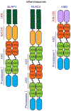
Fig. 1.. Types of inflammasomes.
Schematic representation of three different types of inflammasomes, showing the modular protein components (domain names in red and black) and full-length proteins (vertical protein names in blue), including the adapter ASC.
Fig. 2.
Fig. 2.. Three-dimensional atomic-resolution solution structure of the inflammasome adapter ASC.
A. Ribbon diagram representing the secondary structure arrangement of the 3D structure of ASC (N and C termini as well as helix numbers are indicated). B. Electrostatic surface of ASC (red and blue denote negatively and positively charged surfaces, respectively). C. Conformational space available to the CARD domain relative to the PYD. D. Conformational space available to the PYD domain relative to the CARD. Image created with MOLMOL [97]. This research was originally published in the Journal of Biological Chemistry. E. de Alba. Structure and Interdomain Dynamics of Apoptosis-associated Speck-like Protein Containing a CARD (ASC). J. Biol. Chem. 2009; 284:32932-32941. © E. de Alba.
Fig. 3.
Fig. 3.. ASC semi-structured linker.
Deviations of 13Cα chemical shits of ASC [94] relative to random coil values [95,96]. Positive values indicate α-helical conformation, whereas negative values indicate the presence of extended conformation. Most residues in the 95–112 fragment are clustered in the extended structure region except G99 and G111. This research was originally published in the Journal of Biological Chemistry. E. de Alba. Structure and Interdomain Dynamics of Apoptosis-associated Speck-like Protein Containing a CARD (ASC). J. Biol. Chem. 2009; 284:32932-32941. © E. de Alba.
Fig. 4.
Fig. 4.. Model for ASC functional oligomerization.
A. Available space for CARD relative to the PYD domain shown in its volume. The type I, II and III interactions are possible for the PYD in the presence of CARD motion, as helices 1 and 4 (red), 2 and 3 (blue), and the loops connecting them are accessible for binding. B. Structural dimer of ASC based on the CARD-CARD dimer complex between Apaf-1 CARD and caspase-1 CARD [104,105]. C. Schematic representation of ASC preferred directionality for functional oligomerization. D. Unfavored modes for ASC self-association.
Fig. 5.
Fig. 5.. ASCPYD interacts with other PYDs through two equivalent interfaces.
Superimposed sofast-HMQC spectra resulting from the titration of 15N-labeled ASCPYD with increasing concentrations of unlabeled ASCPYD (A) and NLRP3PYD (B). Some signals (e. g., L12 NH amide) remain unperturbed, whereas others show significant changes in δ upon protein interaction. This research was originally published in the Journal of Biological Chemistry. J. Oroz et al. ASC Pyrin Domain Self-associates and Binds NLRP3 Protein Using Equivalent Binding Interfaces. J. Biol. Chem. 2016; 291:19487–19501. © J. Oroz et al.
Fig. 6.
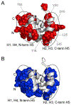
Fig. 6.. ASCPYD self-association and interaction with NLRP3PYD is mediated by similar opposing binding surfaces.
ASCPYD structure showing exposed residues with δ changes above the threshold value upon ligand binding for self-association (A) and interaction with NLRP3PYD (B). (ASC structure PDB code 2KN6 [92]). This research was originally published in the Journal of Biological Chemistry. J. Oroz et al. ASC Pyrin Domain Self-associates and Binds NLRP3 Protein Using Equivalent Binding Interfaces. J. Biol. Chem. 2016; 291:19487–19501. © J. Oroz et al.
Fig. 7.
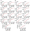
Fig. 7.. Dissociation constant values indicate stronger affinity of ASCPYD for NLRP3PYD vs. self-association.
Changes in chemical shift vs. protein concentration for ASCPYD self-association (A) and ASCPYD-NLRP3PYD interaction (B). KD values are shown for each graph. This research was originally published in the Journal of Biological Chemistry. J. Oroz et al. ASC Pyrin Domain Self-associates and Binds NLRP3 Protein Using Equivalent Binding Interfaces. J. Biol. Chem. 2016; 291:19487–19501. © J. Oroz et al.
Fig. 8.
Fig. 8.. Influence of NaCl and pH on ASCPYD interacting mode.
A. sofast-HMQC spectra of ASCPYD at different concentrations in the absence and presence of NaCl. Top: In blue 50 μM 15N ASCPYD; green: 50 μM 15N ASCPYD + 500 μM ASCPYD; red: 50 μM 15N ASCPYD/100 mM NaCl. Bottom: In red: 50 μM 15N ASCPYD/100 mM NaCl (same as in top), black: 50 μM 15N ASCPYD + 250 μM ASCPYD/100 mM NaCl. B. Chemical shift perturbation data mapped onto the 3D structure for the ASCPYD-ASCPYD and ASCPYD-NLRP3PYD interaction at pH 5.8 (In red: ASCPYD-ASCPYD; in blue: ASCPYD-NLRP3PYD). This research was originally published in the Journal of Biological Chemistry. J. Oroz et al. ASC Pyrin Domain Self-associates and Binds NLRP3 Protein Using Equivalent Binding Interfaces. J. Biol. Chem. 2016; 291:19487–19501. © J. Oroz et al.
Fig. 9.
Fig. 9.. Structural perturbations in ASCPYD caused by the L25A mutation.
A. Chemical shift differences between wild type and mutant L25A ASCPYD vs. residue number. B. Largest chemicals shift variations mapped onto the ASCPYD structure. L25 is shown in red. The largest structural effects (shown in blue) involve H2–H3. This research was originally published in the Journal of Biological Chemistry. J. Oroz et al. ASC Pyrin Domain Self-associates and Binds NLRP3 Protein Using Equivalent Binding Interfaces. J. Biol. Chem. 2016; 291:19487–19501. © J. Oroz et al.
Fig. 10.
Fig. 10.. NLRP3PYD interacts with ASCPYD via similar interfaces.
A. Superimposed sofast-HMQC spectra acquired in the titration of 15N-labeled NLRP3PYD with ASC. NLRP3PYD severe aggregation precluded titration at molar ratios > 1:1. NMR signal changes upon titration with ASC match those observed at high NLRP3PYD concentration, indicating that self-association of the latter and its interaction with ASCPYD are mediated by identical interfaces. B. Chemical shift perturbation data for the NLRP3PYD-ASCPYD interaction. C. Mapping of NLRP3PYD residues undergoing above-threshold δ changes upon ASC binding. This research was originally published in the Journal of Biological Chemistry. J. Oroz et al. ASC Pyrin Domain Self-associates and Binds NLRP3 Protein Using Equivalent Binding Interfaces. J. Biol. Chem. 2016; 291:19487–19501. © J. Oroz et al.
Fig. 11.
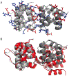
Fig. 11.. Electrostatic complementarity of the ASCPYD dimer compared with proposed dimer based on mutational studies.
A. Structural model of the ASCPYD dimer from NMR-based docking experiments. Side chains of residues involved in the interaction are shown in blue (positive charge) and red (negative charge). The interaction interface and the free interfaces show complementary charges for the polymerization. B. Comparison of dimer in A with dimer proposed on the basis of mutational studies [122] (in red). Protomers at the left are superimposed to show the different orientation of the remaining protomer in both dimer models. This research was originally published in the Journal of Biological Chemistry. J. Oroz et al. ASC Pyrin Domain Self-associates and Binds NLRP3 Protein Using Equivalent Binding Interfaces. J. Biol. Chem. 2016; 291:19487–19501. © J. Oroz et al.
Fig. 12.
Fig. 12.. ASCPYD fibril formation based on NMR data.
The polymerization of the ASCPYD dimeric complex formed by the type I interaction results in a helical fibril. The ribbon diagram represents the dimer. The side chains of the residues that drive the docking calculation are shown. The colored circles represent the type I interaction taking place for fibril formation. Each replicate is axially rotated ~38°. This research was originally published in the Journal of Biological Chemistry. J. Oroz et al. ASC Pyrin Domain Self-associates and Binds NLRP3 Protein Using Equivalent Binding Interfaces. J. Biol. Chem. 2016; 291:19487–19501. © J. Oroz et al.
Fig. 13.
Fig. 13.. Death Domain interaction types in the cryo-EM structure of the helical tube formed by ASCPYD polymerization.
Top and lateral views of human ASCPYD helical tube [124] with the 15 ASCPYD domains of the PDB entry (PDB 3j63). Each group of 5 ASCPYD domains is colored in yellow, red and blue to show the helical arrangement. Bottom figure shows the interaction types that participate in polymerization.
Fig. 14.
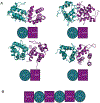
Fig. 14.. ASCPYD-NLRP3PYD oligomeric structures.
A. Models of four different complexes between ASC and NLRP3 PYDs based on NMR data: ASC H1–H4/NLRP3 H5, ASC H1–H4/NLRP3 H1–H4, ASC H2–H3/NLRP3 H5 and ASC H2–H3/NLRP3 H1–H4. ASCPYD and NLRP3PYD are shown as ribbon diagrams, colored in green and purple, respectively. Side chains of residues used in the docking are highlighted. ASCPYD and NLRP3PYD are depicted as green circles and purple squares, respectively, showing the corresponding type of interaction with numbers. B. Combination of alternative interactions based on the dimeric structures shown in A. This research was originally published in the Journal of Biological Chemistry. J. Oroz et al. ASC Pyrin Domain Self-associates and Binds NLRP3 Protein Using Equivalent Binding Interfaces. J. Biol. Chem. 2016; 291:19487–19501. © J. Oroz et al.
Fig. 15.
Fig. 15.. Macromolecular complex formed by ASCPYD-NLRP3PYD oligomerization.
Top (left) and side (right) views of the hexameric ring modeled using as template the resulting docking pairs of the different dimeric units shown in Fig. 14. ASCPYD and NLRP3PYD are depicted in green and purple, respectively. ASCCARD is shown in blue. The C-terminal domains of NLRP3 point to the bottom of the side view of the ring (right). The inner and outer dimensions of the ring are shown. This research was originally published in the Journal of Biological Chemistry. J. Oroz et al. ASC Pyrin Domain Self-associates and Binds NLRP3 Protein Using Equivalent Binding Interfaces. J. Biol. Chem. 2016; 291:19487–19501. © J. Oroz et al.
Fig. 16.
Fig. 16.. ASCCARD self-associates into filaments and filament bundles.
ASCCARD forms ~0.5–1 μm long bundles with preferred directionality (A, B). Narrower bundles branch out from the thicker bundles. ASCCARD forms thin filaments or fibrils of ~4 nm width (labeled as type A) and wider filaments likely composed of two narrower filaments (labeled as type B) (C, D, E). This research was originally published in the Journal of Biological Chemistry. R.J.T. Nambayan et al. The inflammasome adapter ASC assembles into filaments with integral participation of its two Death Domains, PYD and CARD. J. Biol. Chem. 2019; 294:439–452. © R.J.T. Nambayan et al.
Fig. 17.
Fig. 17.. Filaments and filament bundles formed by ASCPYD.
A, B, C, Representative micrographs with scale bar of 200 nm, and D, 0.5 μm. This research was originally published in the Journal of Biological Chemistry. R.J.T. Nambayan et al. The inflammasome adapter ASC assembles into filaments with integral participation of its two Death Domains, PYD and CARD. J. Biol. Chem. 2019; 294:439–452. © R.J.T. Nambayan et al.
Fig. 18.
Fig. 18.. Interacting surfaces in ASCCARD self-association.
ASCCARD structure [92] is shown as ribbon diagram in two different views. The amino acids that are exposed to the solvent and with the largest chemical shift perturbations are depicted in purple and labeled in black. Helices are numbered in purple. This research was originally published in the Journal of Biological Chemistry. R.J.T. Nambayan et al. The inflammasome adapter ASC assembles into filaments with integral participation of its two Death Domains, PYD and CARD. J. Biol. Chem. 2019; 294:439–452. © R.J.T. Nambayan et al.
Fig. 19.
Fig. 19.. ASCCARD self-associates with KD ~50 μM.
Chemical shift perturbations of 13C, 15N-uniformly labeled ASCCARD vs. unlabeled ASCCARD concentration. Representative residues exposed to the solvent with largest perturbations are shown, together with the corresponding KD values. This research was originally published in the Journal of Biological Chemistry. R.J.T. Nambayan et al. The inflammasome adapter ASC assembles into filaments with integral participation of its two Death Domains, PYD and CARD. J. Biol. Chem. 2019; 294:439–452. © R.J.T. Nambayan et al.
Fig. 20.
Fig. 20.. Filaments formed by ASCCARD mutant 2.
Mutations include E130A, W131A, and D134A. This research was originally published in the Journal of Biological Chemistry. R.J.T. Nambayan et al. The inflammasome adapter ASC assembles into filaments with integral participation of its two Death Domains, PYD and CARD. J. Biol. Chem. 2019; 294:439–452. © R.J.T. Nambayan et al.
Fig. 21.
Fig. 21.. Dimensions of the ASC dimer.
Contact surface of the ASC dimer structure derived from NMR-based protein docking [136]. The PYD and CARD domains are labeled. Dimensions were calculated with MOLMOL [97]. This research was originally published in the Journal of Biological Chemistry. R.J.T. Nambayan et al. The inflammasome adapter ASC assembles into filaments with integral participation of its two Death Domains, PYD and CARD. J. Biol. Chem. 2019; 294:439–452. © R.J.T. Nambayan et al.
Fig. 22.
Fig. 22.. Full-length ASC forms filament bundles with individual filaments formed by the stacking of ring-shaped particles of the size of ASC dimers.
A. Full-length ASC filaments. Rectangles show zoomed areas that compose panels (B), bottom rectangle and (C), upper rectangle. Arrows point to ring-shaped structures (possibly ASC dimers) laterally attached to pre-existing filaments. D. An additional zoom of the upper-central region of (C). The circle shows stacked rings forming the filaments. This research was originally published in the Journal of Biological Chemistry. R.J.T. Nambayan et al. The inflammasome adapter ASC assembles into filaments with integral participation of its two Death Domains, PYD and CARD. J. Biol. Chem. 2019; 294:439–452. © R.J.T. Nambayan et al.
Fig. 23.
Fig. 23.. Dimensions of stacked filaments of full-length human ASC deviate from the model based on the cryo-EM structure of truncated ASCPYD.
A&B. Top and lateral views of human ASCPYD helical tube (cryo-EM PDB 3j63) with the dimension reported in the published work [124] and containing the 15 ASCPYD domains of the PDB entry. Each group of 5 ASCPYD domains is colored in yellow, red and blue to show the helical arrangement (A, B). Top (C) and side (D) views of the ASCPYD tube with protruding ASCCARD domains and linker to show CARD availability for potential interactions. The PYDs of 15 full-length NMR structures of ASC molecules ([92]; PDB 2KN6) are superimposed to the 15 PYD domains of the cryo-EM structure. After superposition, the NMR PYDs are deleted and only the cryo-EM PYDs and the NMR CARDs are shown. One side of the helical tube is colored in red and the corresponding CARDs in orange. Dimension shown was calculated with MOLMOL [97] (C, D). E Modeled bundle of an arrangement of four ASCPYD helical filaments with protruding CARDs and linker from D. The red side of the PYD tube and corresponding CARDs in orange (from C and D) are shown. CARD domains in filament interfaces are assumed to stack vertically (overlapping as worse-case scenario), thus occupying approximately the dimension of the ASCCARD (~3.5 nm). The dimensions shown are; 1) the minimum width of the 4-filament bundle, which is estimated by assuming a filament width of 9 nm from the cryo-EM structure; 2) the estimated width range according to the model shown in (D) for the 4-filament bundle (E), which is calculated assuming a filament width of 6–7 nm from our TEM studies. This research was originally published in the Journal of Biological Chemistry. R.J.T. Nambayan et al. The inflammasome adapter ASC assembles into filaments with integral participation of its two Death Domains, PYD and CARD. J. Biol. Chem. 2019; 294:439–452. © R.J.T. Nambayan et al.
Fig. 24.
Fig. 24.. Protomer spacing in bundled human ASC filaments.
A. Subsection of TEM micrograph of ASC filaments shown in panel A of Fig. 22. Line cuts of 3pxl (corresponding to 3 nm) using ImageJ along the long axis of each filament in the region of interest. B. Line cut along filament number 3 is shown in cyan. C. Average values of intensity profiles over the 3 nm width. Red arrowheads indicate locations of pixel intensity values above the mean intensity along the filament (green-dashed line). D. Mean values for the separation of maximum peaks from averaged intensity profiles for each of the filaments/lines cuts across a total number of 22 filaments. The mean protomer separation is 5.0 nm ± 0.6 nm. Scale bar in (A) is 100 nm. This research was originally published in the Journal of Biological Chemistry. R.J.T. Nambayan et al. The inflammasome adapter ASC assembles into filaments with integral participation of its two Death Domains, PYD and CARD. J. Biol. Chem. 2019; 294:439–452. © R.J.T. Nambayan et al.
Fig. 25.
Fig. 25.. Directionality model for ASC-dependent inflammasome assembly.
ASC dimer shown as the minimal building block. The ASC filament presents two interacting sides, one for recruiting procaspase 1 CARD and the other for interaction with the PYD of NLRs sensor proteins. The binding direction propagated by the ASC filament results in micrometer-size inflammasomes with a concentric distribution of inflammasome components; NLRs, ASC, and procaspase 1.

References

    1. Martinon F, Burns K, Tschopp J, The inflammasome: a molecular platform triggering activation of inflammatory caspases and processing of proIL-beta, Mol. Cell 10 (2002) 417–426. - PubMed
    1. Schroder K, Tschopp J, The inflammasomes, Cell 140 (2010) 821–832. - PubMed
    1. Guo H, Callaway JB, Ting JP, Inflammasomes: mechanism of action, role in disease, and therapeutics, Nat. Med 21 (2015) 677–687. - PMC - PubMed
    1. Broz P, Dixit Inflammasomes VM, Mechanism of assembly, regulation and signaling, Nat. Rev. Immunol 16 (2016) 407–420. - PubMed
    1. He Y, Hara H, Núñez G, Mechanism and regulation of NLRP3 inflammasome activation, Trends Biochem. Sci 41 (2016) 1012–1021. - PMC - PubMed

Publication types

LinkOut - more resources