. 2019 Jun 1;24(1):39.
doi: 10.1186/s12199-019-0794-8.

cVEMP correlated with imbalance in a mouse model of vestibular disorder

Affiliations

cVEMP correlated with imbalance in a mouse model of vestibular disorder

Reina Negishi-Oshino et al. Environ Health Prev Med. .

Abstract

Background: Cervical vestibular evoked myogenic potential (cVEMP) testing is a strong tool that enables objective determination of balance functions in humans. However, it remains unknown whether cVEMP correctly expresses vestibular disorder in mice.

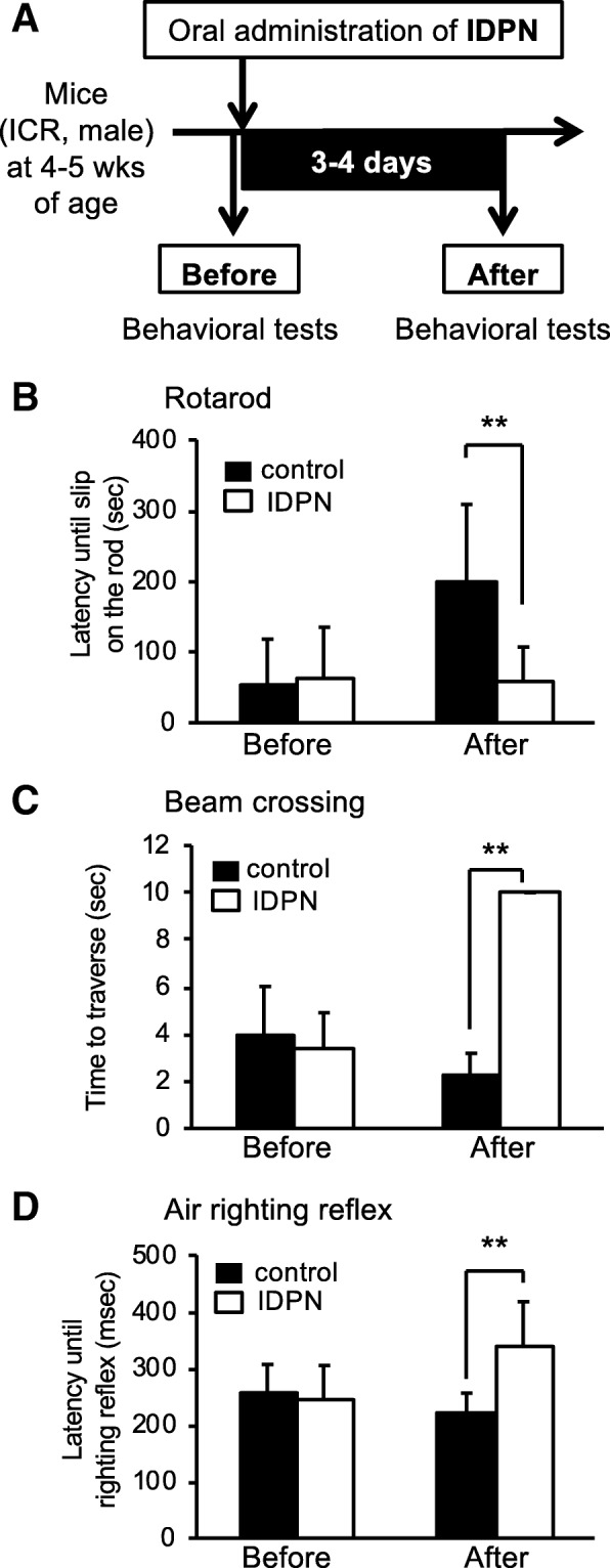
Objective: In this study, correlations of cVEMP with scores for balance-related behavior tests including rotarod, beam, and air-righting reflex tests were determined in ICR mice with vestibular disorder induced by 3,3'-iminodipropiontrile (IDPN) as a mouse model of vestibular disorder.

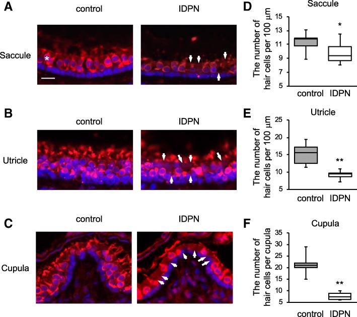
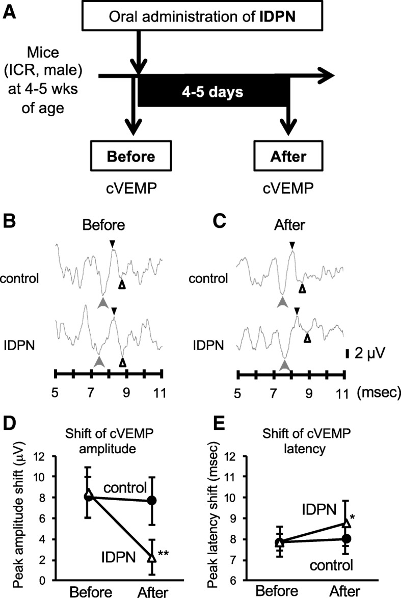
Methods: Male ICR mice at 4 weeks of age were orally administered IDPN in saline (28 mmol/kg body weight) once. Rotarod, beam crossing, and air-righting reflex tests were performed before and 3-4 days after oral exposure one time to IDPN to determine balance functions. The saccule and utricles were labeled with fluorescein phalloidin. cVEMP measurements were performed for mice in the control and IDPN groups. Finally, the correlations between the scores of behavior tests and the amplitude or latency of cVEMP were determined with Spearman's rank correlation coefficient. Two-tailed Student's t test and Welch's t test were used to determine a significant difference between the two groups. A difference with p < 0.05 was considered to indicate statistical significance.

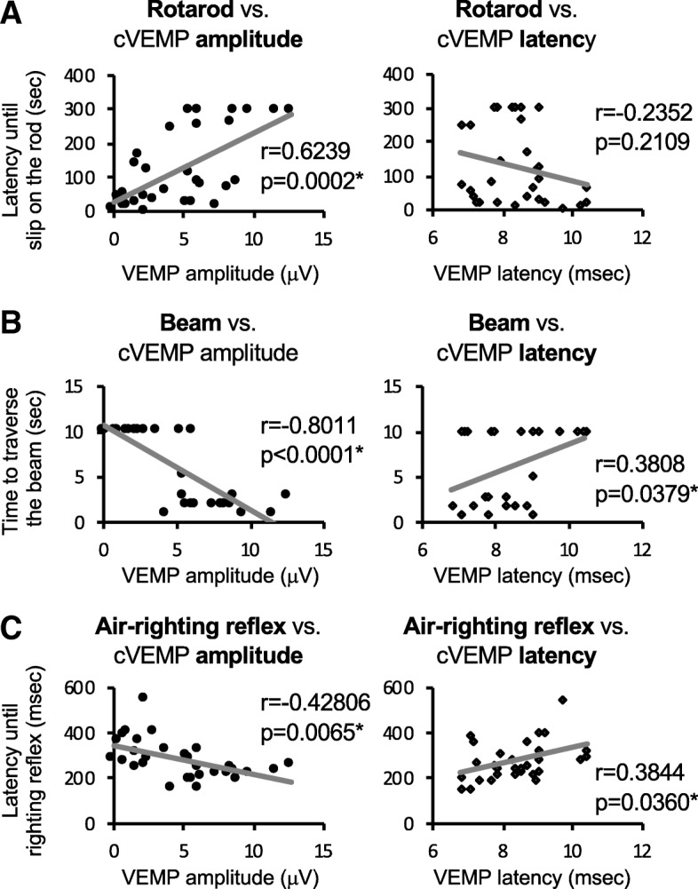
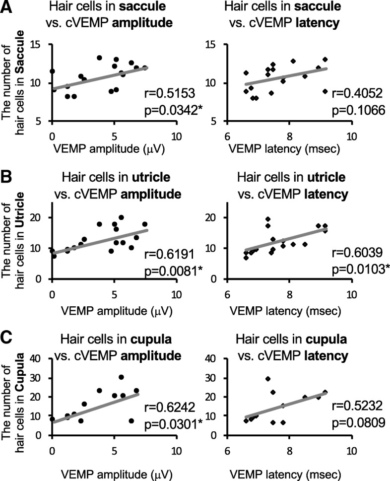
Results: After oral administration of IDPN at 28 mmol/kg, scores of the rotarod, beam, and air-righting reflex tests in the IDPN group were significantly lower than those in the control group. The numbers of hair cells in the saccule, utricle, and cupula were decreased in the IDPN group. cVEMP in the IDPN group was significantly decreased in amplitude and increased in latency compared to those in the control group. cVEMP amplitude had significant correlations with the numbers of hair cells as well as scores for all of the behavior tests in mice.

Conclusions: This study demonstrated impaired cVEMP and correlations of cVEMP with imbalance determined by behavior tests in a mouse model of vestibular disorder.

Keywords: Balance; Hair cells; IDPN; Vestibule; cVEMP.


Conflict of interest statement

The authors declare that they have no competing interests.

Figures

Fig. 1
Fig. 1
Balance impairments of mice treated with IDPN and a scheme of experiments. a Scheme of experiments. Behavioral tests were performed with 4–5 aged male mice following oral administration of IDPN. The control group (n = 5) and the IDPN group (n = 5) were subjected to behavioral tests after administration of IDPN. b A rotarod test, c a beam crossing test, and d an air-righting reflex test were performed before and 3–4 days after administration of IDPN (28 mmol/kg) to wild-type mice having an ICR background. b Latency until falling or slipping on the rotarod (seconds, mean ± SD), c time to traverse (seconds, mean ± SD), and d latency until righting reflex (milliseconds) were recorded for the control group (black bars, n = 5) and the IDPN group (white bars, n = 5). For the beam crossing test, a score of 10 s was given for mice that could not walk on the beam anymore because of complete loss of balance. Significant differences (**p < 0.01) between the two groups were analyzed by two-tailed Student’s t test
Fig. 2
Fig. 2
Vestibular hair cell loss in the saccule, utricle, and cupula of mice treated with IDPN. ac After administration of IDPN, hair cells were stained with anti-MyosinVIIa antibody. Equivalent positions in the saccule (a), utricle (b), and cupula (c) from mice in the control group (left panels) and mice in the exposure group (right panels) are shown with the same scale (scale bar 20 μm in a). An asterisk shows an example of normal hair cells (a), and arrows indicate loss of hair cells (ac, right panels). d, e Numbers of hair cells per 100 μm (mean ± SD) in the saccules (d) and utricles (e) from 3 mice in the control group (gray) and 3 mice in the IDPN group (white) are shown. f Numbers of hair cells in the cupula (means ± SD) from three mice in the control group (gray) and 3 mice in the IDPN group (white) are shown. Significant differences (*p < 0.05, **p < 0.01) between the two groups were analyzed by Welch’s t test
Fig. 3
Fig. 3
Impairments of cVEMP in the IDPN group and a scheme of experiments. a Scheme of experiments. cVEMP recordings were performed following oral administration of IDPN in male mice in the control group (n = 5) and the IDPN group (n = 5). cVEMP recording was performed after the administration of IDPN. b, c cVEMPs in the control group (upper waves) and the IDPN group (lower waves) before (b) and after administration of IDPN (c) are shown. cVEMPs with a positive peak (p1, indicated by black triangles) and those with a negative peak (n1, indicated by white triangles) were identified by the appearance of negative waves (indicated by gray arrows) just before p1 indicated by black triangles. d, e Graphs of cVEMP amplitudes [microvolts (μV), mean ± SD in e] and latencies [milliseconds (msec), mean ± SD in e] in the control group (black circles, n = 5) and the IDPN group (white triangles, n = 5) before and after administration of IDPN are presented. Significant differences (**p < 0.01; *p < 0.05) between the two groups were analyzed by Welch’s t test
Fig. 4
Fig. 4
Correlations between scores of the behavior tests and cVEMP results. ac Correlations of a rotarod test score [latency until slipping on the rod, seconds (sec)], b beam crossing test score [time to traverse the beam, seconds (sec)], and c air-righting reflex test score [latency until righting reflex, milliseconds (msec)] with cVEMP amplitude [microvolts (μV), left graphs] and VEMP latency (msec, right graphs) are shown. Triplicated measurements for each test with a total of 10 mice including 5 mice for the control group and 5 mice for the IDPN group were performed. Spearman’s rank correlation coefficients and significance differences (*p < 0.05) were analyzed for a rotarod vs. cVEMP amplitude [r = 0.6239, p = 0.0002] and cVEMP latency [r = − 0.2352, p = 0.2109], b beam vs. cVEMP amplitude [r = − 0.8011, p < 0.0001] and cVEMP latency [r = 0.3808, p = 0.0379], and c air-righting reflex vs. cVEMP amplitude [r = − 0.42806, p = 0.0065] and cVEMP latency [r = 0.3844, p = 0.0360]
Fig. 5
Fig. 5
Correlations between the numbers of hair cells and cVEMP results. ac Correlations of the numbers of hair cells in the saccule (a), utricle (b), and cupula (c) with cVEMP amplitude [microvolts (μV), left graphs] and VEMP latency (msec, right graphs) are shown. Triplicated measurements for cVEMP with a total of 6 mice including 3 mice for the control group and 3 mice for the IDPN group were performed. Spearman’s rank correlation coefficients and significance differences (*p < 0.05) were analyzed for a hair cells in the saccule vs. cVEMP amplitude [r = 0.5153, p = 0.0342] and cVEMP latency [r = 0.4052, p = 0.1066], b hair cells in the utricle vs. cVEMP amplitude [r = 0.6191, p = 0.0081] and cVEMP latency [r = 0.6039, p = 0.0103], and c hair cells in the cupula vs. cVEMP amplitude [r = 0.6242, p = 0.0301] and cVEMP latency [r = 0.5232, p = 0.0809]

Similar articles

Cited by

References

    1. Rosengren SM, Welgampola MS, Colebatch JG. Vestibular evoked myogenic potentials: past, present and future. Clin Neurophysiol. 2010;121:636–651. doi: 10.1016/j.clinph.2009.10.016. - DOI - PubMed
    1. Chiarovano E, Darlington C, Vidal PP, Lamas G, de Waele C. The role of cervical and ocular vestibular evoked myogenic potentials in the assessment of patients with vestibular schwannomas. PLoS One. 2014;9:e105026. doi: 10.1371/journal.pone.0105026. - DOI - PMC - PubMed
    1. Maheu M, Alvarado-Umanzor JM, Delcenserie A, Champoux F. The clinical utility of vestibular-evoked myogenic potentials in the diagnosis of Ménière’s disease. Front Neurol. 2017;8:415. doi: 10.3389/fneur.2017.00415. - DOI - PMC - PubMed
    1. Maes L, De Kegel A, Van Waelvelde H, Dhooge I. Rotatory and collic vestibular evoked myogenic potential testing in normal- hearing and hearing-impaired children. Ear Hear. 2014;35:e21–e32. doi: 10.1097/AUD.0b013e3182a6ca91. - DOI - PubMed
    1. Zhaoa X, Jonesb SM, Yamoahc EN, Lundberg YW. Otoconin-90 deletion leads to imbalance but normal hearing: a comparison with other otoconia mutants. Neuroscience. 2008;153:289–299. doi: 10.1016/j.neuroscience.2008.01.055. - DOI - PMC - PubMed

MeSH terms

LinkOut - more resources