. 2019 Jun 1;20(1):16.
doi: 10.1186/s12867-019-0134-y.

Giant group I intron in a mitochondrial genome is removed by RNA back-splicing

Affiliations

Giant group I intron in a mitochondrial genome is removed by RNA back-splicing

Sylvia Ighem Chi et al. BMC Mol Biol. .

Abstract

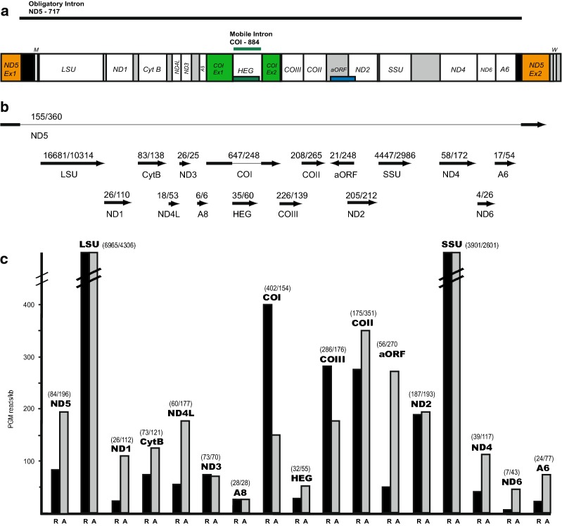
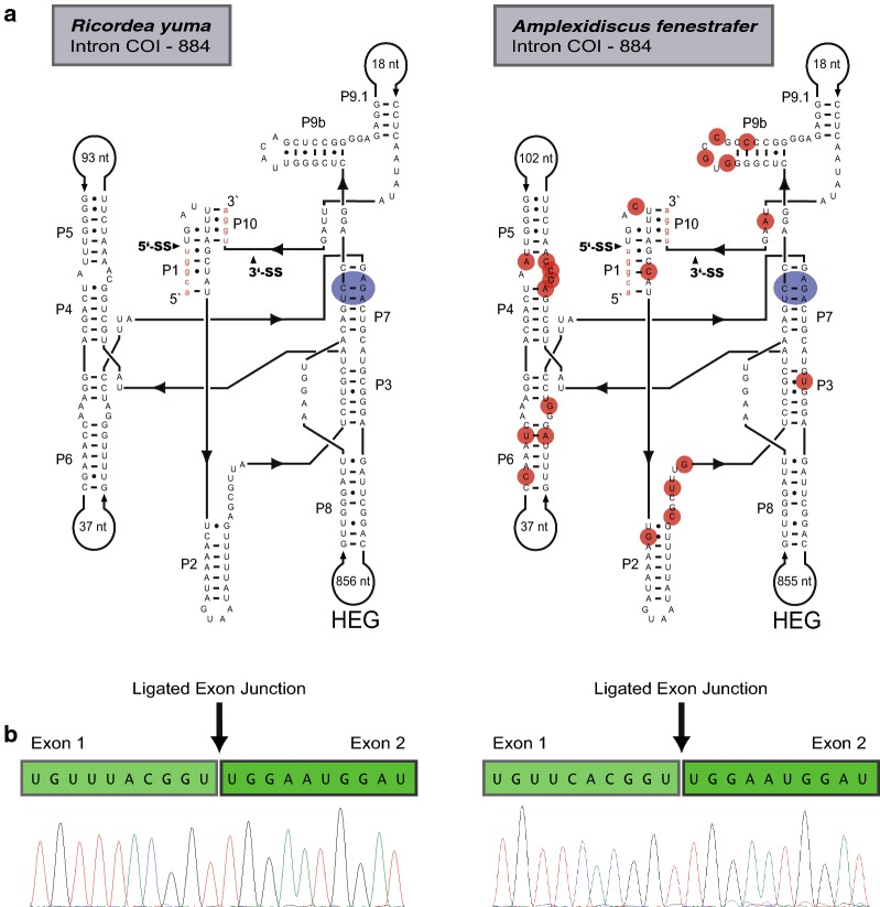
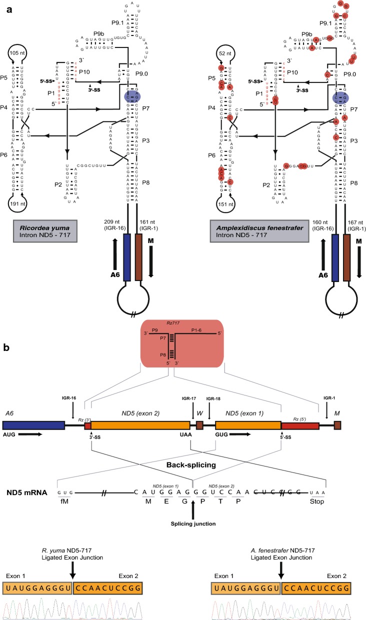
Background: The mitochondrial genomes of mushroom corals (Corallimorpharia) are remarkable for harboring two complex group I introns; ND5-717 and COI-884. How these autocatalytic RNA elements interfere with mitochondrial RNA processing is currently not known. Here, we report experimental support for unconventional processing events of ND5-717 containing RNA.

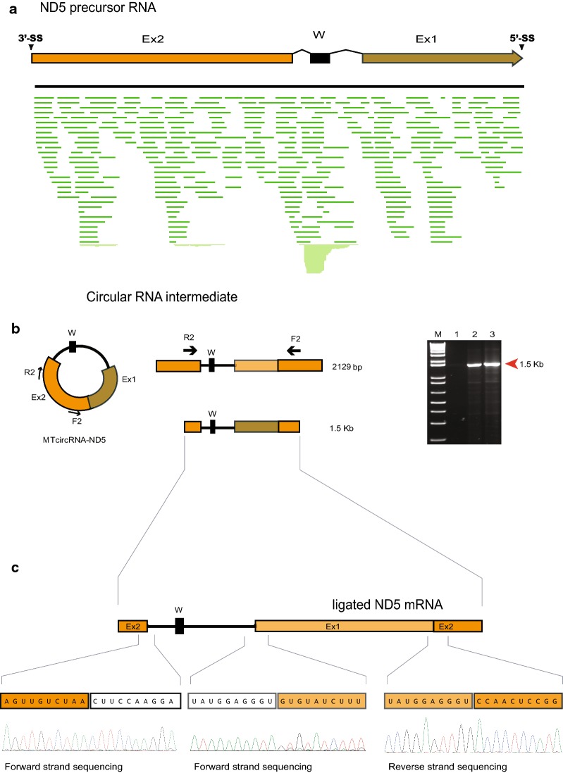
Results: We obtained the complete mitochondrial genome sequences and corresponding mitochondrial transcriptomes of the two distantly related corallimorpharian species Ricordea yuma and Amplexidiscus fenestrafer. All mitochondrial genes were found to be expressed at the RNA-level. Both introns were perfectly removed by autocatalytic splicing, but COI-884 excision appeared more efficient than ND5-717. ND5-717 was organized into giant group I intron elements of 18.1 kb and 19.3 kb in A. fenestrafer and R. yuma, respectively. The intron harbored almost the entire mitochondrial genome embedded within the P8 peripheral segment.

Conclusion: ND5-717 was removed by group I intron splicing from a small primary transcript that contained a permutated intron-exon arrangement. The splicing pathway involved a circular exon-containing RNA intermediate, which is a hallmark of RNA back-splicing. ND5-717 represents the first reported natural group I intron that becomes excised by back-splicing from a permuted precursor RNA. Back-splicing may explain why Corallimorpharia mitochondrial genomes tolerate giant group I introns.

Keywords: Amplexidiscus; Back-splicing; Catalytic RNA; Group I intron; Intron retention; Mitochondrial RNA; Ribozyme; Ricordea.


Conflict of interest statement

The authors declare that they have no competing interests.

Figures

Fig. 1
Fig. 1
Mitochondrial genome and transcripts in Ricordea yuma and Amplexidiscus fenestrafer. a Schematic view of mitochondrial genome content and organization of circular mtDNA (presented as a linear map). Obligatory ND5-717 and mobile-like COI-884 introns are indicated. ND5 exons and COI exons are shown in orange and green colors, respectively. Abbreviations: SSU and LSU, mitochondrial small- and large-subunit ribosomal RNA genes. ND1-6, NADH dehydrogenase subunit 1 to 6 genes. COI-III, cytochrome c oxidase subunit I to III genes. A6 and A8, ATPase subunit 6 and 8 genes. CytB, cytochrome B gene; HEG, homing endonuclease gene; aORF, antisense open reading frame. W and M; tRNA genes for Trp and Met, respectively, indicated by the standard one-letter symbols for amino acids. b Mapped transcript reads generated by Ion Torrent PGM from protein coding and rRNA coding regions. Presented below is read coverage per gene region (R. yuma/A. fenestrafer). c Histograms representing estimated normalized read numbers for R. yuma (R; black) and A. fenestrafer (A; grey). Three separate PGM runs were performed for each species. The PGM transcript number of each RNA was normalized to the size of gene coding regions (PGM reads/kb). The estimated normalized read numbers vary between approximately 7 reads/kb and 6965 reads/kb
Fig. 2
Fig. 2
COI gene features in Ricordea yuma and Amplexidiscus fenestrafer. a Secondary structure diagrams of corallimorpharian COI-884 group I introns (R. yuma, left; A. fenestrafer, right). The ten conserved paired segments of the catalytic core (P1 to P10) are shown, and flanking COI exons sequences are in red letters. The P8 extension containing the HEG is indicated and in small red circles are nucleotides substitutions in A. fenestrafer compared to R. yuma. The blue circles show the catalytic core sequences at the G-binding site. b Representation of COI mRNA sequences consisting of ligated exons. The ligation junctions are indicated, and below are the sequencing chromatograms
Fig. 3
Fig. 3
Mitochondrial ND5 gene organization and ND5-717 intron secondary structure in Ricordea yuma and Amplexidiscus fenestrafer. a Secondary structure diagrams of ND5-717 group I introns in R. yuma (left) and A. fenestrafer (right). The ten conserved paired segments of the catalytic core (P1 to P10) are shown and flanking ND5 exons sequences are in red letters. The P8 extensions containing mitochondrial genes are indicated. The blue circles indicate catalytic core sequences at the G-binding site and in small red circles are nucleotides substitutions in A. fenestrafer compared to R. yuma. b Organization map of mitochondrial ND5 gene in Corallimorpharian. A schematic view of the ND5-717 ribozyme is presented above the map. The splice sites are distant apart, but brought in proximity in a permuted intron–exon order due to the circular organization of the mtDNAs. Thus, ND5 exon 2 is presented upstream of ND5 exon 1. ND5 mRNA consisting of the ligated exons with the splicing junction indicated is shown below. Chromatograms of the exon ligation sequences are shown for R. yuma (left) and A. fenestrafer (right)
Fig. 4
Fig. 4
RNA back-splicing by ND5-717 group I intron in corallimorpharians. a In silico mapping of Ion PGM transcriptome reads to the ND5 precursor RNA. The R. yuma map is shown. The map also corresponds to the 2.13 kb circular RNA intermediate (MTcircRNA-ND5). Ex1, ND5 exon 1; Ex2, ND5 exon 2; W, tRNA Trp. b Map of circular RNA intermediate. Primers F2/R2 binds to exon 2, but in opposite directions. Right panel—the 1.5 kb F2/R2 amplicon from R. yuma (lane 2) and A. fenestrafer (lane 3). M, 1 kb ladder marker; lane 1, negative control. c Chromatogram of the 1.5 kb amplicon (exemplified in R. yuma)

Similar articles

Cited by

References

    1. Medina M, Collins AG, Takaoka TL, Kuehl JV, Boore JL. Naked corals: skeleton loss in scleractinia. Proc Natl Acad Sci USA. 2006;103:9096–9100. doi: 10.1073/pnas.0602444103. - DOI - PMC - PubMed
    1. Kitahara MV, Lin MF, Foret S, Huttley G, Miller DJ, Chen CA. The ‘naked’ coral hypothesis revisited—evidence for and against scleractinian monophyly. PLoS ONE. 2014;9:e94774. doi: 10.1371/journal.pone.0094774. - DOI - PMC - PubMed
    1. Fautin DG. Catalog to families, genera, and species of orders Actiniaria and Corallimorpharia (Cnidaria: Anthozoa) Zootaxa. 2016;2016(4145):1–449. doi: 10.11646/zootaxa.4145.1.1. - DOI - PubMed
    1. Emblem Å, Karlsen BO, Evertsen J, Johansen SD. Mitogenome rearrangement in the cold-water scleractinian coral Lophelia pertusa (Cnidaria, Anthozoa) involves a long-term evolving group I intron. Mol Phylogenet Evol. 2011;61:495–503. doi: 10.1016/j.ympev.2011.07.012. - DOI - PubMed
    1. Lin MF, Kitahara MV, Luo H, Tracey D, Geller J, Fukami H, Miller DJ, Chen CA. Mitochondrial genome rearrangements in the scleractinia/corallimorpharia complex: implications for coral phylogeny. Genome Biol Evol. 2014;6:1086–1095. doi: 10.1093/gbe/evu084. - DOI - PMC - PubMed

Publication types

LinkOut - more resources