. 2019 Jul 17;103(2):242-249.e4.
doi: 10.1016/j.neuron.2019.04.039. Epub 2019 May 29.

Temporally and Spatially Distinct Thirst Satiation Signals

Affiliations

Temporally and Spatially Distinct Thirst Satiation Signals

Vineet Augustine et al. Neuron. .

Abstract

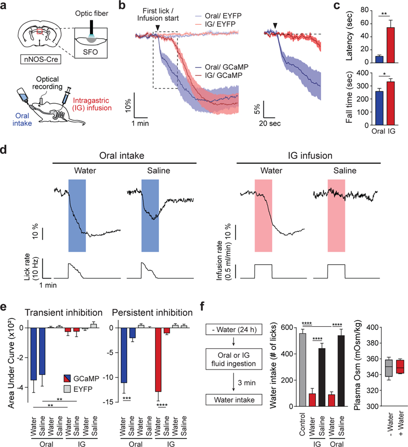
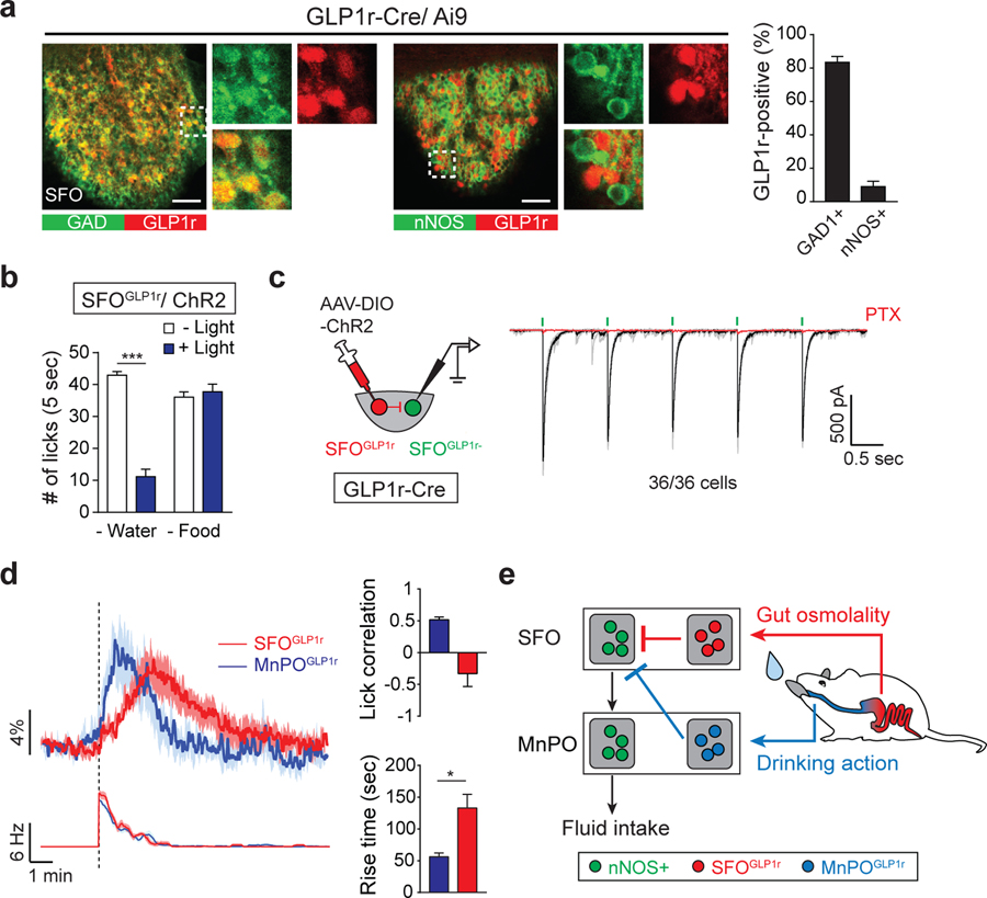
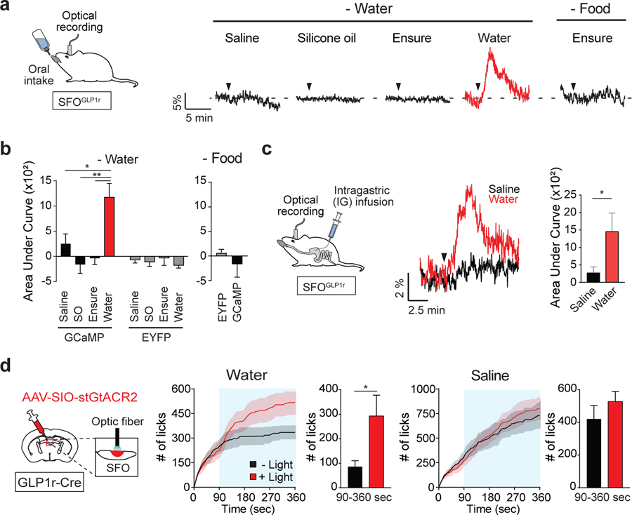
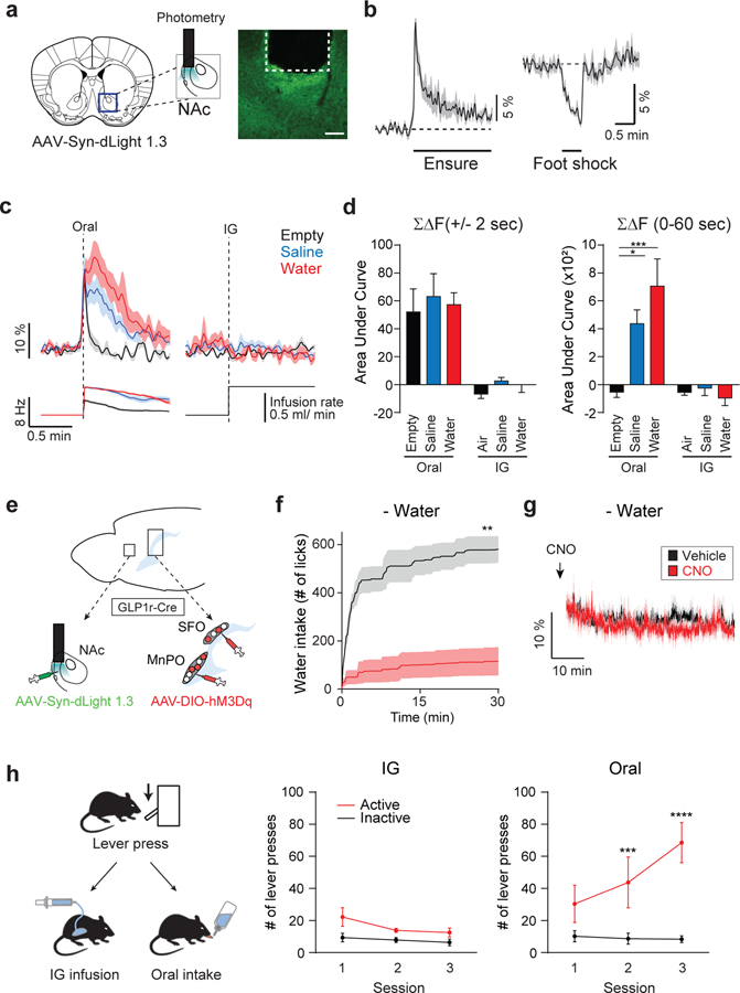
For thirsty animals, fluid intake provides both satiation and pleasure of drinking. How the brain processes these factors is currently unknown. Here, we identified neural circuits underlying thirst satiation and examined their contribution to reward signals. We show that thirst-driving neurons receive temporally distinct satiation signals by liquid-gulping-induced oropharyngeal stimuli and gut osmolality sensing. We demonstrate that individual thirst satiation signals are mediated by anatomically distinct inhibitory neural circuits in the lamina terminalis. Moreover, we used an ultrafast dopamine (DA) sensor to examine whether thirst satiation itself stimulates the reward-related circuits. Interestingly, spontaneous drinking behavior but not thirst drive reduction triggered DA release. Importantly, chemogenetic stimulation of thirst satiation neurons did not activate DA neurons under water-restricted conditions. Together, this study dissected the thirst satiation circuit, the activity of which is functionally separable from reward-related brain activity.

Keywords: appetite; gut-brain axis; homeostasis; reward circuit; satiation; thirst.


Conflict of interest statement

DECLARATION OF INTERESTS

The declare no competing interests.

Figures

Figure 1.
Figure 1.. Thirst Circuits Receive Temporally Distinct Inhibitory Signals after Water Intake
(A) A diagram of optical recording of GCaMP6s signals from SFOnNOS neurons. Fluid was given either orally or via IG infusion. (B) Temporally distinct inhibition of SFOnNOS neurons by ad lib oral intake or IG infusion of water (0.5 mL/min for 2 min; n = 8 mice for GCaMP6s; n = 4 and 6 mice for enhanced yellow fluorescent protein (EYFP) for oral and IG administration, respectively). (C) IG water infusion induced significantly slower onset of inhibition compared to oral water intake (latency). Fall time is defined as the time to maximum inhibition from first lick or infusion onset (n = 8 mice for GCaMP6s). (D) Representative traces of calcium dynamics during oral intake or IG infusion of water and saline (1 out of 8 mice). Lick and infusion rates are indicated below calcium traces. (E) Quantified responses of SFOnNOS neurons. Signals were quantified during (transient) and after (persistent) liquid ingestion or infusion; n = 8 mice for GCaMP6s; n = 6 mice for EYFP). (F) Drinking-induced satiation after oral or IG water administration. Animals were given access to water after oral intake or IG infusion (0.5 mL/min) of fluid for 2 min. Water consumption was measured for 10 min (left, n = 11 mice for control [no pre-ingestion] and for pre-IG; n = 7 mice for pre-oral). Note that the systemic osmolality was unchanged after oral water intake (right, n = 4 mice). *p < 0.05, **p < 0.01, ***p < 0.001, and ****p < 0.0001 by two-tailed paired t test; two-way repeated-measures ANOVA (Bonferroni’s multiple comparisons) or one-way ANOVA (Tukey’s multiple comparisons). Data are presented as mean ± SEM. Boxplots show median, quartiles (boxes), and range (whiskers). See also Figure S1.
Figure 2.
Figure 2.. GLP1r-Positive SFO Neurons Monosynaptically Inhibit Thirst-Driving Neurons
(A) GLP1r is specifically expressed in GABAergic neurons of the SFO. Immunohistological staining shows that a majority of GLP1r-positive neurons (labeled by Ai9) overlapped with glutamic acid decarboxylase (GAD; left panels). These neurons did not overlap with glutamatergic nNOS-positive neurons (middle panels). Quantification of the percentage of GLP1r-positive neurons that coexpressed GAD or nNOS is shown (n = 3 mice; representative images are from 1 out of 3 mice). (B) Optogenetic stimulation of SFOGLP1r neurons selectively suppresses water intake, but not liquid food intake (n = 5 mice). (C) The SFOGLP1r → SFOnon-GLP1r monosynaptic connections. All GLP1r-negative neurons (36/36 cells) in the SFO received monosynaptic inhibitory inputs from SFOGLP1r neurons. (D) Two inhibitory populations in the LT exhibit temporally distinct response to drinking behavior. Calcium dynamics of SFOGLP1r and MnPOGLP1r neurons upon water drinking and lick rate (left) is shown. Quantification of calcium dynamics is shown. MnPOGLP1r neurons have significantly faster activation kinetics compared to SFOGLP1r neurons (right). MnPOGLP1r, but not SFOGLP1r, neurons have a positive correlation with lick timing (n = 6 mice). Rise time is defined as the time to maximum excitation from first lick. For MnPOGLP1r neurons, we re-analyzed the data from the previous report (Augustine et al., 2018a). (E) A possible model of thirst-quenching signals. Liquid gulping signals are mediated by MnPOGLP1r neurons, which provide rapid and transient suppression of SFOnNOS neurons. Subsequently, SFOGLP1r neurons are activated by gastrointestinal hypo-osmotic stimuli to mediate slower inhibitory signals. *p < 0.05; ***p < 0.001 by two-tailed paired or unpaired (Welch’s correction) t test. Data are presented as mean ± SEM. Scale bars, 50 μm. See also Figure S2.
Figure 3.
Figure 3.. SFOGLP1r Neurons Are Activated by Hypo-osmotic Stimuli in the Gut
(A) Representative traces showing calcium responses of SFOGLP1r neurons upon ingestion of different fluids. SFOGLP1r neurons were selectively activated by water, but not by other fluids. Black triangles indicate the onset of licking (1 out of 6 mice). (B) Quantification of responses of GCaMP6s and EYFP signals during 5 min after the first lick (n = 6 and 3 mice for GCaMP6s for water and food restriction, respectively, and n = 6 mice for EYFP). SO, silicone oil. (C) Responses of SFOGLP1r neurons upon intragastric fluid infusion. A diagram of IG infusion and fiber photometry is shown (left panel). Representative traces are shown for IG water (red) or isotonic saline (black) infusion. A total of 1 mL (0.5 mL/min) of water or saline was infused. Black triangle indicates the onset of infusion (middle panel, traces are from 1 of 5 mice). Quantification of calcium responses to water or isotonic saline is shown (right panel, n = 5 mice). (D) Optogenetic inhibition of SFOGLP1r neurons selectively increases water intake, but not isotonic saline intake. SFO neurons of GLP1r-Cre mice were infected with AAV-SIO-stGtACR2. Continuous illumination was performed from 90 to 360 s (blue shaded area; n = 5 mice). *p < 0.05 and **p < 0.01 by two-tailed paired t test or one-way repeated-measures ANOVA (Dunnett’s multiple comparisons). Data are presented as mean ± SEM. See also Figure S3.
Figure 4.
Figure 4.. Activity of the Reward Circuits Is Separable from Thirst Satiation Signals
(A) A diagram of optical recording of DA release by dLight1.3. A representative image of dLight expression is shown. (B) DA release is induced by appetitive (Ensure) stimulus and suppressed by aversive (footshock) stimulus (n = 7 mice). (C) dLight fluorescence changes are shown during oral ad lib intake and IG infusion (n = 7 mice). Spontaneous drinking induced robust DA release in the NAc compared to empty control regardless of liquid type (left). For empty control experiments, DA release was observed transiently prior to lick due to reward expectation. By contrast, IG infusion of fluid had no effect on DA release (right, n = 7 mice). (D) Quantified data of dLight responses during 4 s around the first lick (left) or 60 s (right) after the first lick or IG infusion (n = 7 mice). (E) A schematic for activating thirst satiation circuits in the LT by hM3Dq while measuring DA release in the NAc by dLight1.3. (F) Chemogenetic stimulation of SFOGLP1r and MnPOGLP1r neurons attenuates water intake under dehydrated conditions (n = 6 mice). (G) By contrast, the same stimulation paradigm did not induce DA release (n = 6 mice). (H) A diagram of operant task. Mice were initially trained to associate lever press and water reward. After extinction sessions (see Figure S4F), animals were subjected to reinstatement paradigms with either IG or oral water reward (left). In IG sessions, animals received water through a gastric catheter on an FR3 schedule (middle). In oral sessions, the same amount of water reward was provided through a spout (right, n = 6 mice). Only oral water intake efficiently reinforced lever press behavior. *p < 0.05, **p < 0.01, ***p < 0.001, and ****p < 0.0001 by two-tailed paired t test; one-way repeated-measures ANOVA (Dunnett’s multiple comparisons) or two-way repeated-measures ANOVA (Bonferroni’s multiple comparisons). Data are presented as mean ± SEM. Scale bar, 50 μm. See also Figure S4.

References

    1. Abbott SB, Machado NL, Geerling JC, and Saper CB. (2016). Reciprocal control of drinking behavior by median preoptic neurons in mice. J. Neurosci 36, 8228–8237. - PMC - PubMed
    1. Allen WE, DeNardo LA, Chen MZ, Liu CD, Loh KM, Fenno LE, Ramakrishnan C, Deisseroth K, and Luo L. (2017). Thirst-associated preoptic neurons encode an aversive motivational drive. Science 357, 1149–1155. - PMC - PubMed
    1. Andermann ML, and Lowell BB. (2017). Toward a wiring diagram understanding of appetite control. Neuron 95, 757–778. - PMC - PubMed
    1. Augustine V, Gokce SK, Lee S, Wang B, Davidson TJ, Reimann F, Gribble F, Deisseroth K, Lois C, and Oka Y. (2018a). Hierarchical neural architecture underlying thirst regulation. Nature 555, 204–209. - PMC - PubMed
    1. Augustine V, Gokce SK, and Oka Y. (2018b). Peripheral and central nutrient sensing underlying appetite regulation. Trends Neurosci 41, 526–539. - PMC - PubMed

Publication types

MeSH terms