Review
. 2019 Sep 20;431(20):3975-4006.
doi: 10.1016/j.jmb.2019.05.042. Epub 2019 May 31.

The Mechanisms of Substrate Selection, Catalysis, and Translocation by the Elongating RNA Polymerase

Affiliations
Review

The Mechanisms of Substrate Selection, Catalysis, and Translocation by the Elongating RNA Polymerase

Georgiy A Belogurov et al. J Mol Biol. .

Abstract

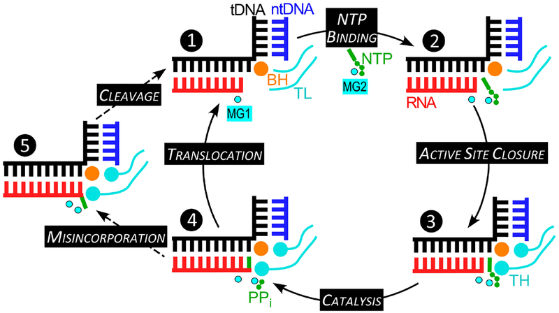
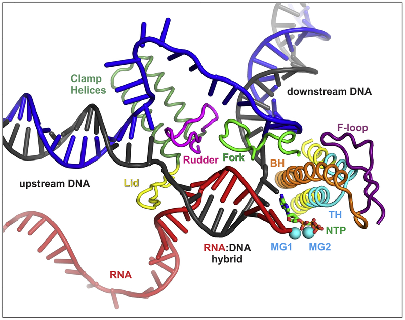
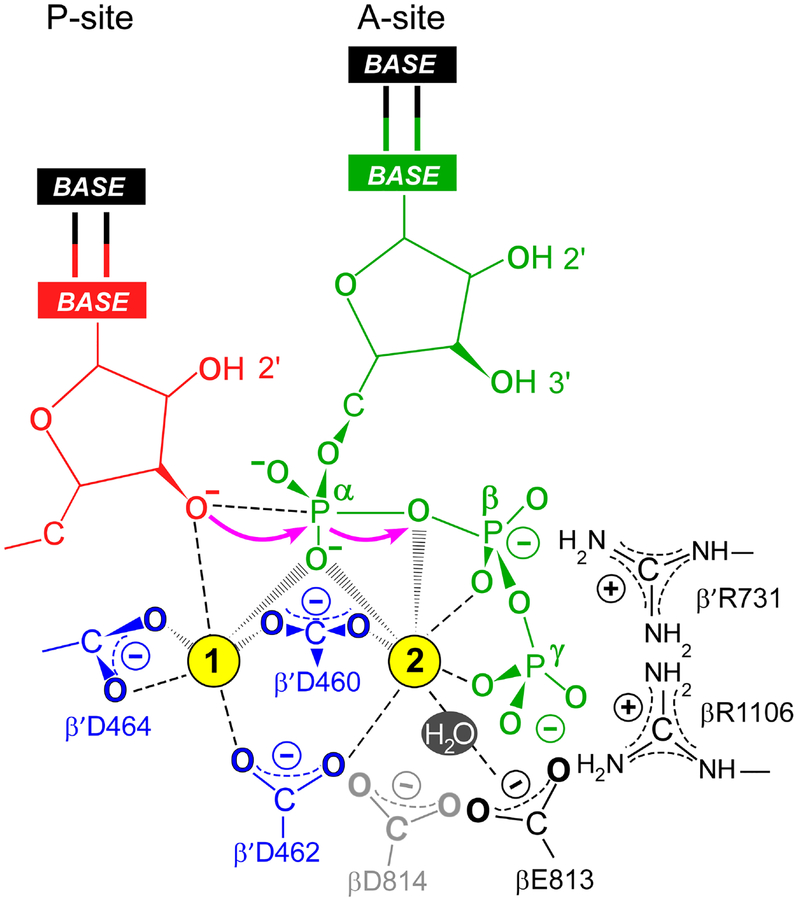
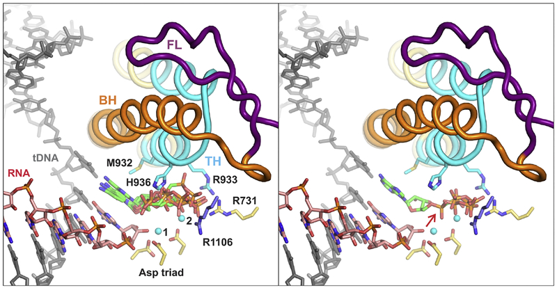
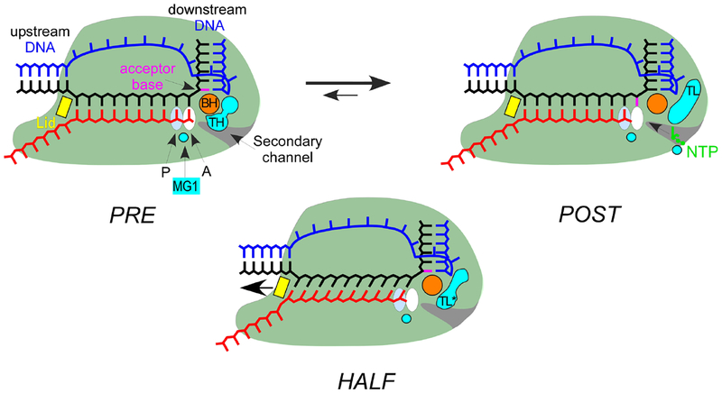
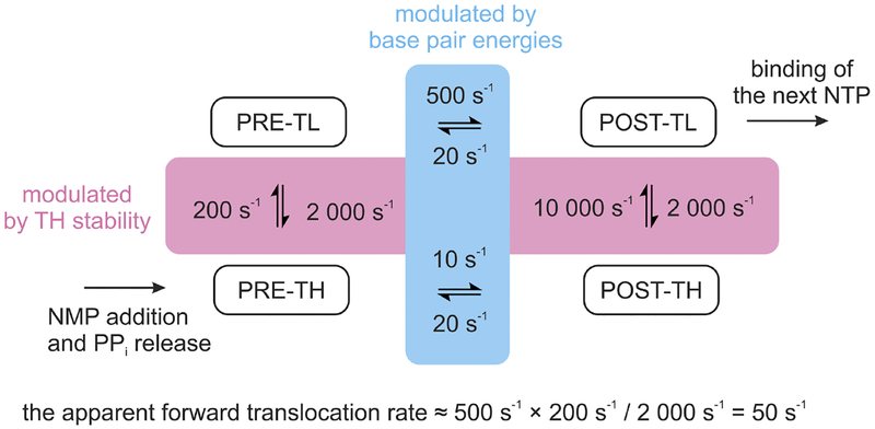
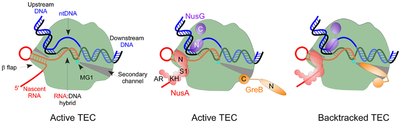
Multi-subunit DNA-dependent RNA polymerases synthesize all classes of cellular RNAs, ranging from short regulatory transcripts to gigantic messenger RNAs. RNA polymerase has to make each RNA product in just one try, even if it takes millions of successive nucleotide addition steps. During each step, RNA polymerase selects a correct substrate, adds it to a growing chain, and moves one nucleotide forward before repeating the cycle. However, RNA synthesis is anything but monotonous: RNA polymerase frequently pauses upon encountering mechanical, chemical and torsional barriers, sometimes stepping back and cleaving off nucleotides from the growing RNA chain. A picture in which these intermittent dynamics enable processive, accurate, and controllable RNA synthesis is emerging from complementary structural, biochemical, computational, and single-molecule studies. Here, we summarize our current understanding of the mechanism and regulation of the on-pathway transcription elongation. We review the details of substrate selection, catalysis, proofreading, and translocation, focusing on rate-limiting steps, structural elements that modulate them, and accessory proteins that appear to control RNA polymerase translocation.

Keywords: RNA polymerase; proofreading; transcription elongation; translocation; trigger loop.


Conflict of interest statement

Declarations of interest: none

Figures

Fig. 1.
Fig. 1.
The nucleotide addition and proofreading cycle. In the TEC, the RNA:DNA hybrid is separated from the downstream DNA, a duplex of the template (t; black) and non-template (nt; blue) DNA strands, by a 90° bend near the active site (indicated by the position of the catalytic MG1 ion) and the β’ bridge helix (BH; orange). The substrate NTP complexed with the low-affinity MG2 ion binds to the post-translocated TEC (❶) to form an inactive, preinsertion intermediate (❷). Upon folding of the tip of the β’ trigger loop (TL; cyan) into the trigger helices (TH), the NTP is repositioned to form the catalytically-competent insertion TEC, in which the active site is closed (❸). During catalysis, a cognate NMP is added to the RNA 3’ end (❹). The release of pyrophosphate (PPi) and the reverse TH→TL transition reset the cycle, with the transcript extended by one nucleotide. If an incorrect NMP is added to the growing chain (❺), RNAP backtracks by one nucleotide and removes two nucleotides from the mismatched 3’ terminus to restore the 3’ end in the active site.
Fig. 2.
Fig. 2.
Key structural features of RNAP. The nucleic acid strands, catalytic Mg2+ ions, the substrate NTP, and the β’ BH and TL elements are colored as in Figure 1. The β Fork loop 2 (green) and the β’ BH are positioned near the downstream edge of the transcription bubble. The TL resides on top of two α-helices (TL base, yellow) and alternates between a loop conformation (ordered in some and disordered in other structures) and a helical conformation called trigger helices (TH; cyan). TH form triple helical bundle with BH and interact with F-loop (FL, purple). The N-terminal arm of TH contacts NTP (green) in the active site. In some bacterial lineages, the C-terminal arm of TL contains a large insertion called SI3 (not shown). The β’ lid (yellow) and rudder (magenta) are placed at the junction of the upstream DNA duplex and the bubble. The figure was prepared from PDB ID 2O5J using PyMOL Molecular Graphics System, Version 2.0 Schrodinger, LLC; the upstream DNA and the single-stranded ntDNA modeled in as described previously [156].
Fig. 3.
Fig. 3.
The SN2 mechanism of nucleotide addition. In the post-translocated TEC, the RNA 3’ hydroxyl (red) is bound in the P-site and the incoming substrate NTP (green) is bound to the A-site through base pairing with the acceptor template base and interactions of basic residues (βR1106 and β’R731) with the β- and γ-phosphates. Two Mg2+ ions (cyan spheres) are required for catalysis. The high-affinity Mg1 is coordinated by the catalytic triad residues (β’Asp460, Asp462, and Asp464; blue). The low-affinity Mg2 ion bound to the β- and γ-phosphates of the NTP is coordinated by β’Asp460 and β’Asp462 and βGlu813; βAsp814 (gray) is likely dispensable for NMP addition but could be involved in RNA cleavage. The electron transfers occurring during the SN2 nucleophilic substitution are indicated with magenta arrows.
Fig. 4.
Fig. 4.
The TL folding into TH biases the substrate NTP from a pre-insertion (left panel) to the insertion pose (right panel) thereby repositioning the α-phosphate and aligning the PPi moiety inline for the attack of the 3’ OH group (right panel, red arrow). The fully closed active site depicted in both panels is drawn using the atomic coordinates of the T. thermophilus TEC with the ATP analogue AMPCPP in the insertion site (PDB ID 2O5J). In the left panel, the insertion pose of the AMPCPP was replaced with AMPCPP from T. thermophilus TEC where the active site closure is inhibited by streptolydigin (PDB ID 2PPB) and four CMPCPPs from partially closed active sites of the de novo initiation complexes (PDB IDs 4OIO, 4Q4Z, 5X22). In the right panel, the insertion pose of the AMPCPP was supplemented with two PPi molecules, one from the fully closed initially transcribing complex of E. coli RNAP (PDB ID 5IPL) and the other from the reiterative transcription complex of T. thermophilus RNAP (PDB ID 5VO8). Selected active site residues are shown as sticks; βArg1106 is colored blue, β’ residues are colored yellow or cyan. The figure was prepared using PyMOL Molecular Graphics System, Version 2.0 Schrodinger, LLC. The heterologous ligands (NTP analogues and PPi) were positioned in the closed active site using β subunits as anchors and “super” command of PyMOL.
Fig. 5.
Fig. 5.
Schematics of half and complete translocations. In the pre-translocated TEC (left), the 3’ hydroxyl is bound in the A-site and the TL is folded into TH, forming the THB. Translocation by one nt generates the post-translocated TEC (right) in which the 3’ OH is bound in the P-site, the tDNA acceptor base (magenta) is positioned in the A-site to pair with the incoming NTP substrate, and the TL is unfolded. In some TECs structures, RNA is fully translocated but tDNA translocates only partially [146] or not at all [37]; in both cases, the acceptor base has not moved to the A-site, blocking substrate binding (center). The tDNA The asynchronous translocation lengthens the RNA:DNA hybrid and changes its tilt, necessitating shifting the β’ lid (yellow) and possibly stabilizing an altered state of the TL (TL*).
Fig. 6.
Fig. 6.
A thermodynamic model integrating the contributions of TH-TL transition and base pair energies to the rate of the forward translocation along the DNA. Rates of the TH-TL transition and the forward translocation of the nucleic acids in the open active site are chosen semi-arbitrarily to result in an apparent forward translocation rate of 50 s−1. The equalities of (i) TL folding rates for pre- and post-translocated states and (ii) backward translocation rates in the closed and open active site are not coincidental. The backward translocation rates correspond to the smallest reported estimate and sequence-dependent variations by several fold are conceivable [46].
Fig. 7.
Fig. 7.
General transcription factors GreB, NusA, and NusG bind to different sites on the TEC to modulate elongation. The NTDs (N) of NusA and NusG interact with RNAP, whereas their CTDs (C) establish interactions with the nascent RNA and S10/Rho, respectively. The GreB CTD interacts with the β’ rim helices domain in active and backtracked TECs; the NTD swings into the secondary channel and activates RNA cleavage in backtracked TEC.

References

    1. Hurwitz J The discovery of RNA polymerase. J Biol Chem. 2005;280:42477–85. - PubMed
    1. Lee F, Squires CL, Squires C, Yanofsky C. Termination of transcription in vitro in the Escherichia coli tryptophan operon leader region. J Mol Biol. 1976;103:383–93. - PubMed
    1. Maizels NM. The nucleotide sequence of the lactose messenger ribonucleic acid transcribed from the UV5 promoter mutant of Escherichia coli. Proc Natl Acad Sci U S A. 1973;70:3585–9. - PMC - PubMed
    1. Kingston RE, Chamberlin MJ. Pausing and attenuation of in vitro transcription in the rrnB operon of E. coli. Cell. 1981;27:523–31. - PubMed
    1. Uptain SM, Kane CM, Chamberlin MJ. Basic mechanisms of transcript elongation and its regulation. Annu Rev Biochem. 1997;66:117–72. - PubMed

Publication types

MeSH terms

LinkOut - more resources