. 2019 Dec;76(24):5011-5025.
doi: 10.1007/s00018-019-03160-y. Epub 2019 Jun 1.

PGC-1α induced browning promotes involution and inhibits lactation in mammary glands

Affiliations

PGC-1α induced browning promotes involution and inhibits lactation in mammary glands

Elena Piccinin et al. Cell Mol Life Sci. 2019 Dec.

Abstract

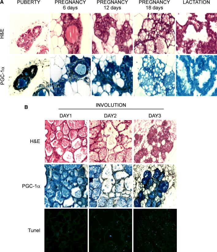
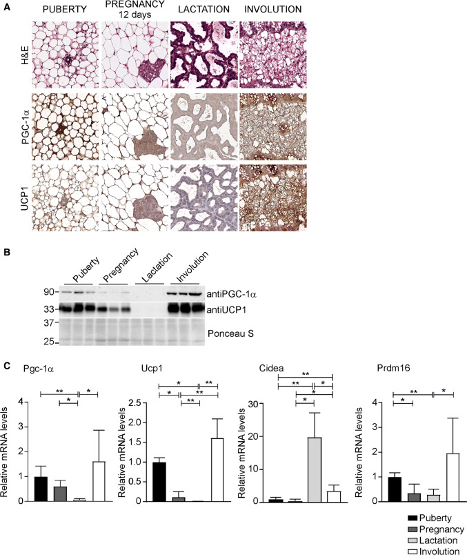
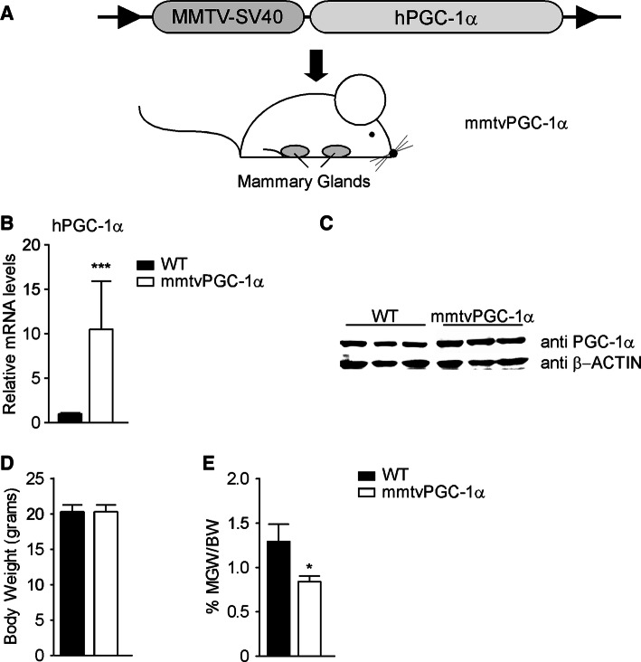
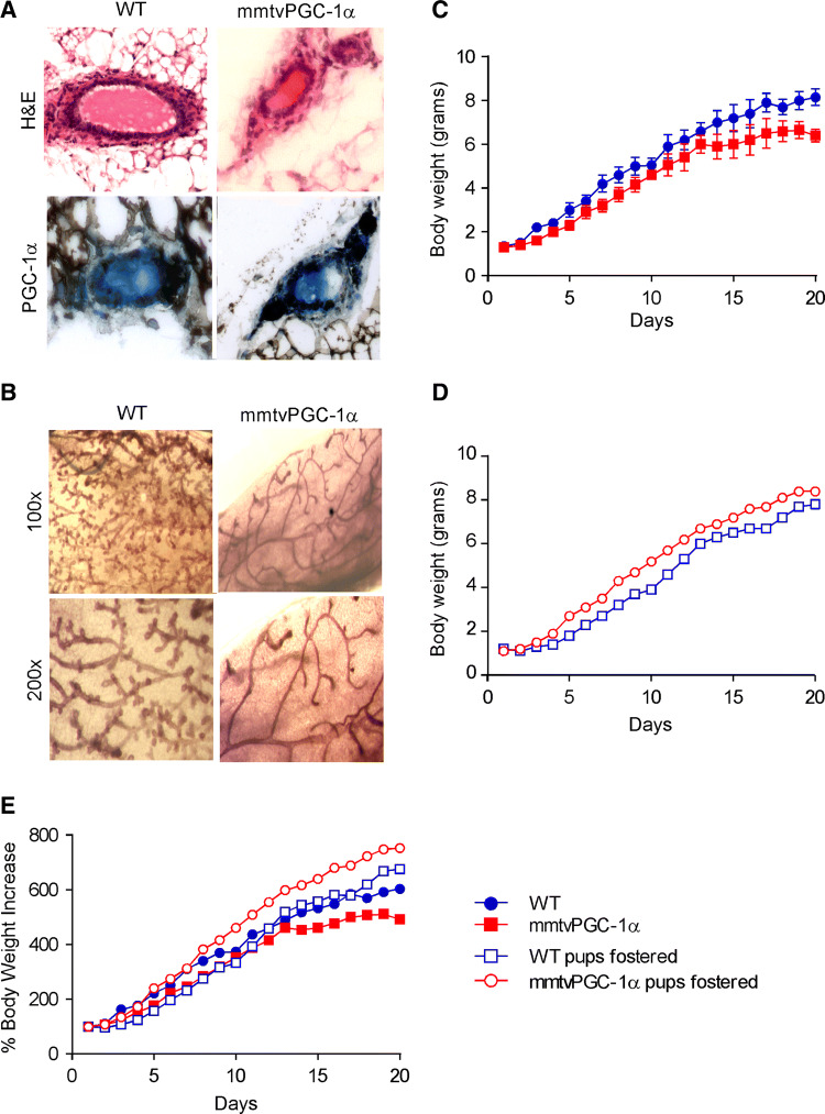
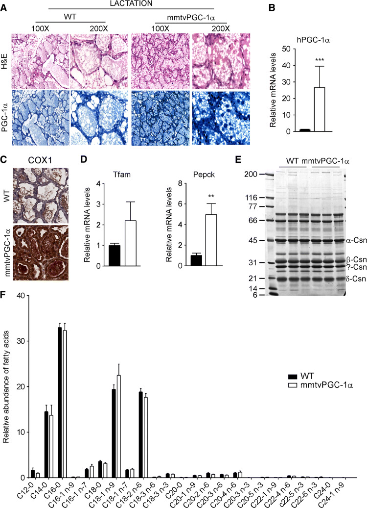
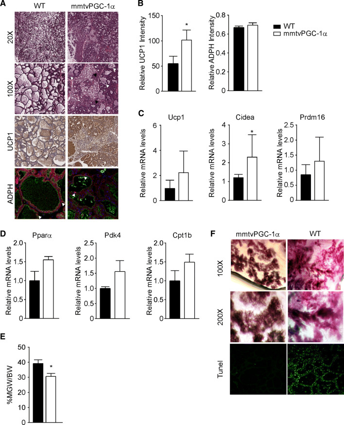
The PPARγ coactivator 1α (PGC-1α) is a transcriptional regulator of mitochondrial biogenesis and oxidative metabolism. Recent studies have highlighted a fundamental role of PGC-1α in promoting breast cancer progression and metastasis, but the physiological role of this coactivator in the development of mammary glands is still unknown. First, we show that PGC-1α is highly expressed during puberty and involution, but nearly disappeared in pregnancy and lactation. Then, taking advantage of a newly generated transgenic mouse model with a stable and specific overexpression of PGC-1α in mammary glands, we demonstrate that the re-expression of this coactivator during the lactation stage leads to a precocious regression of the mammary glands. Thus, we propose that PGC-1α action is non-essential during pregnancy and lactation, whereas it is indispensable during involution. The rapid preadipocyte-adipocyte transition, together with an increased rate of apoptosis promotes a premature mammary glands involution that cause lactation defects and pup growth retardation. Overall, we provide new insights in the comprehension of female reproductive cycles and lactation deficiency, thus opening new roads for mothers that cannot breastfeed.

Keywords: Adipocytes; Coactivator; Development; Involution; Mammary glands; Nuclear receptor.


Conflict of interest statement

The authors declare no competing financial interests.

Figures

Fig. 1
Fig. 1
Dynamic expression of PGC-1α protein levels during mammary glands development and involution. Highly expressed during puberty and in the first 12 days of pregnancy, the PGC-1α levels slightly disappear in the last days of pregnancy and during lactation. On the first day after cessation of lactation, PGC-1α starts to be re-expressed, reaching its steady state on day 3 of involution. a Staining of mammary glands sections from WT mice at different development stages with H&E (upper panel) and PGC-1α immunohistochemistry (lower panel) (n = 3 mice per stage; magnification ×200). b Mammary glands prepared from WT mice on the indicated days of involution and stained for H&E, PGC-1α immunohistochemistry, and TUNEL (n = 3 mice per stage; magnification ×100)
Fig. 2
Fig. 2
PGC-1α expression in mammary glands is associated with UCP1 induction. a Staining of mammary glands sections from WT mice at different development stages with H&E (upper panel) and PGC-1α (middle panel) and UCP1 immunohistochemistry (lower panel) (n = 5 mice per stage; magnification ×200). b Western blot analysis of PGC-1α and UCP1 on mammary glands samples throughout different developmental stages. Ponceau stain was used as loading control. c Relative expression of PGC-1α and fat-browning related genes, Ucp1, Cidea and Prdm16, at different stages of mammary glands development evaluated by real time qPCR. TBP was used as housekeeping gene. Comparison of different groups (n = 6, 7) was performed using Kruskal–Wallis test. Data are expressed as mean ± SEM (*p < 0.05; **p < 0.01)
Fig. 3
Fig. 3
Generation of mouse model with stable specific overexpression of human PGC-1α in mammary glands. A transgenic mouse model, mmtvPGC-1α, was generated by cloning the human sequence of PGC-1α downstream the mmtv promoter. Analysis of 8 weeks-old wild type and mmtvPGC-1α virgin mice shows a specific PGC-1α overexpression in the mammary glands of transgenic mice. a Scheme of the mmtvPGC-1α transgenic mouse model generated: the hPGC-1α coding sequence was cloned downstream of the mmtv promoter of mmtv-SV40-Bssk plasmid and then injected into the pronuclei of the fertilized eggs of the FVB/N mice. b Relative mRNA expression of hPGC-1α in mammary glands isolated from mmtvPGC-1α and WT control mice measured by real time qPCR, using TBP as housekeeping gene and WT mice as calibrator. c Western blot analysis of PGC-1α on mammary glands samples isolated from transgenic and WT mice. d Body weight and e inguinal mammary glands weight/body weight ratio (MGW/BW) of wild type and mmtvPGC-1α mice. Results are expressed as mean ± SEM (***p < 0.001, *p < 0.05). Comparison of wild type and transgenic mice (n = 6, 7) was performed using Mann–Whitney U test
Fig. 4
Fig. 4
Maternal PGC-1α overexpression decreases lobulogenesis in mammary glands during early development and results in growth retardation in the nursing neonates. Examination of WT and mmtvPGC-1α mammary glands from 8 weeks-old virgin mice reveals that PGC-1α overexpression in transgenic mice is associated with an impairment of ductal growth and ductal branching. a H&E staining and PGC-1α immunohistochemistry of mammary glands sections from WT and mmtvPGC-1α mice (magnification ×200). b Whole mount of the inguinal mammary glands in a virgin WT and a mmtvPGC-1α mouse. Developmental time line is depicted as the average body weight (± SEM) of pups from each of four independent litters on each postnatal day, nursed by the c biological mother and the d foster one. e Comparison of the four different group depicted as percentage of body weight increase. Blue color indicates WT pups, whereas red one specifies mmtvPGC-1α newborns. Circle symbol is used for WT breastfeeding mother and square one for mmtvPGC-1α breastfeeding mother. Closed circle indicates that pups are fed by biological mother, while open circle is used to denote pups fed by foster mother
Fig. 5
Fig. 5
PGC-1α overexpression during lactation does not affect milk quality. Analysis of WT and mmtvPGC-1α mammary glands from lactating females on day 10 of lactation shows that PGC-1α overexpression is preserved in transgenic mice during lactation. a H&E staining (left) and PGC-1α immunohistochemistry (right) of mammary glands sections from WT and mmtvPGC-1α female mice on 10th day of lactation. b Relative expression of human PGC-1α in lactating mammary glands evaluated by real time qPCR demonstrating that the transgene is not lost during lactation. TBP was used as housekeeping gene to normalize data and WT mice was used as calibrators c COX1 immunohistochemistry of mammary glands sections from WT and mmtvPGC-1α female mice on 10th day of lactation. d Relative expression of PGC-1α target genes, Tfam and Pepck, in lactating mammary glands evaluated by real-time qPCR. TBP was used as housekeeping gene. e A 12% SDS–polyacrylamide gel analysis of different types of casein in milk derived from lactating WT and mmtvPGC-1α mice. The sizes of the protein molecular weight markers are indicated in lane 1. f Fatty acids composition of milk harvested from lactating WT and mmtvPGC-1α females. Fatty acids were analyzed as fatty acid methyl esters (FAMEs) by gas–liquid chromatography. Comparison of WT and transgenic mice (n = 6, 7) was performed using Mann–Whitney U test. Results are expressed as mean ± SEM (*p < 0.05; ***p < 0.001)
Fig. 6
Fig. 6
Stable overexpression of PGC-1α during lactation promotes apoptosis and mammary glands regression. a Mammary tissue sections from 10 days lactating WT and mmtvPGC-1α females mice stained with H&E (different magnification), with UCP1 immunohistochemistry, and ADPH immunofluorescence. In ×100 H&E, white arrows indicate the shedding of epithelial cells into the alveolar lumen; and black arrows represent brown adipocytes. Immunolocalization of ADPH was performed using Alexa 594-conjugated antibodies against the N-terminus of mouse ADPH (red, arrowed). Luminal borders of mammary alveoli were identified by staining with Alexa 488-conjugated WGA (green). Nuclei were stained with TO-PRO-3 (blue). b Quantification of UCP1 and ADPH immunostaining. Relative expression of c fat-browning related genes, Ucp1, Cidea and Prdm16, and d β-oxidation genes, Pparα, Pdk4 and Cpt1b, in lactating mammary glands evaluated by real time qPCR. TBP was used as housekeeping gene. e Inguinal mammary glands weight to body weight ratio (MGW/BW) of WT and mmtvPGC-1α lactating mice. f Low and high magnification of whole mount staining together with TUNEL staining of mammary glands isolated from lactating mmtvPGC-1α and WT mice. Comparison of wild-type and transgenic mice (n = 6, 7) was performed using Mann–Whitney U test. Data are expressed as mean ± SEM (*p < 0.05; **p < 0.01)

Similar articles

Cited by

References

    1. Watson CJ, Kreuzaler PA. Remodeling mechanisms of the mammary gland during involution. Int J Dev Biol. 2011;55:757–762. - PubMed
    1. Neville MC. Physiology of lactation. Clin Perinatol. 1999;26(251–79):v. - PubMed
    1. Neville MC, Picciano MF. Regulation of milk lipid secretion and composition. Annu Rev Nutr. 1997;17:159–183. - PubMed
    1. Allen JC, Keller RP, Archer P, Neville MC. Studies in human lactation: milk composition and daily secretion rates of macronutrients in the first year of lactation. Am J Clin Nutr. 1991;54:69–80. - PubMed
    1. Schwertfeger KL, McManaman JL, Palmer CA, Neville MC, Anderson SM. Expression of constitutively activated Akt in the mammary gland leads to excess lipid synthesis during pregnancy and lactation. J Lipid Res. 2003;44:1100–1112. - PubMed

Substances

LinkOut - more resources