

Indirect visualization of endogenous nuclear actin by correlative light and electron microscopy (CLEM) using an actin-directed chromobody
- PMID: 31154480
- PMCID: PMC6675784
- DOI: 10.1007/s00418-019-01795-3
Indirect visualization of endogenous nuclear actin by correlative light and electron microscopy (CLEM) using an actin-directed chromobody
Abstract
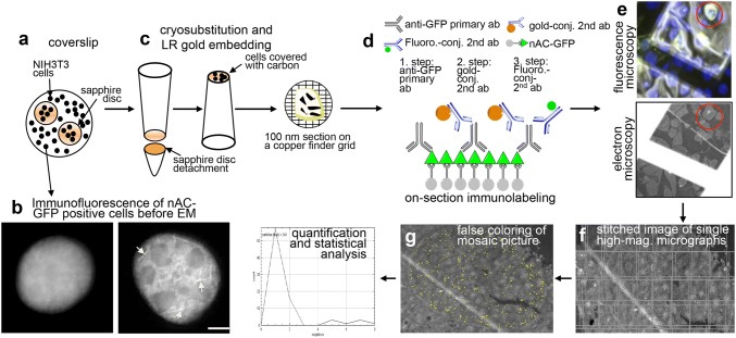
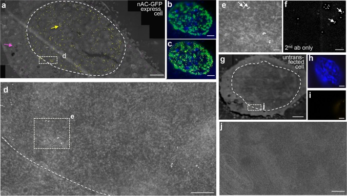
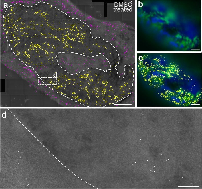
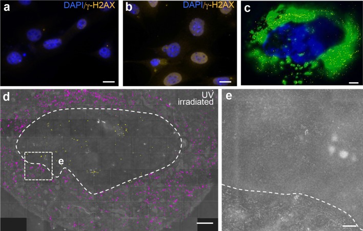
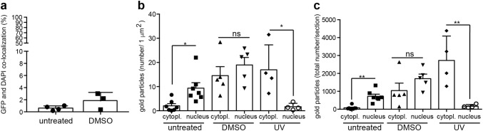
Actin fulfills important cytoplasmic but also nuclear functions in eukaryotic cells. In the nucleus, actin modulates gene expression and chromatin remodeling. Monomeric (G-actin) and polymerized actin (F-actin) have been analyzed by fluorescence microscopy in the nucleus; however, the resolution at the ultrastructural level has not been investigated in great detail. We provide a first documentation of nuclear actin in mouse fibroblasts by electron microscopy (EM). For this, we employed correlative light and electron microscopy on the same section using actin-directed nanobodies recognizing endogenous monomeric and polymeric actin proteins (so-called nuclear Actin-chromobody-GFP; nAC-GFP). Indeed, using this strategy, we could identify actin proteins present in the nucleus. Here, immunogold-labeled actin proteins were spread throughout the entire nucleoplasm. Of note, nuclear actin was complementarily localized to DAPI-positive areas, the latter marking preferentially transcriptionally inactive heterochromatin. Since actin aggregates in rod structures upon cell stress including neurodegeneration, we analyzed nuclear actin at the ultrastructural level after DMSO or UV-mediated cell damage. In those cells the ratio between cytoplasmic and nuclear gold-labeled actin proteins was altered compared to untreated control cells. In summary, this EM analysis (i) confirmed the presence of endogenous nuclear actin at ultrastructural resolution, (ii) revealed the actin abundance in less chromatin-dense regions potentially reflecting more transcriptionally active euchromatin rather than transcriptionally inactive heterochromatin and (iii) showed an altered abundance of actin-associated gold particles upon cell stress.
Keywords: Actin; EM; Immunogold; Nanobody; Neurodegeneration; Nucleus.
Conflict of interest statement
The authors declare no competing interests.
Figures
References
-
- Baarlink C, Plessner M, Sherrard A, Morita K, Misu S, Virant D, Kleinschnitz EM, Harniman R, Alibhai D, Baumeister S, Miyamoto K, Endesfelder U, Kaidi A, Grosse R. A transient pool of nuclear F-actin at mitotic exit controls chromatin organization. Nat Cell Biol. 2017;19:1389–1399. doi: 10.1038/ncb3641. - DOI - PubMed
MeSH terms
Substances
Grants and funding
LinkOut - more resources
Full Text Sources
