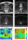
Evaluation of diffusion kurtosis and diffusivity from baseline staging MRI as predictive biomarkers for response to neoadjuvant chemoradiation in locally advanced rectal cancer
- PMID: 31154482
- PMCID: PMC7457148
- DOI: 10.1007/s00261-019-02073-5
Evaluation of diffusion kurtosis and diffusivity from baseline staging MRI as predictive biomarkers for response to neoadjuvant chemoradiation in locally advanced rectal cancer
Abstract
Purpose: To evaluate the role of diffusion kurtosis and diffusivity as potential imaging biomarkers to predict response to neoadjuvant chemoradiation therapy (CRT) from baseline staging magnetic resonance imaging (MRI) in locally advanced rectal cancer (LARC).
Materials and methods: This retrospective study included 45 consecutive patients (31 male/14 female) who underwent baseline MRI with high b-value sequences (up to 1500 mm/s2) for LARC followed by neoadjuvant chemoradiation and surgical resection. The mean age was 57.4 years (range 34.2-72.9). An abdominal radiologist using open source software manually segmented T2-weighted images. Segmentations were used to derive diffusion kurtosis and diffusivity from diffusion-weighted images as well as volumetric data. These data were analyzed with regard to tumor regression grade (TRG) using the four-tier American Joint Committee on Cancer (AJCC) classification, TRG 0-3. Proportional odds regression was used to analyze the four-level ordinal outcome. A sensitivity analysis was performed using univariable logistic regression for binary TRG groups, TRG 0/1 (> 90% response), or TRG 2/3 (< 90% response). p < 0.05 was considered significant throughout.
Results: In the univariable proportional odds regression analysis, higher diffusivity summary (Dsum) values were observed to be significantly associated with higher odds of being in one or more favorable TRG group (TRG 0 or 1). In other words, on average, patients with higher Dsum values were more likely to be in a more favorable TRG group. These results are mostly consistent with the sensitivity analysis, in which higher values for most Dsum values [all but region of interest (ROI)-max D median (p = 0.08)] were observed to be significantly associated with higher odds of being TRG 0 or 1. Tumor volume of interest (VOI) and ROI volume, ROI kurtosis mean and median, and VOI kurtosis mean and median were not significantly associated with TRG.
Conclusion: Diffusivity derived from the baseline staging MRI, but not diffusion kurtosis or volumetric data, is associated with TRG and therefore shows promise as a potential imaging biomarker to predict the response to neoadjuvant chemotherapy in LARC.
Clinical relevance statement: Diffusivity shows promise as a potential imaging biomarker to predict AJCC TRG following neoadjuvant CRT, which has implications for risk stratification. Patients with TRG 0/1 have 5-year disease-free survival (DFS) of 90-98%, as opposed to those who are TRG 2/3 with 5-year DFS of 68-73%.
Keywords: Diffusion kurtosis; Diffusivity; Imaging biomarker; Neoadjuvant chemoradiation; Rectal cancer; Tumor regression grade.
Conflict of interest statement
Compliance with Ethical Standards
Conflict of Interest: The authors declare that they have no relevant conflicts of interest.
Figures
References
-
- Benson AB 3rd, Venook AP, Al-Hawary MM, Cederquist L, Chen YJ, Ciombor KK, Cohen S, Cooper HS, Deming D, Engstrom PF, Grem JL, Grothey A, Hochster HS, Hoffe S, Hunt S, Kamel A, Kirilcuk N, Krishnamurthi S, Messersmith WA, Meyerhardt J, Mulcahy MF, Murphy JD, Nurkin S, Saltz L, Sharma S, Shibata D, Skibber JM, Sofocleous CT, Stoffel EM, Stotsky-Himelfarb E, Willett CG, Wuthrick E, Gregory KM, Gurski L, Freedman-Cass DA (2018) Rectal Cancer, Version 2.2018, NCCN Clinical Practice Guidelines in Oncology. J Natl Compr Canc Netw 16 (7):874–901. doi: 10.6004/jnccn.2018.0061 - DOI - PMC - PubMed
-
- Trakarnsanga A, Gonen M, Shia J, Nash GM, Temple LK, Guillem JG, Paty PB, Goodman KA, Wu A, Gollub M, Segal N, Saltz L, Garcia-Aguilar J, Weiser MR (2014) Comparison of tumor regression grade systems for locally advanced rectal cancer after multimodality treatment. J Natl Cancer Inst 106 (10). doi: 10.1093/jnci/dju248 - DOI - PMC - PubMed
MeSH terms
Substances
Grants and funding
LinkOut - more resources
Full Text Sources
Research Materials


