. 2019 Jul 23;140(4):319-335.
doi: 10.1161/CIRCULATIONAHA.118.039345. Epub 2019 Jun 3.

Novel Role for the AnxA1-Fpr2/ALX Signaling Axis as a Key Regulator of Platelet Function to Promote Resolution of Inflammation

Affiliations

Novel Role for the AnxA1-Fpr2/ALX Signaling Axis as a Key Regulator of Platelet Function to Promote Resolution of Inflammation

Elena Y Senchenkova et al. Circulation. .

Erratum in

Abstract

Background: Ischemia reperfusion injury (I/RI) is a common complication of cardiovascular diseases. Resolution of detrimental I/RI-generated prothrombotic and proinflammatory responses is essential to restore homeostasis. Platelets play a crucial part in the integration of thrombosis and inflammation. Their role as participants in the resolution of thromboinflammation is underappreciated; therefore we used pharmacological and genetic approaches, coupled with murine and clinical samples, to uncover key concepts underlying this role.

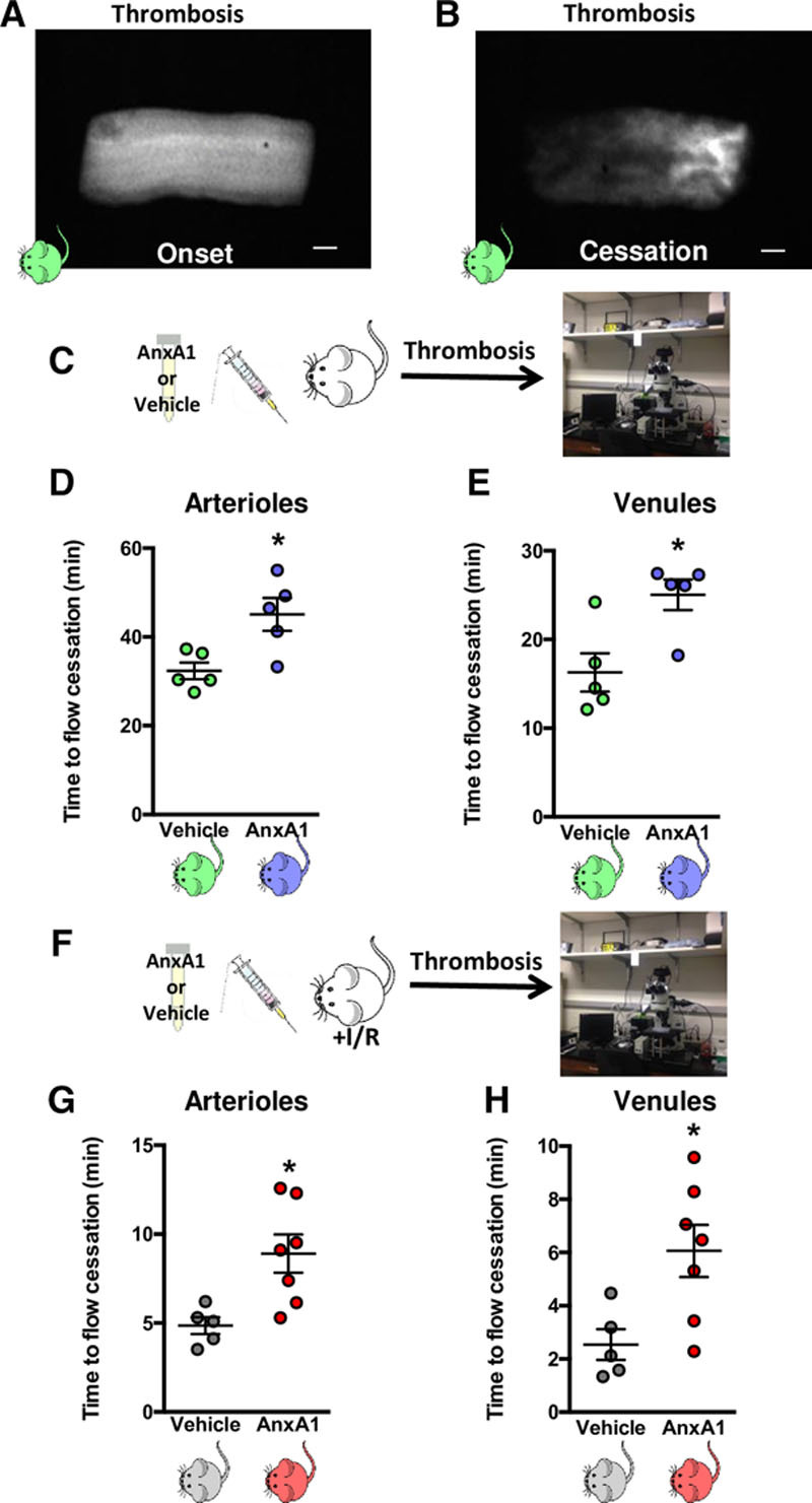
Methods: Middle cerebral artery occlusion with reperfusion was performed in wild-type or annexin A1 (AnxA1) knockout (AnxA1-/-) mice. Fluorescence intravital microscopy was used to visualize cellular trafficking and to monitor light/dye-induced thrombosis. The mice were treated with vehicle, AnxA1 (3.3 mg/kg), WRW4 (1.8 mg/kg), or all 3, and the effect of AnxA1 was determined in vivo and in vitro.

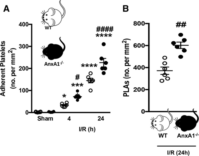
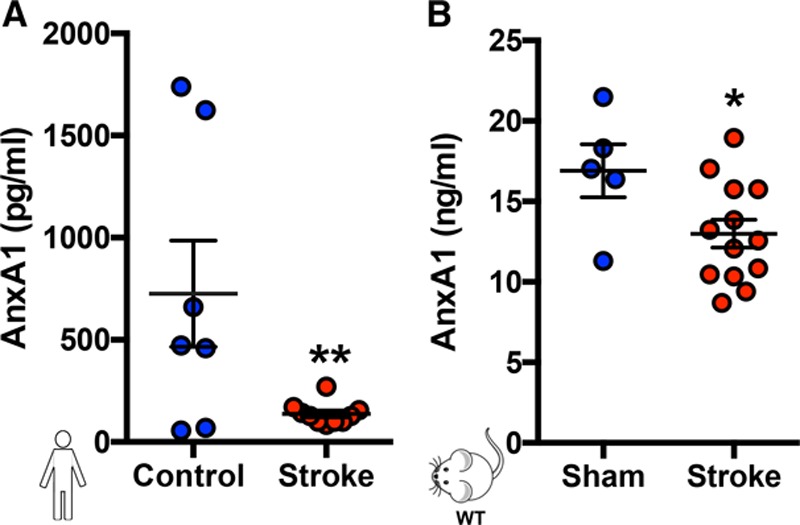
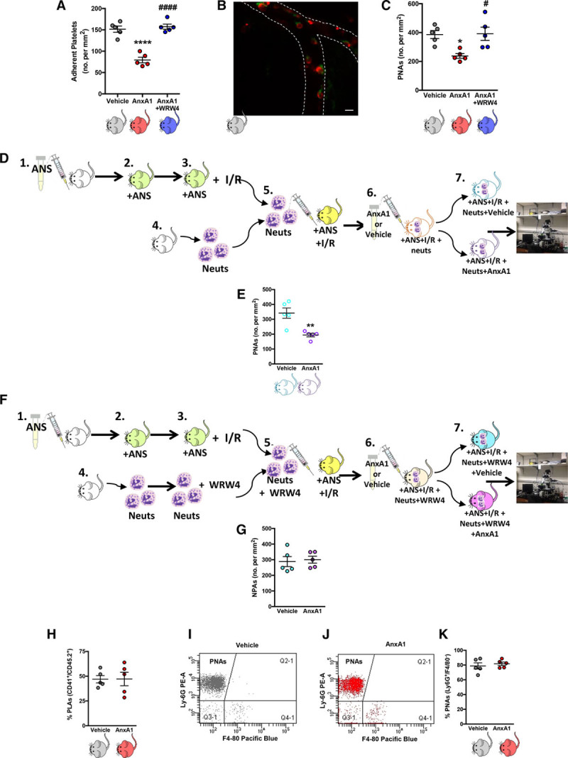
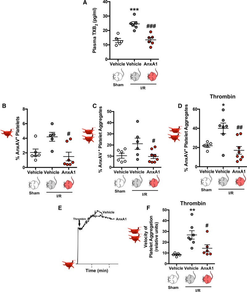
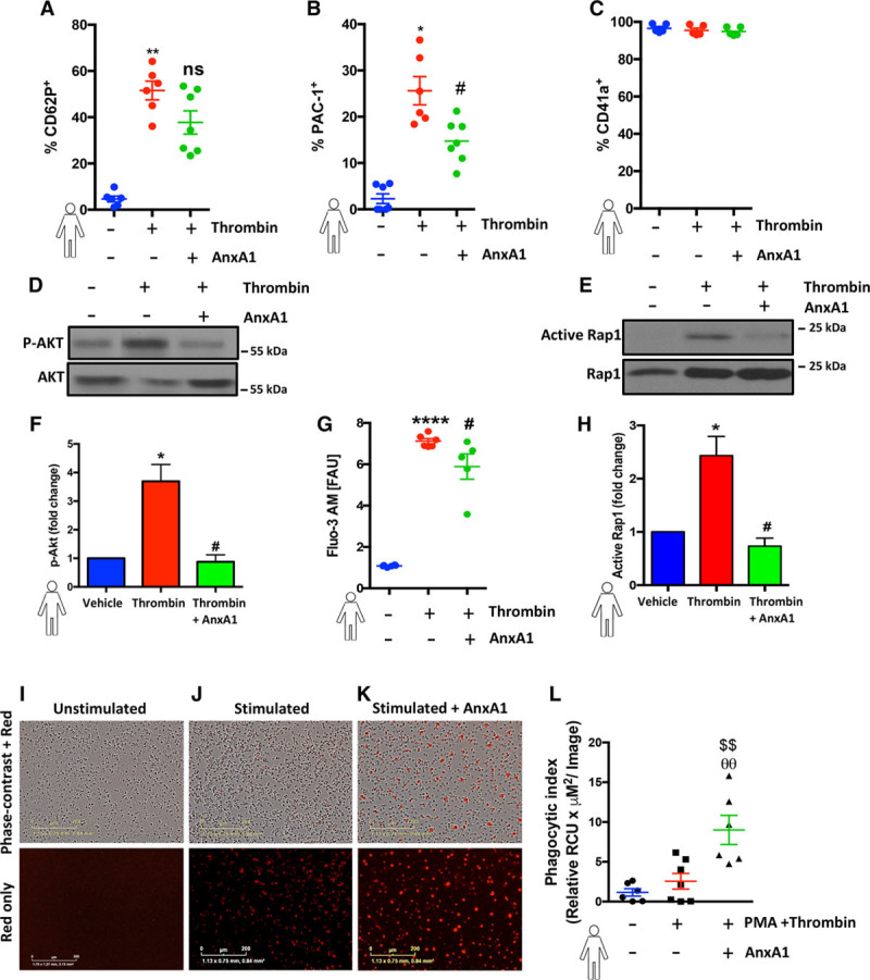
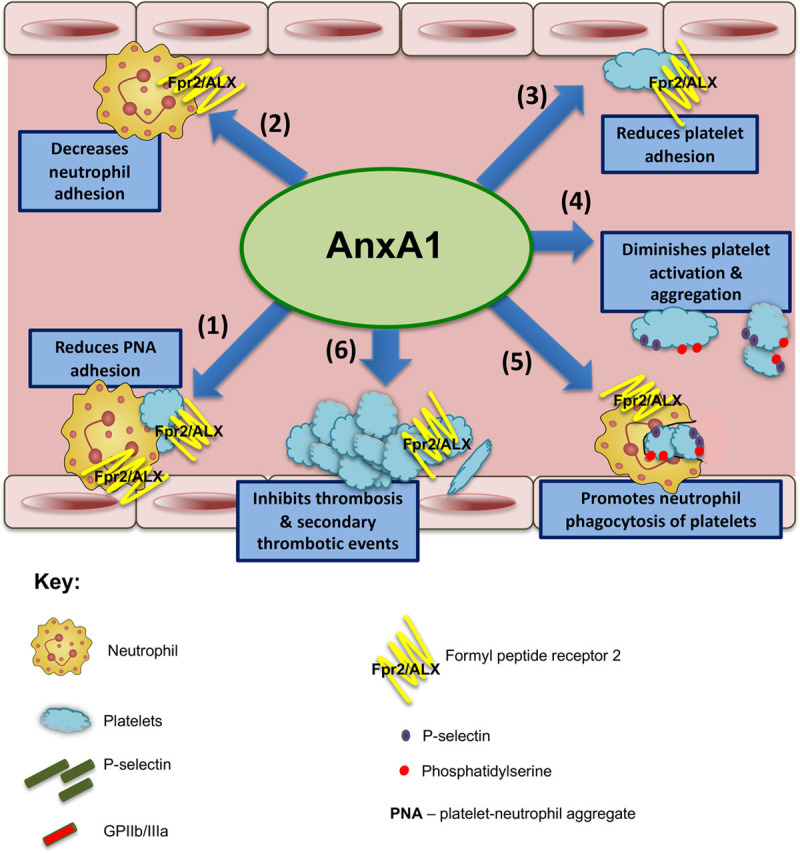
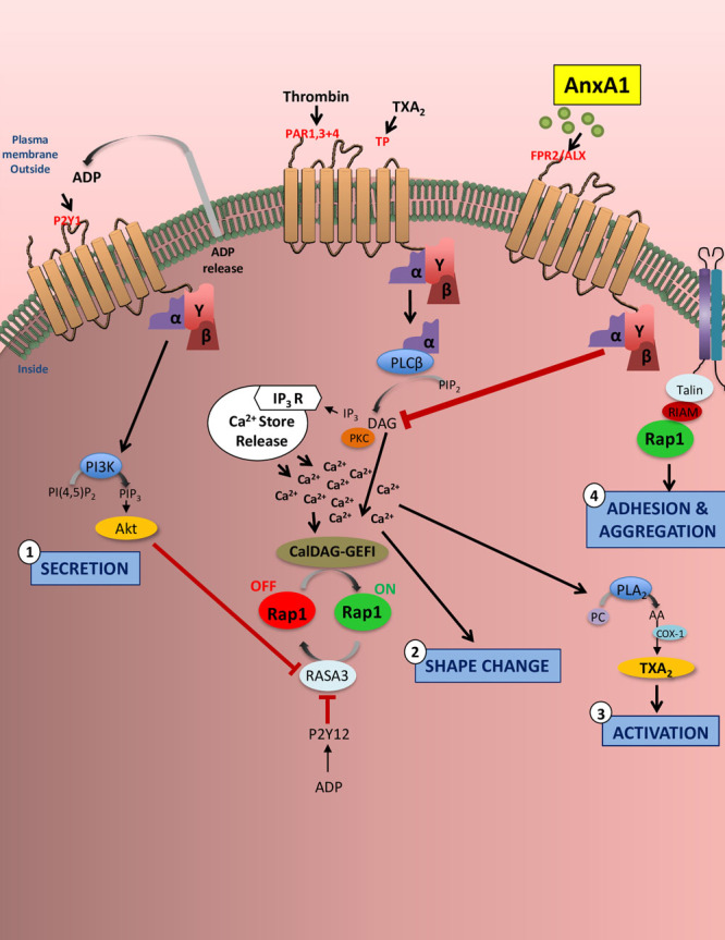
Results: Intravital microscopy revealed heightened platelet adherence and aggregate formation post I/RI, which were further exacerbated in AnxA1-/- mice. AnxA1 administration regulated platelet function directly (eg, via reducing thromboxane B2 and modulating phosphatidylserine expression) to promote cerebral protection post-I/RI and act as an effective preventative strategy for stroke by reducing platelet activation, aggregate formation, and cerebral thrombosis, a prerequisite for ischemic stroke. To translate these findings into a clinical setting, we show that AnxA1 plasma levels are reduced in human and murine stroke and that AnxA1 is able to act on human platelets, suppressing classic thrombin-induced inside-out signaling events (eg, Akt activation, intracellular calcium release, and Ras-associated protein 1 [Rap1] expression) to decrease αIIbβ3 activation without altering its surface expression. AnxA1 also selectively modifies cell surface determinants (eg, phosphatidylserine) to promote platelet phagocytosis by neutrophils, thereby driving active resolution. (n=5-13 mice/group or 7-10 humans/group.) Conclusions: AnxA1 affords protection by altering the platelet phenotype in cerebral I/RI from propathogenic to regulatory and reducing the propensity for platelets to aggregate and cause thrombosis by affecting integrin (αIIbβ3) activation, a previously unknown phenomenon. Thus, our data reveal a novel multifaceted role for AnxA1 to act both as a therapeutic and a prophylactic drug via its ability to promote endogenous proresolving, antithromboinflammatory circuits in cerebral I/RI. Collectively, these results further advance our knowledge and understanding in the field of platelet and resolution biology.

Keywords: annexin A1; formyl peptide receptor; inflammation; integrins; stroke; thrombosis.


Figures

Figure 1.
Figure 1.
Platelet and platelet–leukocyte interactions in the cerebral microcirculation are heightened in annexin A1 knockout (AnxA1−/−) mice after ischemia reperfusion injury (I/R). Wild-type (WT) and AnxA1−/− mice were subjected to transient middle cerebral artery occlusion for 60 min, followed by 4- or 24-h reperfusion. Intravital fluorescence microscopy was performed to assess cellular interactions in the cerebral microcirculation (pial vessels) of mice subjected to cerebral I/R. Platelets were labeled with carboxyfluorescein succinimidyl ester (90 µmol/L), and leukocytes were labeled with rhodamine 6G (0.02%). Platelet interactions were quantified in terms of numbers (no.) of adherent platelets on the endothelium (A, cells stationary for ≥2 s) and platelets interacting directly with adherent leukocytes on the endothelium (B), termed platelet–leukocyte aggregates (PLAs). The data are means±SEM of 6 mice per group with 2 to 3 vessels per mouse and assessed by ANOVA with Bonferroni post hoc test (A) or Mann–Whitney test (B). *P<0.05, ***P<0.001, and ****P<0.0001 vs sham control of the same genotype. #P<0.05, ##P<0.01, and ####P<0.0001 vs a different genotype at the same time point.
Figure 2.
Figure 2.
Plasma levels of AnxA1 are reduced in human and murine stroke. Plasma was collected from control volunteers and stroke patients (A) and mice with and without stroke (B, transient middle cerebral artery occlusion for 60 min followed by 24 h of reperfusion). Annexin A1 (AnxA1) levels were assessed by ELISA. The data are means±SEM of 7 to 13, with 10 humans per group (8 samples were collected from control volunteers, but 1 outlier [defined as at least 2 standard deviations] was removed; 11 samples were collected from stroke patients, but 1 outlier was removed) and 5 to 13 mice per group and assessed by a Student t test (A and B). *P<0.05 and **P<0.01 vs control. WT indicates wild type.
Figure 3.
Figure 3.
Administration of exogenous annexin A1 (AnxA1) moderates platelet interactions in the brain microcirculation after ischemia reperfusion injury (I/R). Wild-type mice were subjected to transient middle cerebral artery occlusion for 60 min, followed by 24 h of reperfusion (tMCAo/R). A, Vehicle, AnxA1 (3.3 mg/kg), or WRW4 (1.8 mg/kg) were administered intravenously at the start of reperfusion and numbers (no.) of platelet adhesion (cells stationary for at least 2 s) were quantified. B, Representative image of cerebral platelet-neutrophil aggregates (PNAs) 24 h after reperfusion as measured by confocal intravital microscopy. Neutrophils (Neuts, arrow) were labeled with eFluor 488 (green)-labeled anti-mouse Ly-6G, 2 μg/mouse. Platelets (arrowhead) were labeled with Dylight 649 (red)-labeled antimouse CD42, 1 μg/mouse. The scale bar indicates 20 μm. C, Quantification of endothelial-PNAs in the cerebral microcirculation. D, Adoptive transfer experiments were performed by injecting neutrophils isolated from donor mice into neutropenic recipient mice. In steps 1 and 2, recipient mice were rendered neutropenic by administration of antineutrophil serum (ANS). In step 3, neutropenic mice were subjected to cerebral I/R by tMCAo/R. In step 4, neutrophils were isolated from donor mice. In step 5, neutrophils were injected into neutropenic recipient mice subjected MCAo/R. In step 6, the mice were then treated with vehicle (saline) or AnxA1 (3.3 mg/kg) for 30 min before intravital microscopy, which is shown in step 7. E, Quantification of PNAs in the cerebral microcirculation from D using intravital microscopy. F, Adoptive transfer experiments were performed by injecting neutrophils isolated from donor mice into neutropenic recipient mice. In steps 1 and 2, recipient mice were rendered neutropenic by administration of ANS. In step 3, neutropenic mice were subjected to cerebral I/R by tMCAo/R. In step 6, the mice were then treated with vehicle (saline) or AnxA1 (3.3 mg/kg) for 30 min before intravital microscopy, which is shown in step 7. G, Quantification of NPAs (neutrophil platelet aggregates) in the cerebral microcirculation from F using intravital microscopy. H through K, Blood samples were taken from wild-type mice subjected to MCAo, followed by a 24-h reperfusion and treatment with vehicle (saline) or AnxA1 and analyzed by flow cytometry to assess systemic aggregate formation: quantification of circulating. H, Platelet–leukocyte aggregates (PLAs, CD45.2+, and CD41+). I and J, Flow cytometry population groups of PNAs, which are denoted within the CD45.2+, CD41+ population as Ly-6G+, and F4/80 aggregates. K, Quantification of circulating PNAs. The data are means±SEM of 5 mice/group and assessed by ANOVA with a Bonferroni post hoc test (A and B), Mann–Whitney test (E), or a Student t test (G, H, and K). *P<0.05 and ****P<0.0001 vs vehicle (saline)–treated control. #P<0.05 and ####P<0.0001 vs AnxA1.
Figure 4.
Figure 4.
Annexin A1 (AnxA1) reduces platelet activation/aggregation postischemia reperfusion injury (I/R). Wild-type mice were subjected to sham or transient middle cerebral artery occlusion for 60 min, followed by 24 h of reperfusion. Vehicle (saline) or AnxA1 (3.3 mg/kg) was administered intravenously at the start of reperfusion. A, At the end of reperfusion, plasma thromboxane B2 (TXB2) levels were measured. B through F, Platelets were isolated for quantification as follows. B through D, Using flow cytometry, phosphatidylserine externalization (using annexin AV [AnxAV] staining, presented as a percentage of AnxAV-positive platelets) was quantified on single platelets (B), platelet–platelet aggregates (C), or platelet–platelet aggregates after further stimulation with thrombin (D, 0.1 U/mL). E, Representative platelet aggregation velocity chart in response to thrombin (0.1 U/mL). F, Velocity of aggregate formation was measured using a low-angle light-scattering technique. The data are means±SEM of 5 to 8 mice per group and assessed by ANOVA with Bonferroni post hoc test (A through D and F). *P<0.05, **P<0.01, and ***P<0.001 vs platelets from vehicle treated sham mice. #P<0.05, ##P<0.001, and ###P<0.001 vs platelets from vehicle-treated I/R mice.
Figure 5.
Figure 5.
Annexin A1 (AnxA1) protects against initial cerebral thrombosis and development of subsequent thrombotic events. A and B, Still images of cerebral pial vessels showing onset (A) and cessation (B) of blood flow in the light/dye-induced thrombus model in wild-type (WT) mice. C through E, Effects of AnxA1 (C, 1 µg/mouse) or saline vehicle injected 20 min before the onset of thrombus formation in cerebral arterioles (D) and venules (E). F, The mice were subjected to transient middle cerebral artery occlusion for 60 min, followed by 24 h of reperfusion. G and H, At the end of reperfusion, the mice were treated with vehicle (saline) or AnxA1 (1 µg/mouse) 20 min before the onset of light/dye-induced thrombus formation in cerebral arterioles (G) and venules (H). The data are means±SEM of 5 to 7 mice/group and assessed by a Student t test (D, E, G, and H). *P<0.05 vs vehicle treated control. The scale bar indicates 10 μm. I/R indicates ischemia reperfusion injury.
Figure 6.
Figure 6.
Annexin A1 (AnxA1) decreases human platelet activation and promotes phagocytosis. A through C, Human platelets (1×10⁶) were isolated, washed, preincubated with 100 ng of AnxA1 (30 min at 37°C), and stimulated with thrombin and using flow cytometry; levels of P-selectin expression (A), activated integrin αIIbβ3 (PAC-1; B), and surface levels of αIIbβ3 (CD41a; C) were quantified. D through H, Freshly isolated platelets (1×10⁶) were treated with AnxA1 at 100 ng and treated with vehicle (saline), thrombin (0.1 U), or thrombin and AnxA1 (100 ng) for 3 min, and cells were subsequently lysed and blotted for phospho-AKT (P-AKT) and total AKT (n=3 individual donors/group; D and F) and active Ras-associated protein 1 (Rap1) and Rap1 (n=5 individual donors/group; E and H) or changes cytosolic Ca2+ in fluo-3-acetoxymethyl ester (Fluo-3 AM)–labeled platelets was determined in the presence or absence of AnxA1 (G). F and H, Akt and Rap1 were quantified by ImageJ. The ratio of p-AKT/AKT and Active Rap1/Rap1 was calculated to get the fold change in reference to the untreated control. G, The graph shows the mean fluorescence intensity (fluorescent arbitrary units [FAU]) at baseline (no stimulation), in stimulated platelets (thrombin 0.1 U/1×106 platelets), and in platelets incubated with AnxA1 (100 ng/1×106 platelets) and stimulated with thrombin. I through K, Representative images of phagocytosis using pHrodo dye from 1 experiment at 1 h. The scale bars indicate 200 μm. I, Human platelets (1×10⁶) preincubated with 100 ng of AnxA1 and 2.5 μmol/L pHrodo Red AM intracellular pH indicator for 20 min, followed by opsonization with 1% human serum, with and without thrombin stimulation, were coincubated with neutrophils (with and without phorbol myristate acetate [PMA]) for 1 h at 37°C in a 96-well flat clear-bottomed black-walled plate, and fluorescence emission was measured in the IncuCyteZOOM (Essen BioScience) imaging platform. The data are means±SEM of 6 to 7 individual donors/group unless otherwise stated (2 outliers [defined as at least 2 standard deviations] were removed for A, and 1 outlier was removed for B and assessed by a Student t test (A and B), Kruskal–Wallis with Dunn’s test (F and G), or ANOVA with Bonferroni post hoc test (C, H, and L). *P<0.05, **P<0.01, and ****P<0.0001 vs unstimulated platelets. #P<0.05, ##P<0.01 vs thrombin-stimulated platelets. θθP<0.01 vs unstimulated neutrophils and platelets. $$P<0.01 vs PMA-stimulated neutrophils and thrombin-stimulated platelets. ns indicates not significant.
Figure 7.
Figure 7.
Annexin A1 (AnxA1) mitigates thromboinflammatory responses and promotes resolution during ischemia reperfusion injury.Schematic overview shows the effects of AnxA1 on both thrombotic and inflammatory events as follows. Step 1 reduces platelet–neutrophil aggregate (PNA) adhesion on endothelial cells in the cerebral microvasculature; steps 2 and 3 decrease neutrophil (step 2) and platelet adhesion (step 3); step 4 diminishes platelet activation and aggregate velocity, promoting a phosphatidylserine and P-selectin–dependent clearance program; step 5 promotes neutrophil phagocytosis of platelets for resolution; and step 6 protects against enhanced intravascular thrombus formation and secondary thrombotic events in the cerebral microvasculature. ALX indicates the lipoxin A4 receptor; and Fpr2, formyl peptide receptor 2.
Figure 8.
Figure 8.
Annexin A1 (AnxA1) reduces platelet activation and integrin activity. In thromboinflammation, platelets are stimulated via G-protein–coupled receptors (eg, protease-activated receptors 1, 3, and 4; human thromboxane A2 receptor [TPs]; P2Y12; P2Y1; and P2X1) and immunoreceptor tyrosine-based activation motif-coupled receptors with agonists such as thrombin, thromboxane A2 (TXA2), and ADP. This stimulation results in downstream signaling events that increase cytosolic Ca2+ levels leading to platelet secretion (step 2), shape change (step 2), activation (step 3), and adhesion and aggregation (step 4). The small GTPase Ras-related protein 1 (RAP1) regulates multiple functional responses in platelets, in particular integrin activation. As with many platelet components, Rap1 regulators CalDAG–GEFI (calcium- and DAG-regulated guanine exchange factor-1) and RASA3 are affected by small changes in the cytosolic Ca2+ concentration, leading to rapid αIIbβ3 integrin activation. To sustain αIIbβ3 activation, ADP (which is rapidly released and secreted from storage granules on increased Ca2+ concentrations) must engage P2Y12 receptors and phosphatidylinositol 3-kinase (PI3K) signaling, which reduces RASA3 activity. AnxA1 acts via platelet formyl peptide receptor 2 (also known as the lipoxin A4 receptor; FPR2/ALX) to affect each of these 4 named platelet events, thereby inhibiting thromboinflammation. In particular, our data showed that AnxA1 reduced Akt expression, Ca2+ levels, Rap1 expression, and the affinity of αIIbβ3 (the most abundant of the β1 integrins and β3 integrins expressed on the platelet surface) for PAC-1. The effects of AnxA1 on platelets point toward a novel and previously undiscovered role for AnxA1 to act as an antithrombotic agent by suppressing integrin activation and thereby reducing platelet activation, a prerequisite for platelet aggregation and thrombosis. AA indicates arachidonic acid; COX-1, cyclooxygenase 1; DAG, diacylglycerol; IP3R, receptor for inositol 1,4,5-trisphosphate; PC, phosphatidylcholine; PKC, protein kinase C; PLA2, phospholipase A2; and PLCβ, phospholipase Cβ.

Similar articles

Cited by

References

    1. Esmon CT. Crosstalk between inflammation and thrombosis. Maturitas. 2008;61:122–131. doi: 10.1016/j.maturitas.2003.10.015. - PubMed
    1. Nieswandt B, Kleinschnitz C, Stoll G. Ischaemic stroke: a thrombo-inflammatory disease? J Physiol. 2011;589:4115–4123. doi: 10.1113/jphysiol.2011.212886. - PMC - PubMed
    1. Li J, Kim K, Barazia A, Tseng A, Cho J. Platelet–neutrophil interactions under thromboinflammatory conditions. Cell Mol Life Sci. 2015;72:2627–2643. doi: 10.1007/s00018-015-1845-y. - PMC - PubMed
    1. Luengo-Fernandez R, Gray AM, Bull L, Welch S, Cuthbertson F, Rothwell PM Oxford Vascular Study. Quality of life after TIA and stroke: ten-year results of the Oxford Vascular Study. Neurology. 2013;81:1588–1595. doi: 10.1212/WNL.0b013e3182a9f45f. - PMC - PubMed
    1. Iadecola C, Anrather J. The immunology of stroke: from mechanisms to translation. Nat Med. 2011;17:796–808. doi: 10.1038/nm.2399. - PMC - PubMed

Publication types