. 2019 Jul 2;27(7):1057-1070.e4.
doi: 10.1016/j.str.2019.04.014. Epub 2019 May 30.

Structure and Mechanism of Acetylation by the N-Terminal Dual Enzyme NatA/Naa50 Complex

Affiliations

Structure and Mechanism of Acetylation by the N-Terminal Dual Enzyme NatA/Naa50 Complex

Sunbin Deng et al. Structure. .

Abstract

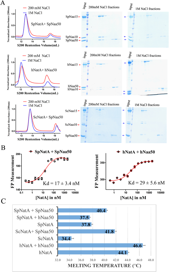
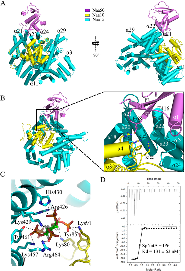
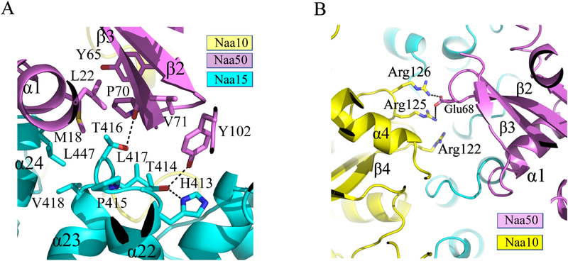
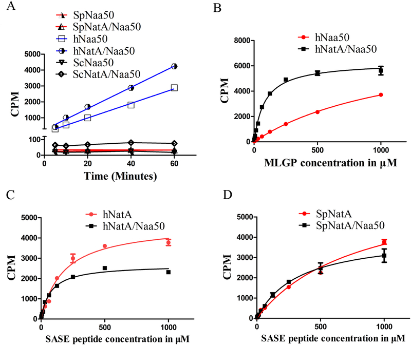
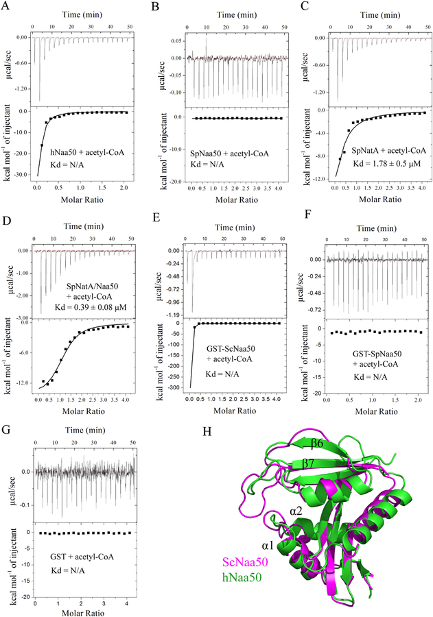
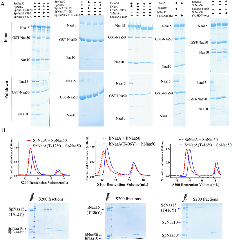
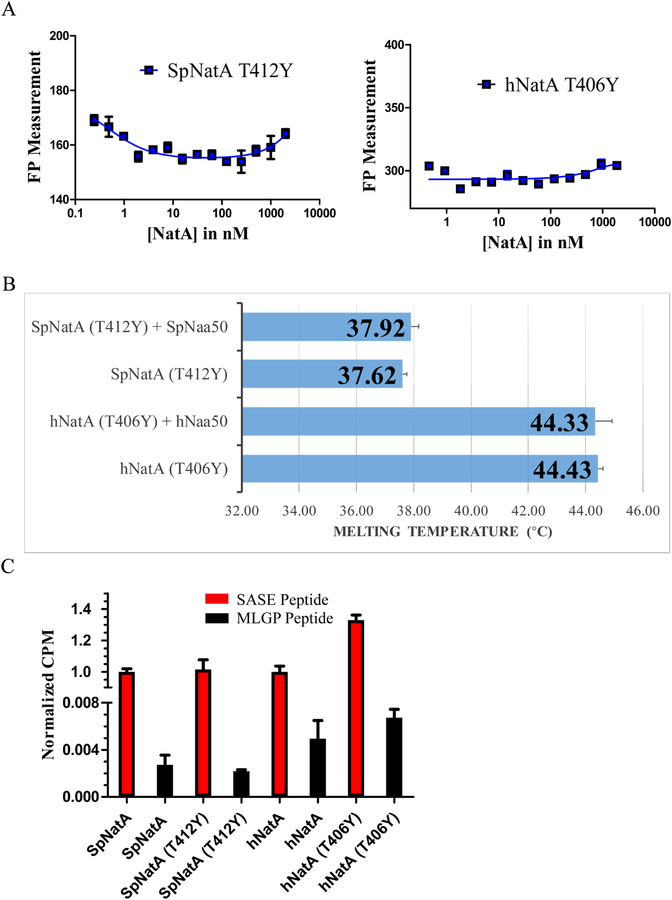
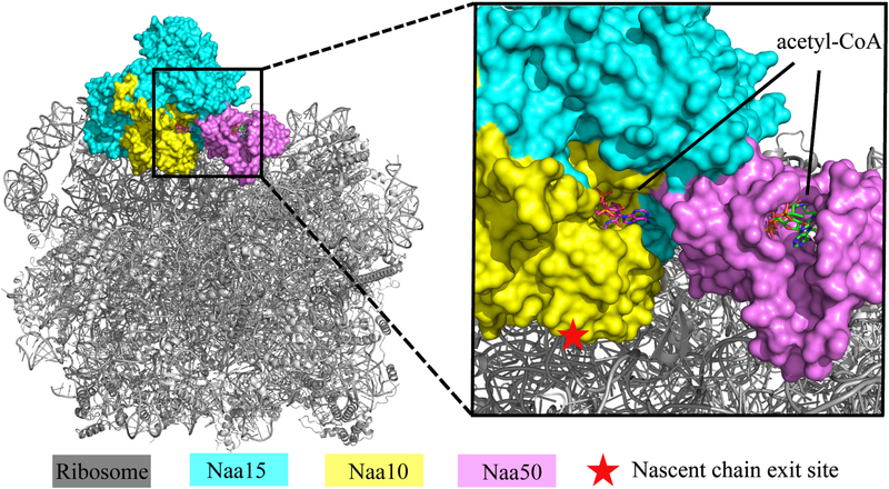
NatA co-translationally acetylates the N termini of over 40% of eukaryotic proteins and can associate with another catalytic subunit, Naa50, to form a ternary NatA/Naa50 dual enzyme complex (also called NatE). The molecular basis of association between Naa50 and NatA and the mechanism for how their association affects their catalytic activities in yeast and human are poorly understood. Here, we determined the X-ray crystal structure of yeast NatA/Naa50 as a scaffold to understand coregulation of NatA/Naa50 activity in both yeast and human. We find that Naa50 makes evolutionarily conserved contacts to both the Naa10 and Naa15 subunits of NatA. These interactions promote catalytic crosstalk within the human complex, but do so to a lesser extent in the yeast complex, where Naa50 activity is compromised. These studies have implications for understanding the role of the NatA/Naa50 complex in modulating the majority of the N-terminal acetylome in diverse species.

Keywords: N-terminal acetylation; Naa50; NatA; NatE; X-ray crystallography; protein complex.


Conflict of interest statement

DECLARATION OF INTERESTS

The authors declare no competing interest.

Figures

Figure 1.
Figure 1.. NatA and Naa50 form stable complexes in yeast and human.
(A) Gel filtration elution profiles of Naa50 with NatA in S. pombe (top),human (middle), and cross-species between yeast S. pombe and S. cerevisiae (bottom), using a Superdex S200 column, with either 200 mM or 1M NaCl in sizing buffer. Coomassie-stained SDS–PAGE of peak fractions are shown to the right of the chromatograms. (B) Fluorescence polarization assays with NatA titrated into fluorescein-5-maleimide labeled Naa50 in both S. pombe (left) and human (right) systems. The data is fit to calculate a dissociation constant between NatA and Naa50. (C) Differential scanning fluorimetry assays of NatA alone or with Naa50 in both yeast and human systems. Recorded melting temperature transitions are indicated.
Figure 2.
Figure 2.. Crystal structure of the ternary NatA/Naa50 complex shows that Naa50 contacts both subunits of NatA.
(A) ScNaa10 (yellow), ScNaa15(cyan), and ScNaa50 (violet) are shown in cartoon. The N-terminal two alpha helices (residues 1–53) of Naa15 are not resolved and not shown in the structure. Several alpha helices of Naa15 that contribute to Naa10 and Naa50 binding are labeled. (B) Zoom-in view of the interface between Naa50, Naa10 and Naa15. Residues that contribute to interactions between Naa10 and Naa50 are shown. (C) Zoom-in view showing key residues involved in interactions with IP6. (D) Representative isothermal titration calorimetry (ITC) of curve of IP6 titrated into SpNatA with the calculated dissociation constant indicated.
Figure 3.
Figure 3.. Hydrophobic interactions dominate the ScNatA-ScNaa50 binding interface.
(A) Zoom-in view of the major hydrophobic binding interface between Naa15 and Naa50 with residues that participate in interaction shown. (B) Zoom-in view of the contacts between Naa10 and Naa50 with residues that participate in interaction shown.
Figure 4.
Figure 4.. The NatA/Naa50 complex promotes catalytic crosstalk.
(A) Time course acetylation activity of SpNaa50, SpNatA/Naa50, hNaa50, hNatA/Naa50, ScNaa50 and ScNatA/Naa50 against the MLGP peptide substrate. (B) Michaelis–Menten kinetic curve of hNaa50 and hNatA/Naa50 against the MLGP peptide substrate. (C) Michaelis–Menten kinetic curve of hNatA and hNatA/Naa50 against the SASE peptide substrate. (D) Michaelis–Menten kinetic curve of SpNatA and SpNatA/Naa50 against the SASE peptide substrate.
Figure 5.
Figure 5.. Weak acetyl-CoA binding activity and a narrow substrate binding grove contributes to the catalytic inactivity of yeast Naa50.
(A) Representative ITC curve of acetyl-CoA titrated into hNaa50. (B) Representative ITC curve of acetyl-CoA titrated into SpNaa50. (C) Representative ITC curve of acetyl-CoA titrated into SpNatA. The calculated dissociation constant is indicated. (D) Representative ITC curve of acetyl-CoA titrated into SpNatA/Naa50. The calculated dissociation constant is indicated. (E) Representative ITC curve of acetyl-CoA titrated into GST-ScNaa50. (F) Representative ITC curve of acetyl-CoA titrated into GST-SpNaa50. (G) Representative ITC curve of acetyl-CoA titrated into free GST. (H) Superimposition of ScNaa50 and hNaa50 (PDB: 3TFY) structures.
Figure 6.
Figure 6.. NatA TY mutants are unable to pull-down Naa50 and co-migrate with Naa50.
(A) GST pull-down assay with NatA/Naa50 mutants to interrogate the contribution of residues in Naa50-NatA association. (B) Gel filtration elution profiles of Naa50 with either wild-type (dotted red line taken from Figure 1A) or TY mutants (blue line) of NatA using a Superdex S200 column. Coomassie-stained SDS–PAGE of peak fractions of Naa50 with NatA TY mutants shown below the chromatograms.
Figure 7.
Figure 7.. NatA TY mutants disrupt NatA/Naa50 complex interactions.
(A) Fluorescence polarization assays of NatA TY mutants and Naa50 in S. pombe and human. (B) Differential scanning fluorimetry assays of NatA TY mutants with and without Naa50. Recorded melting temperature transitions are indicated. (C) Comparison of acetylation activity of NatA wild-type and TY mutants. Activities are normalized to WT protein activity level.
Figure 8.
Figure 8.. Docking of our ScNatA/Naa50 crystal structure onto the ribosome-NatA/Naa50 Cryo-EM structure.
The ScNatA/Naa50 crystal structure is aligned to the NatA/Naa50-ribosome structure (PDB: 6HD7). The magnified view shows that Naa10 is most proximal to the ribosome peptide exit tunnel, while the acetyl-CoA binding sites of Naa10 and Naa50 are facing the same side.

Comment in

References

    1. ABUL KALAM S & SANCHITA B 2012. IP6 (Inositol Hexaphosphate) as a Signaling Molecule. Current Signal Transduction Therapy, 7, 289–304.
    1. ADAMS PD, AFONINE PV, BUNKOCZI G, CHEN VB, DAVIS IW, ECHOLS N, HEADD JJ, HUNG L-W, KAPRAL GJ,GROSSE-KUNSTLEVE RW, MCCOY AJ, MORIARTY NW, OEFFNER R, READ RJ, RICHARDSON DC, RICHARDSON JS, TERWILLIGER TC & ZWART PH 2010. PHENIX: a comprehensive Python-based system for macromolecular structure solution. Acta Crystallographica Section D, 66, 213–221. - PMC - PubMed
    1. AKSNES H, DRAZIC A, MARIE M & ARNESEN T 2016. First Things First: Vital Protein Marks by N-Terminal Acetyltransferases. Trends in Biochemical Sciences, 41, 746–760. - PubMed
    1. AKSNES H, GORIS M, STROMLAND O, DRAZIC A, WAHEED Q, REUTER N & ARNESEN T 2017. Molecular determinants of the N-terminal acetyltransferase Naa60 anchoring to the Golgi membrane. J Biol Chem, 292, 6821–6837. - PMC - PubMed
    1. AKSNES H, VAN DAMME P, GORIS M, STARHEIM KK, MARIE M, STOVE SI, HOEL C, KALVIK TV, HOLE K, GLOMNES N, FURNES C, LJOSTVEIT S, ZIEGLER M, NIERE M, GEVAERT K & ARNESEN T 2015. An organellar nalpha-acetyltransferase, naa60, acetylates cytosolic N termini of transmembrane proteins and maintains Golgi integrity. Cell Rep, 10, 1362–74. - PubMed

Publication types

MeSH terms

LinkOut - more resources