. 2019 Apr;31(2):223-258.
doi: 10.21147/j.issn.1000-9604.2019.02.01.

Chinese guidelines for diagnosis and treatment of esophageal carcinoma 2018 (English version)

Chinese guidelines for diagnosis and treatment of esophageal carcinoma 2018 (English version)

National Health Commission Of The People's Republic Of China. Chin J Cancer Res. 2019 Apr.
No abstract available


Figures

1
1
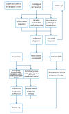
Standardized diagnosis and treatment process for esophageal cancer. EMR, endoscopic mucosal resection; ESD, endoscopic submucosal dissection.
2
2
Diagram of the Chinese version of naming and grouping of mediastinal lymph nodes in esophageal cancer: “C” represents Chinese nomenclature, and “2” represents thoracic lymph nodes. Station C201, right recurrent laryngeal nerve nodes; Station C202, left recurrent laryngeal nerve nodes; Station C203, upper thoracic paraesophageal lymph nodes; Station C204, right thoracic paratracheal lymph nodes; Station C205, subcarinal lymph nodes; Station C206, middle thoracic paraesophageal lymph nodes; Station C207, lower thoracic paraesophageal lymph nodes; Station C208, inferior pulmonary ligament lymph nodes; Station C209, paradiaphragmatic lymph nodes.
3
3
Areas of lymphadenectomy. (A) Incomplete two-field lymphadenectomy (light thoracic approach); (B) Total two-field lymphadenectomy (right thoracic approach); (C) Total three-field lymphadenectomy.
4
4
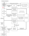
Flowchart of standardized diagnosis for esophagus cancer. CT, computed tomography; EMR, endoscopic mucosal resection; VATS, video-assisted thoracoscopic surgery; LN, lymph node.
5
5
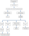
Flowchart of esophageal cancer screening. NBI, narrow band imaging; *, Biopsy pathological examination mainly aims at a certain point, but it has some limitations to reflect the whole picture of lesion. Endoscopic resection for diagnosis can be performed if necessary after careful assessment.
6
6
Endoscopic classification of early esophageal cancer (Paris classification, 2005).
7
7
Endoscopic mucosal resection with a cap (EMRC) procedure. (A) Rough and erosive esophageal mucosa is showed endoscopically. Biopsy result reveals high-grade dysplasia; (B) The lesion is dark brown under narrow band imaging (NBI) mode; (C) Positive iodine staining; (D) After marking; (E) After submucosa injection; (F) Endoscopic mucosal resection with transparent cap method; (G) Wound surface after resection; (H) Re-iodine staining after resection. Positive lesions are absent around artificial ulceration; (I) Resected specimen.
8
8
Multi-band mucosectomy (MBM) procedure. (A) Esophageal mucosal erosion is shown on white-light endoscopy; (B) Narrow band imaging (NBI) image of the lesion; (C) Positive iodine staining of the lesion; (D) The range of the lesion is marked; (E) The lesion is removed with a snare; (F) Wound surface after resection; (G) All of the resected specimens are pieced together into the probable shape.
9
9
Endoscopic submucosal dissection (ESD) procedure. (A) The white light endoscopy shows that the esophageal mucosa of the lesion was rough, and the dendrite vascular network disappeared; (B) White light magnifying endoscopy for intrapapillary capillary loops (IPCL) classification of lesion; (C) Narrow band imaging (NBI) was used to observe the IPCL classification of the lesion, which was IV type (Inoue Haruyo classification); (D) Positive iodine staining; (E) Endoscopic ultrasonography reveals that the lesions are mainly located in the mucosal layer; (F) Observe the boundaries of lesion carefully; (G) after marking; (H) A mucosal defect is made after submucosal injection; (I) Remove the lesion completely and examine the wound surface carefully; (J) The resected specimens.
10
10
Flowchart of endoscopic therapy process for early stage esophageal cancer. EP, epithelium; LPM, lamina propria mucosa; ESD, endoscopic submucosal dissection; EMR, endoscopic mucosal resection; MBM, multiband mucosectomy; RFA, radiofrequency ablation.
11
11
Radiofrequency ablation (RFA) procedure. (A) Esophageal squamous epithelium with high-grade dysplasia is about 4 cm long and nearly circumferential; (B) Narrow band imaging (NBI) image of the lesion; (C) Positive iodine staining; (D, E) The proximal and distal end of treatment range are marked; (E) RFA balloon is positioned at the position of proximal mark (6 o’clock position); (F) Appearance of mucosa after first RFA; (G) Appearance of mucosa after removal of the ablated lesion; (H, I) Appearance of proximal (H) and distal (I) mucosa after the second RFA.

References

LinkOut - more resources