. 2018 Nov;21(9):951-959.
doi: 10.1016/j.mattod.2018.04.001. Epub 2018 Apr 27.

Rapid continuous 3D printing of customizable peripheral nerve guidance conduits

Affiliations

Rapid continuous 3D printing of customizable peripheral nerve guidance conduits

Wei Zhu et al. Mater Today (Kidlington). 2018 Nov.

Abstract

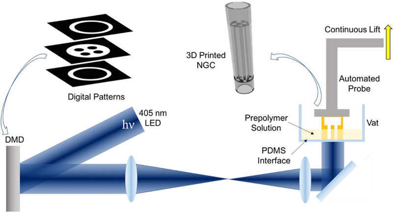
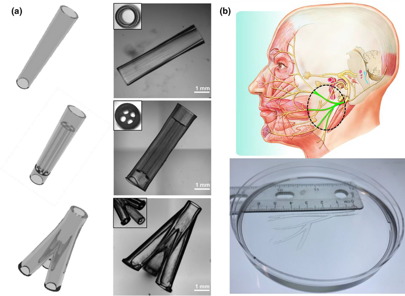
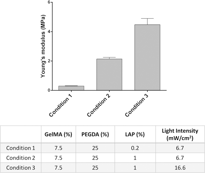
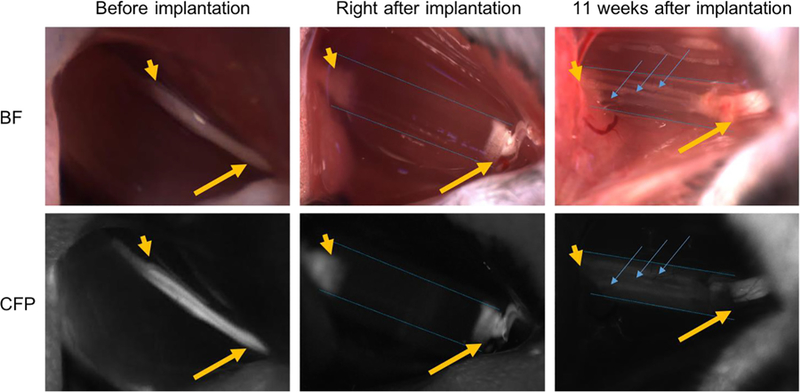
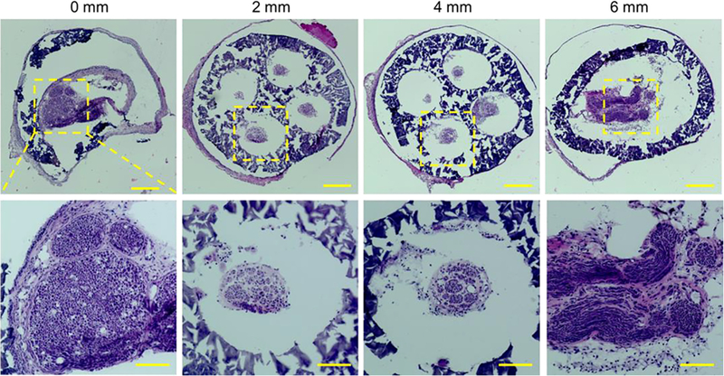
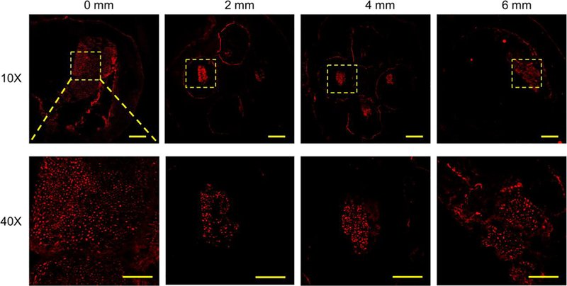
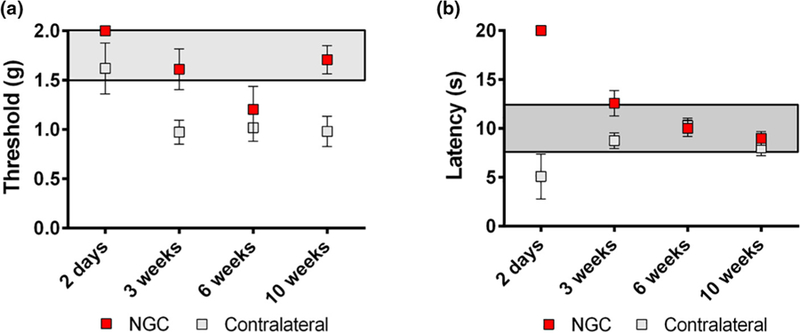
Engineered nerve guidance conduits (NGCs) have been demonstrated for repairing peripheral nerve injuries. However, there remains a need for an advanced biofabrication system to build NGCs with complex architectures, tunable material properties, and customizable geometrical control. Here, a rapid continuous 3D-printing platform was developed to print customizable NGCs with unprecedented resolution, speed, flexibility, and scalability. A variety of NGC designs varying in complexity and size were created including a life-size biomimetic branched human facial NGC. In vivo implantation of NGCs with microchannels into complete sciatic nerve transections of mouse models demonstrated the effective directional guidance of regenerating sciatic nerves via branching into the microchannels and extending toward the distal end of the injury site. Histological staining and immunostaining further confirmed the progressive directional nerve regeneration and branching behavior across the entire NGC length. Observational and functional tests, including the von Frey threshold test and thermal test, showed promising recovery of motor function and sensation in the ipsilateral limbs grafted with the 3D-printed NGCs.


Figures

FIGURE 1
FIGURE 1
Schematic of the rapid continuous 3D-printing system for printing customizable NGCs.
FIGURE 2
FIGURE 2
(a) Various NGC CAD designs (left column) and the corresponding 3D-printed NGCs (right column). (b) 3D-printed human life-size NGC; the facial nerve schematic on top was adapted from Atlas of Human Anatomy (p. 124, 6th Ed.), by F. H. Netter, 2014, Philadelphia, PA: Saunders. Copyright 2014 by Saunders, an imprint of Elsevier Inc. [27].
FIGURE 3
FIGURE 3
Young’s modulus of the 3D-printed constructs prepared with three different conditions.
FIGURE 4
FIGURE 4
Intraoperative photographs of NGCs (bright field/CFP fluorescence) at the surgical nerve repair site. Immediately before implantation, the sciatic nerve is exposed approximately 1.5 cm at its exit from the pelvis (yellow arrow: proximal segment; yellow arrowhead: distal segment). Following transection and implantation of the NGC (blue dotted outlines), the proximal and distal segments of the sciatic nerve is delicately inserted into the NGC and secured into position with tissue glue. 11 weeks after implantation, the proximal segment of the nerve can be seen to connect through the microchannels within the NGC (blue arrow). Note: All images were obtained at the same magnification. Apparent differences in nerve and NGC sizes at different time points are due to the surgical orientation of the animal at the time of surgery.
FIGURE 5
FIGURE 5
H&E staining of the sections of the regenerated sciatic nerve and the NGC with four microchannels at different locations from the proximal end to the distal end at 11 weeks post-implantation. Scale bars: 200 mm (top row), 100 mm (bottom row).
FIGURE 6
FIGURE 6
Confocal images of the sections of the regenerated sciatic nerve and the NGC with four microchannels at different locations from the proximal end to the distal end at 11 weeks post-implantation. SMI 312 was used to stain the neurofilaments. Scale bars: 200 mm (top row), 50 mm (bottom row).
FIGURE 7
FIGURE 7
Functional recovery evaluation by (a) von Frey threshold testing and (b) thermal testing.

References

    1. Kehoe S, Zhang XF, Boyd D, Injury 43 (2012) 553–572. - PubMed
    1. Ichihara S, Inada Y, Nakamura T, Injury 39 (2008) 29–39. - PubMed
    1. Dinis TM et al. , J. Mech. Behav. Biomed. Mater 41 (2015) 43–55. - PubMed
    1. Bellamkonda RV, Biomaterials 27 (2006) 3515–3518. - PubMed
    1. Kim YT et al. , Biomaterials 29 (2008) 3117–3127. - PMC - PubMed