. 2019 May 7:10:291.
doi: 10.3389/fpsyt.2019.00291. eCollection 2019.

The Effects of Transcranial Direct Current Stimulation on the Cognitive and Behavioral Changes After Electrode Implantation Surgery in Rats

Affiliations

The Effects of Transcranial Direct Current Stimulation on the Cognitive and Behavioral Changes After Electrode Implantation Surgery in Rats

Jooyoung Oh et al. Front Psychiatry. .

Abstract

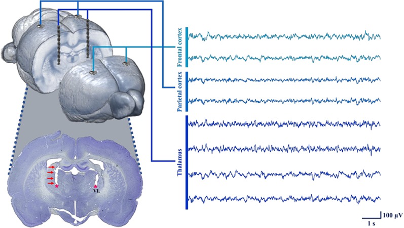
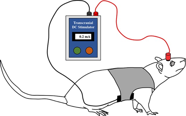
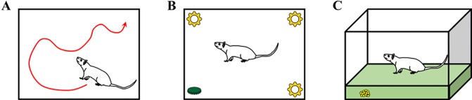
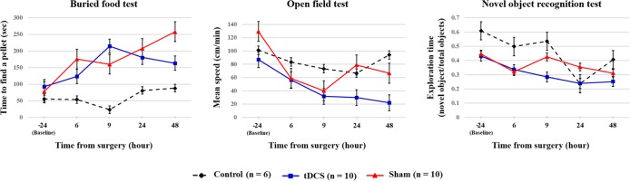
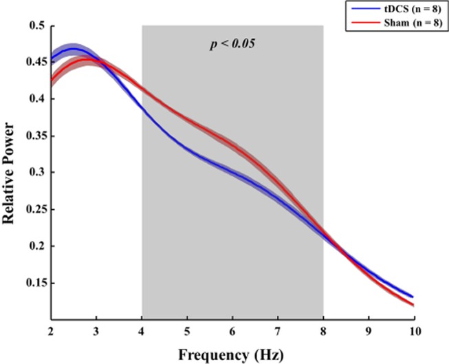
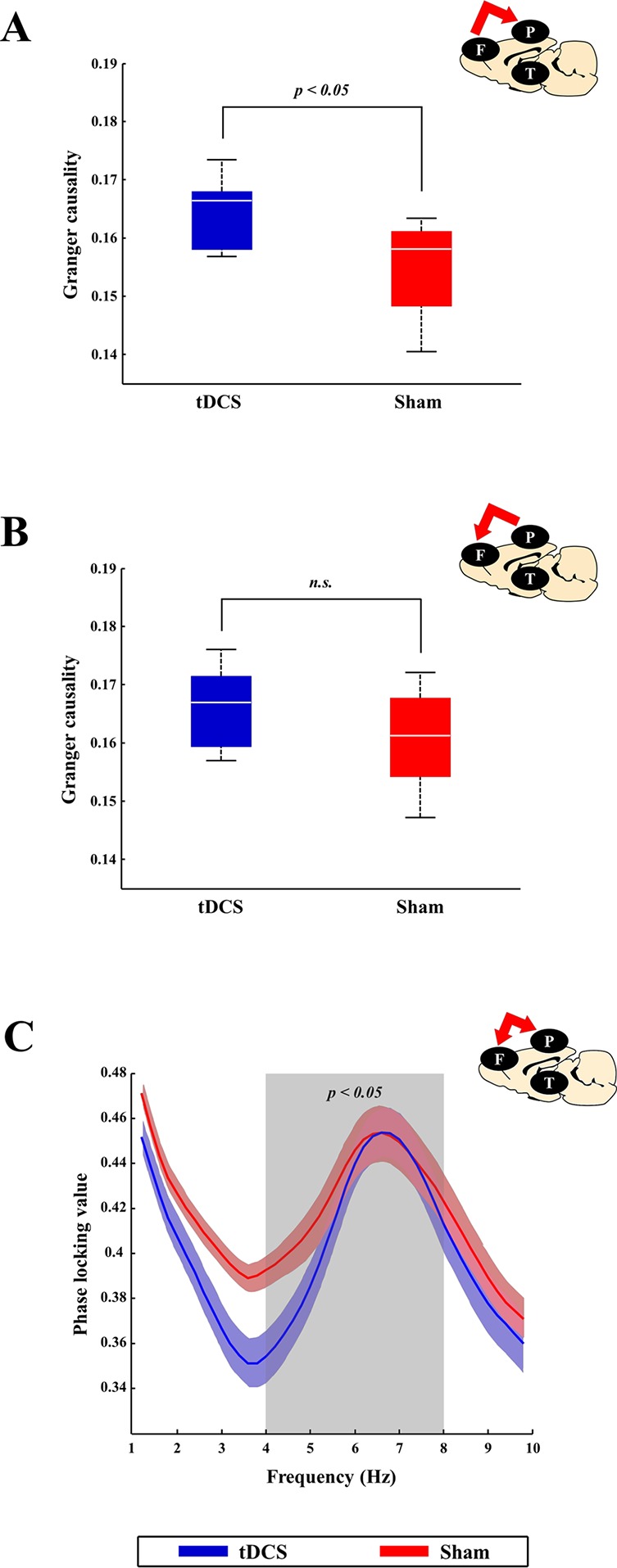
Postoperative delirium can lead to increased morbidity and mortality, and may even be a potentially life-threatening clinical syndrome. However, the neural mechanism underlying this condition has not been fully understood and there is little knowledge regarding potential preventive strategies. To date, investigation of transcranial direct current stimulation (tDCS) for the relief of symptoms caused by neuropsychiatric disorders and the enhancement of cognitive performance has led to promising results. In this study, we demonstrated that tDCS has a possible effect on the fast recovery from delirium in rats after microelectrode implant surgery, as demonstrated by postoperative behavior and neurophysiology compared with sham stimulation. This is the first study to describe the possible effects of tDCS for the fast recovery from delirium based on the study of both electroencephalography and behavioral changes. Postoperative rats showed decreased attention, which is the core symptom of delirium. However, anodal tDCS over the right frontal area immediately after surgery exhibited positive effects on acute attentional deficit. It was found that relative power of theta was lower in the tDCS group than in the sham group after surgery, suggesting that the decrease might be the underlying reason for the positive effects of tDCS. Connectivity analysis revealed that tDCS could modulate effective connectivity and synchronization of brain activity among different brain areas, including the frontal cortex, parietal cortex, and thalamus. It was concluded that anodal tDCS on the right frontal regions may have the potential to help patients recover quickly from delirium.

Keywords: brain stimulation; connectivity; delirium; electrophysiology; rat; transcranial direct current stimulation.


Figures

Figure 1
Figure 1
Flowchart of the whole experiment. Microelectrode implantation surgery was performed under isoflurane anesthesia. Immediately after surgery, transcranial direct current stimulation (tDCS) or sham stimulation was applied to the right frontal area for 20 min. All rats in the control and surgery groups underwent behavioral tests at every time point (before and after surgery). Local field potential (LFP) data were only acquired in the surgery group 6, 9, 24, and 48 h after surgery. The behavioral tests and LFP recording were sequentially performed in the order indicated in the flowchart. The white and gray boxes indicate the control and surgery groups, respectively. O = open field test, N = novel object recognition test, F = buried food test, and E = EEG measurement.
Figure 2
Figure 2
Process used for neural signal acquisition at each target point using the inserted electrodes. To measure LFPs, microelectrodes were implanted into the frontal and parietal cortices. In four animals (two from tDCS group, two from sham group), depth electrodes were also inserted in the thalamus. The depth electrodes successfully targeted the VL (ventrolateral nucleus of the thalamus), as shown in the histology image. All of the neural signals were reliably obtained under our recording conditions.
Figure 3
Figure 3
Illustration of the anodal tDCS stimulation in the rats. tDCS (0.2 mA for 20 min) or sham stimulation was applied to the rats in each group immediately after surgery. The anodal (active) electrode was placed over the right frontal area, and the cathodal (reference) electrode was placed on the ventral torso. The active electrode was filled with conductive gel, and the reference electrode was fixed using a saline-soaked sponge pad and corset.
Figure 4
Figure 4
Descriptions of the three behavioral tests: (A) open field test, (B) novel object recognition test, and (C) buried food test. Rats in the control group (without surgery and stimulation), sham group (with surgery and sham stimulation), and tDCS group (with surgery and stimulation) underwent three different behavioral tests. The tests were performed at all time points (24 h before surgery and 6, 9, 24, and 48 h after surgery). Some of the rats (n = 10, 6 from the control group and 4 from the surgery group with depth electrodes) underwent behavioral testing at an additional time point (1 week after surgery).
Figure 5
Figure 5
Summary of the behavioral test results of each group. The black dotted, blue, and red lines indicate control group, tDCS group, and sham group, respectively. The values are mean and standard errors.
Figure 6
Figure 6
Comparison of the relative power between the tDCS and sham groups. Forty-eight hours after surgery, rats in the tDCS group (blue line) exhibited lower relative theta power in the right frontal lobe than those in the sham group (red line) (p = 0.023). The bold line indicates the mean value, and the shaded bands show the standard error. Mann–Whitney test was utilized.
Figure 7
Figure 7
Comparison of connectivity between the tDCS and sham groups at the cortex level. At the cortex level, brain connectivity in the tDCS group tended to increase bottom-up attention (which is known to be related to gamma frequency band), which, in turn, led to recovery of awareness. tDCS stimulation helped increase bottom-up attention by enhancing gamma band effective connectivity between the frontal and parietal lobes. (A and B) The blue (tDCS group) and red (sham group) boxes represent the spectral GC values between the frontal and parietal lobes 24 h after surgery in the gamma frequency band. Effective gamma band connectivity between the frontal and parietal lobes seemed to be higher in the tDCS group than in the sham group, although only the causal relationship from the frontal cortex to the parietal cortex has a statistically significant difference (p = 0.047). (C) The blue (tDCS group) and red (sham group) lines indicated the phase locking values (PLVs) between the frontal and parietal lobes 24 h after surgery in the theta frequency band (which is known to be related to top-down attention). Rats in the sham group displayed more synchronization than those in the tDCS group (p = 0.016). The bold line indicates the mean value, and shaded bands show the standard error. Mann–Whitney test was utilized in all analyses.

Similar articles

Cited by

References

    1. McGuire BE, Basten CJ, Ryan CJ, Gallagher J. Intensive care unit syndrome. Arch Intern Med (2000) 160:906. 10.1001/archinte.160.7.906 - DOI - PubMed
    1. Fong TG, Tulebaev SR, Inouye SK. Delirium in elderly adults: diagnosis, prevention and treatment. Nat Rev Neurol (2009) 5:210–20. 10.1038/nrneurol.2009.24 - DOI - PMC - PubMed
    1. Girard TD, Pandharipande PP, Ely EW. Delirium in the intensive care unit. Crit Care (2008) 12:S3. 10.1186/cc6149 - DOI - PMC - PubMed
    1. Francis J, Martin D, Kapoor WN. A prospective study of delirium in hospitalized elderly. JAMA J Am Med Assoc (1990) 263:1097. 10.1001/jama.1990.03440080075027 - DOI - PubMed
    1. Marcantonio ER, Goldman L, Mangione CM, Ludwig LE, Muraca B, Haslauer CM, et al. A clinical prediction rule for delirium after elective noncardiac surgery. JAMA J Am Med Assoc (1994) 271:134. 10.1001/jama.1994.03510260066030 - DOI - PubMed

LinkOut - more resources