Even non-experts identify non-dysplastic lesions in inflammatory bowel disease via chromoendoscopy: results of a screening program in real-life
- PMID: 31157291
- PMCID: PMC6524993
- DOI: 10.1055/a-0839-4514
Even non-experts identify non-dysplastic lesions in inflammatory bowel disease via chromoendoscopy: results of a screening program in real-life
Abstract
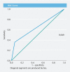
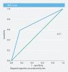
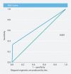
Background and study aims Chromoendoscopy with targeted biopsy is the technique of choice for colorectal cancer screening in longstanding inflammatory bowel disease. We aimed to analyze results of a chromoendoscopy screening program and to assess the possibility of identifying low-risk dysplastic lesions by their endoscopic appearance in order to avoid histological analysis. Materials and methods We retrospectively reviewed chromoendoscopies performed between February 2011 and June 2017 in seven Spanish hospitals in a standardized fashion. We analyzed the findings and the diagnostic yield of the Kudo pit pattern for predicting dysplasia. Results A total of 709 chromoendoscopies (569 patients) were reviewed. Median duration of disease was 16.7 years (SD 8.1); 80.4 % had ulcerative colitis. A total of 2025 lesions (3.56 lesions per patient) were found; two hundred and thirty-two lesions were neoplastic (11.5 %) (223 were LGD (96.1 %), eight were HGD (3.4 %), and one was colorectal cancer (0.5 %). The correlation between dysplasia and Kudo pit patterns predictors of dysplasia (≥ III) was low, with an area under the curve of 0.649. Kudo I and II lesions were correctly identified with a high negative predictive value (92 %), even by non-experts. Endoscopic activity, Paris 0-Is classification, and right colon localization were risk factors for dysplasia detection, while rectum or sigmoid localization were protective against dysplasia. Conclusions Chromoendoscopy in the real-life setting detected 11 % of dysplastic lesions with a low correlation with Kudo pit pattern. A high negative predictive value would prevent Kudo I and, probably, Kudo II biopsies in the left colon, reducing procedure time and avoiding complications.
Conflict of interest statement
Figures


References
-
- Jess T, Gamborg M, Matzen P et al.Increased risk of intestinal cancer in Crohn’s disease: a meta-analysis of population-based cohort studies. Am J Gastroenterol. 2005;100:2724–2729. - PubMed
-
- Castaño-Milla C, Chaparro M, Gisbert J P. Systematic review with meta-analysis: the declining risk of colorectal cancer in ulcerative colitis. Aliment Pharmacol Ther. 2014;39:645–659. - PubMed
-
- Magro F, Gionchetti P, Eliakim R et al.European Crohn’s and Colitis Organisation. Third European Evidence-based Consensus on Diagnosis and Management of Ulcerative Colitis. Part 1: Definitions, Diagnosis, Extra-intestinal Manifestations, Pregnancy, Cancer Surveillance, Surgery, and Ileo-anal Pouch Disorders. J Crohn Colitis. 2017;11:649–670. - PubMed
-
- Loftus E V., JrDoes monitoring prevent cancer in inflammatory bowel disease? J Clin Gastroenterol 20033605S79–83.; discussion S94-96 - PubMed
-
- Collins P D, Mpofu C, Watson A J et al.Strategies for detecting colon cancer and/or dysplasia in patients with inflammatory bowel disease. Cochrane Database Syst Rev. 2006:CD000279. - PubMed
LinkOut - more resources
Full Text Sources

