. 2019 May 15:(147):10.3791/59219.
doi: 10.3791/59219.

Using the Open-Source MALDI TOF-MS IDBac Pipeline for Analysis of Microbial Protein and Specialized Metabolite Data

Affiliations

Using the Open-Source MALDI TOF-MS IDBac Pipeline for Analysis of Microbial Protein and Specialized Metabolite Data

Chase M Clark et al. J Vis Exp. .

Abstract

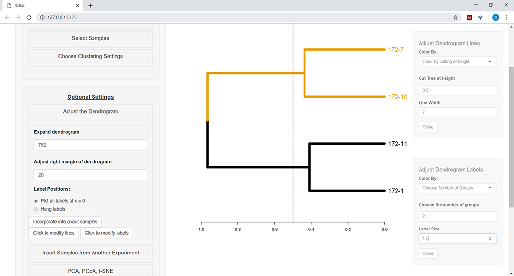
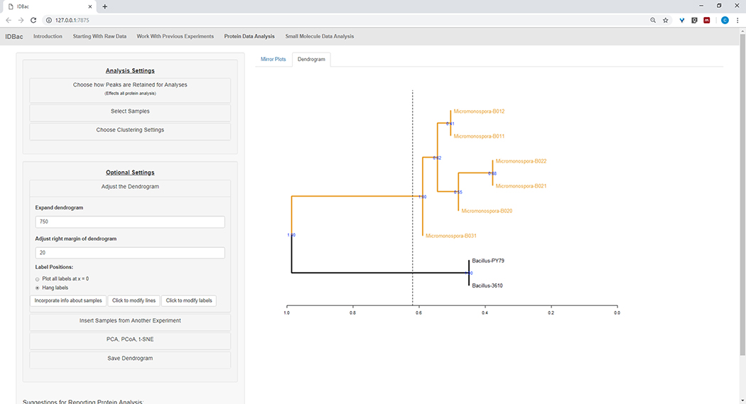
In order to visualize the relationship between bacterial phylogeny and specialized metabolite production of bacterial colonies growing on nutrient agar, we developed IDBac-a low-cost and high-throughput matrix-assisted laser desorption/ionization time-of-flight mass spectrometry (MALDI-TOF MS) bioinformatics pipeline. IDBac software is designed for non-experts, is freely available, and capable of analyzing a few to thousands of bacterial colonies. Here, we present procedures for the preparation of bacterial colonies for MALDI-TOF MS analysis, MS instrument operation, and data processing and visualization in IDBac. In particular, we instruct users how to cluster bacteria into dendrograms based on protein MS fingerprints and interactively create Metabolite Association Networks (MANs) from specialized metabolite data.


Figures

Figure 1:
Figure 1:. MALDI-target plate showing two different isolates before adding formic acid and MALDI matrix (top 3 spots – Bacillus sp.; bottom 3 spots – Streptomyces sp.).
For both, column 3 represents excess sample; column 2 represents the appropriate amount of sample; column 1 represents insufficient sample for MALDI analysis.
Figure 2:
Figure 2:. Example protein spectra displaying the effect of modifying laser power and detector gain.
Spectra quality is best in panel A, and decreases until insufficient spectra quality in panels C and D. While the spectrum in panel B may result in useable peaks, panel A displays optimal data.
Figure 3:
Figure 3:. Example specialized metabolite spectra displaying the effect of modifying laser power and detector gain.
Spectra quality is best in panel A and decreases until insufficient spectra quality in panels C and D. While the spectrum in panel B may result in useable peaks, panel A displays optimal data.
Figure 4:
Figure 4:. IDBac data conversion and preprocessing step.
IDBac converts raw spectra into the open mzML format and stores mzML, peak lists, and sample information in a database for each experiment.
Figure 5:
Figure 5:. “Work with Previous Experiments” page.
Use IDBac’s “Work with Previous Experiments” page to select an experiment to analyze or modify.
Figure 6:
Figure 6:. Input sample information.
Within the “Work with Previous Experiments” page users can input information about samples such as taxonomic identity, collection location, isolation conditions, etc.
Figure 7:
Figure 7:. Transfer data.
The “Work with Previous Experiments” page contains the option to transfer data between existing experiments and to new experiments.
Figure 8:
Figure 8:. Choose how peaks are retained for analysis.
After selecting an experiment to analyze, visiting the “Protein Data Analysis” page and subsequently opening the “Choose how Peaks are Retained for Analysis” menu allows users to choose settings like signal-to-noise ratio for retaining peaks. The displayed mirror plot (or dendrogram) will automatically update to reflect the chosen settings.
Figure 9:
Figure 9:
Select samples from the chosen experiment to include within the displayed dendrogram.
Figure 10:
Figure 10:. Adjust the dendrogram.
IDBac provides a few options for modifying how the dendrogram looks, these may be found within the menu “Adjust the Dendrogram”. This includes coloring branches and labels by k-means, or by “cutting” the dendrogram at a user-provided height.
Figure 11:
Figure 11:. Incorporate info about samples.
Within the “Adjust the Dendrogram” menu is the option “Incorporate info about samples”. Selecting this will allow plotting information about samples next to the dendrogram. Sample information is input within the “Work with Previous Experiments” page.
Figure 12:
Figure 12:. Insert Samples from Another Experiment menu.
Sometimes it is helpful to compare samples from another experiment. Use the “Insert Samples from Another Experiment” menu to choose samples to include within the currently-displayed dendrogram.
Figure 13:
Figure 13:. Small Molecule Data Analysis” page.
If a dendrogram was created from protein spectra, it will be displayed within the “Small Molecule Data Analysis” page. This page will also display Metabolite Associate Networks (MANs) and Principle Components Analysis (PCA) for small molecule data.
Figure 14:
Figure 14:. Spectra processing.
Downloaded Bruker autoFlex spectra were converted and processed using IDBac.
Figure 15:
Figure 15:. Combined IDBac experiment.
Because the Micromonospora and Bacillus spectra were collected on different MALDI target plates, the two experiments were subsequently combined into a single experiment-“Bacillus_Micromonsopora”. This was done within the “Work with Previous Experiments” tab, following directions within the menu “Transfer samples from previous experiments to new/other experiments”.
Figure 16:
Figure 16:. Comparison.
Micromonspora and Bacillus spectra were compared using the mirror plots within the “Protein Data Analysis” page. Ultimately, default peak settings were chosen.
Figure 17:
Figure 17:. Hierarchical clustering.
Hierarchical clustering, using default settings, correctly grouped Bacillus and Micromonospora isolates. The dendrogram was colored by “cutting” the dendrogram at an arbitrary height (displayed as a dashed-line) and 100 bootstraps used to show confidence in branching.
Figure 18:
Figure 18:
MAN created by selecting the Bacillus sp. strains from the protein dendrogram showed differential production of specialized metabolites.
Figure 19:
Figure 19:
MAN created by selecting the six Micromonospora sp. strains from the protein dendrogram showed differential production of specialized metabolites.
Figure 20:
Figure 20:
MAN of Bacillus sp. and Micromonospora sp. strains showing a differential production of specialized metabolites.

References

    1. Sandrin TR, Goldstein JE, Schumaker S MALDI TOF MS profiling of bacteria at the strain level: A review. Mass Spectrometry Reviews 32, (3) 188–217 (2013). - PubMed
    1. Cain TC, Lubman DM, Weber WJ, Vertes A Differentiation of bacteria using protein profiles from matrix-assisted laser desorption/ionization time-of-flight mass spectrometry. Rapid Communications in Mass Spectrometry 8, (12) 1026–1030 (1994).
    1. Holland RD, Wilkes JG, et al. Rapid identification of intact whole bacteria based on spectral patterns using matrix-assisted laser desorption/ionization with time-of-flight mass spectrometry. Rapid Communications in Mass Spectrometry 10, (10) 1227–1232 (1996). - PubMed
    1. Rahi P, Prakash O, Shouche YS Matrix-assisted laser desorption/ionization time-of-flight mass-spectrometry (MALDI-TOF MS) based microbial identifications: challenges and scopes for microbial ecologists. Frontiers in Microbiology 7, 1359 (2016). - PMC - PubMed
    1. Silva R, Lopes NP, Silva DB Application of MALDI mass spectrometry in natural products analysis. Planta Medica 82, 671–689 (2016). - PubMed

Publication types

MeSH terms

Substances